Review
. 2018 Jan 14:2018:8917804.
doi: 10.1155/2018/8917804. eCollection 2018.

Macrophage Polarization in Chronic Inflammatory Diseases: Killers or Builders?

Affiliations
Review

Macrophage Polarization in Chronic Inflammatory Diseases: Killers or Builders?

Luca Parisi et al. J Immunol Res. .

Abstract

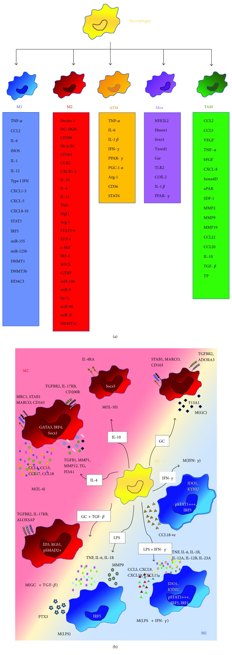
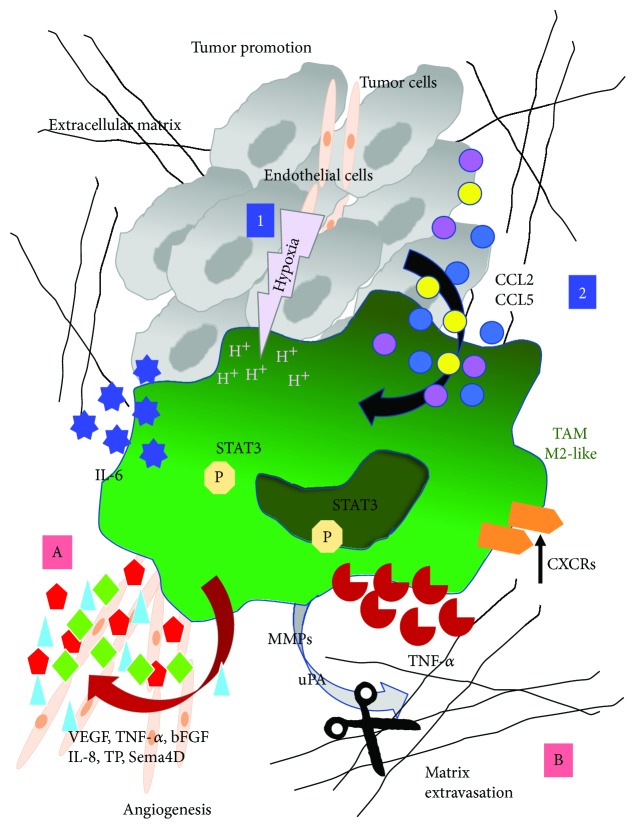
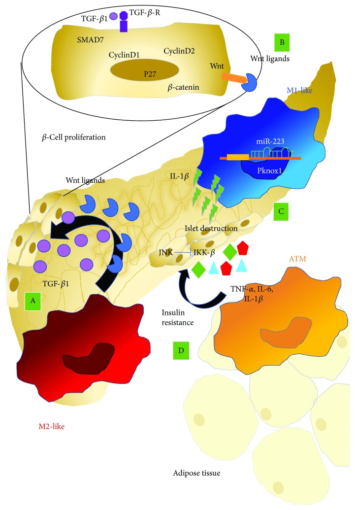
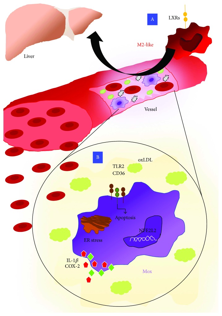
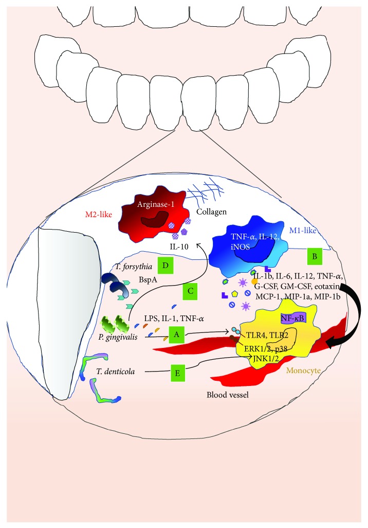
Macrophages are key cellular components of the innate immunity, acting as the main player in the first-line defence against the pathogens and modulating homeostatic and inflammatory responses. Plasticity is a major feature of macrophages resulting in extreme heterogeneity both in normal and in pathological conditions. Macrophages are not homogenous, and they are generally categorized into two broad but distinct subsets as either classically activated (M1) or alternatively activated (M2). However, macrophages represent a continuum of highly plastic effector cells, resembling a spectrum of diverse phenotype states. Induction of specific macrophage functions is closely related to the surrounding environment that acts as a relevant orchestrator of macrophage functions. This phenomenon, termed polarization, results from cell/cell, cell/molecule interaction, governing macrophage functionality within the hosting tissues. Here, we summarized relevant cellular and molecular mechanisms driving macrophage polarization in "distant" pathological conditions, such as cancer, type 2 diabetes, atherosclerosis, and periodontitis that share macrophage-driven inflammation as a key feature, playing their dual role as killers (M1-like) and/or builders (M2-like). We also dissect the physio/pathological consequences related to macrophage polarization within selected chronic inflammatory diseases, placing polarized macrophages as a relevant hallmark, putative biomarkers, and possible target for prevention/therapy.


Figures

Figure 1
Figure 1
Past and new concept in macrophage polarization. (a) Schematic overview of the different stimuli that can induce the diverse macrophage polarization state. M1: classically activated phenotype; M2: alternatively activated macrophages; ATM: adipose tissue-derived macrophages; Mox: atherosclerosis-associated macrophages; TAMs: tumour-associated macrophages. (b) The polarization landscape of macrophages. According to the different stimulation conditions, macrophages can acquire peculiar M1 or M2 phenotype, governed by the different surface antigen expressions, including scavenger receptors, chemokine, matrix-associated protein and cytokine release, and different patterns of transcription factors and metabolic pathway activated. The driver stimuli include IL-4, IL-10, glucocorticoids (GC) with TGF-β, glucocorticoids alone, LPS, LPS and IFN-γ, and IFN-γ alone.
Figure 2
Figure 2
Macrophage polarization in tumour progression. Macrophage recruitment in tumours and their polarization are regulated by several factors. Among all, hypoxia can induce the differentiation of monocytic myeloid-derived suppressor cells (M-MDSCs) via upregulation of CD45 tyrosine phosphatase activity (1). Further, soluble factors, such as CCL2 and CCL5 that are produced by the cancer cells and stroma cells, can increase macrophage infiltrate (2). In the TME, infiltrating associated to tumours (TAM/M2-like macrophages) can orchestrate tumour progression by several mechanisms including the release of cytokine, chemokines, and tissue remodelling proteins. Hypoxia increases the expression of CXCRs in TAMs and promotes tumour angiogenesis by enhancing the production of VEGF, TNF-α, bFGF, IL-8, TP, and Sema4D that can induce endothelial cell proliferation, sprouting and migration, tube formation, and maturation of new vessel, followed by its stabilization by attaching mural cells (A). TAMs can regulate the extracellular matrix degradation by producing different types of enzymes and proteases, such as matrix metalloproteinases (MMPs), in particular MMP2, MMP9, plasmin, urokinase plasminogen activator (uPA) and cathepsins acting on connective tissue surrounding the tumour, and allow tumour cells to detach from the mass of origin and to disseminate, leading to the formation of distant metastases (B).
Figure 3
Figure 3
Macrophage polarization in type 2 diabetes. Macrophage within pancreatic tissues can be switched toward different functionalities according to the environment stimuli. M2-like macrophage supports B-cell proliferation by several trophic factors like TGF-β1 which directly induce upregulation of SMAD7 and increases of cyclinD1, cyclinD2, and p27 (A). Moreover, M2-like macrophages release Wnt ligands, thus activating the Wnt signaling pathway, and β-catenin, supporting β-cell replication (B). M1-like macrophage in pancreatic tissues can secrete IL-1b, inhibiting insulin secretion, followed by islet destruction (C). Adipose-derived macrophages (ATM) can release proinflammatory cytokines, including TNF-α, IL-6, and IL-1β that decrease insulin sensitivity through the activation of Jun N-terminal kinase (JNK), inhibitor of IKκB kinase (IKK-β), and other serine kinases in insulin target cells (D).
Figure 4
Figure 4
Macrophage polarization in atherosclerosis. Macrophages are crucial players involved in the atherosclerosis development due to their ability to regulate cholesterol efflux. In this context, the upregulation of LXRs in M2 macrophages has been found to exert a protective role. Indeed, LRXs reduce peripheral tissue excess cholesterol that is returned to the liver by releasing HDL in the plasma (A). Apart from M1 and M2 polarization, a third macrophage state has been described in the atherosclerosis context that is termed Mox. Macrophages exposed to oxidized phospholipids display reduced phagocytic and chemotactic abilities compared with M1- and M2-like macrophages and are characterized by the expression of the transcription factor NFE2L2 as far as Hmox1, Srxn1, Txnrd1, and Gsr genes. Mox macrophages also activate TLR2­dependent mechanisms in response to oxidized lipids leading to an increase of IL­1β and COX-2 (B).
Figure 5
Figure 5
Macrophage polarization in periodontitis. Macrophages that have been found in the gingival epithelium can be activated by several microorganisms able to induce macrophage polarization toward M1- or M2-like phenotype. P. gingivalis releases LPS, IL-1, and TNF-α that promote the proinflammatory M1 macrophage polarization (A). Moreover, Pg infection enhances the secretion of IL-1β, IL-6, IL-12, TNF-α, G-CSF, GM-CSF, and the chemokines eotaxin, MCP1, MIP-1α, and MIP-1β from macrophages, reflecting a M1-like proinflammatory response (B). In spite of this, it has also been reported that Pg infection can also be associated with the increase of IL10, supporting M2 macrophage and increasing arginase-1 production and collagen deposition, leading to periodontitis (C). T. forsythia releases BspA and other ligands that induce TLR2 signaling favouring the development of Th2-type inflammatory responses (D). T. denticola induces TLR2 signaling that stimulates the prolonged activation of both ERK1/2 p38 and JNK1/2 in monocytes (E).

References

    1. Geissmann F., Manz M. G., Jung S., Sieweke M. H., Merad M., Ley K. Development of monocytes, macrophages, and dendritic cells. Science. 2010;327(5966):656–661. doi: 10.1126/science.1178331. - DOI - PMC - PubMed
    1. Ginhoux F., Greter M., Leboeuf M., et al. Fate mapping analysis reveals that adult microglia derive from primitive macrophages. Science. 2010;330(6005):841–845. doi: 10.1126/science.1194637. - DOI - PMC - PubMed
    1. Gordon S., Taylor P. R. Monocyte and macrophage heterogeneity. Nature Reviews Immunology. 2005;5(12):953–964. doi: 10.1038/nri1733. - DOI - PubMed
    1. Davies L. C., Jenkins S. J., Allen J. E., Taylor P. R. Tissue-resident macrophages. Nature Immunology. 2013;14(10):986–995. doi: 10.1038/ni.2705. - DOI - PMC - PubMed
    1. Dey A., Allen J., Hankey-Giblin P. A. Ontogeny and polarization of macrophages in inflammation: blood monocytes versus tissue macrophages. Frontiers in Immunology. 2014;5:p. 683. doi: 10.3389/fimmu.2014.00683. - DOI - PMC - PubMed