. 2018 Feb 21;4(2):e1701729.
doi: 10.1126/sciadv.1701729. eCollection 2018 Feb.

Allele-specific long-distance regulation dictates IL-32 isoform switching and mediates susceptibility to HIV-1

Affiliations

Allele-specific long-distance regulation dictates IL-32 isoform switching and mediates susceptibility to HIV-1

Robert-Jan Palstra et al. Sci Adv. .

Abstract

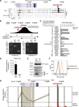
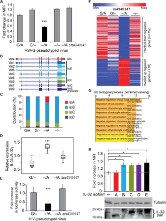
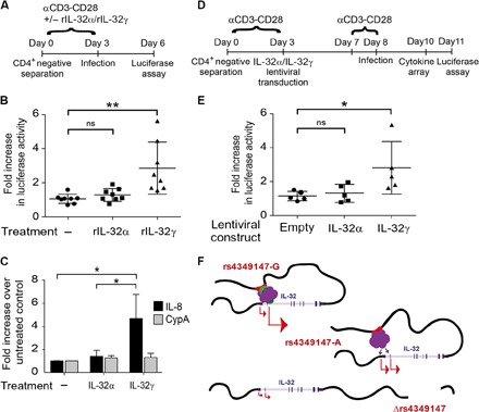
We integrated data obtained from HIV-1 genome-wide association studies with T cell-derived epigenome data and found that the noncoding intergenic variant rs4349147, which is statistically associated with HIV-1 acquisition, is located in a CD4+ T cell-specific deoxyribonuclease I hypersensitive region, suggesting regulatory potential for this variant. Deletion of the rs4349147 element in Jurkat cells strongly reduced expression of interleukin-32 (IL-32), approximately 10-kb upstream, and chromosome conformation capture assays identified a chromatin loop between rs4349147 and the IL-32 promoter validating its function as a long-distance enhancer. We generated single rs4349147-A or rs4349147-G allele clones and demonstrated that IL-32 enhancer activity and interaction with the IL-32 promoter are strongly allele dependent; rs4349147 -/A cells display reduced IL-32 expression and altered chromatin conformation as compared to rs4349147 G/- cells. Moreover, RNA sequencing demonstrated that rs4349147 G/- cells express a lower relative ratio of IL-32α to non-α isoforms than rs4349147 -/A cells and display increased expression of lymphocyte activation factors rendering them more prone to infection with HIV-1. In agreement, in primary CD4+ T cells, both treatment with recombinant IL-32γ (rIL-32γ) but not rIL-32α, and exogenous lentiviral overexpression of IL-32γ or IL-32β but not IL-32α resulted in a proinflammatory T cell cytokine environment concomitant with increased susceptibility to HIV infection. Our data demonstrate that rs4349147-G promotes transcription of non-IL-32α isoforms, generating a proinflammatory environment more conducive to HIV infection. This study provides a mechanistic link between a HIV-associated noncoding DNA variant and the expression of different IL-32 isoforms that display discrete anti-HIV properties.


Figures

Fig. 1
Fig. 1. DNA region encompassing rs4349147 is a strong enhancer element for the IL-32 gene in T cells.
(A) UCSC genome browser tracks show that the HIV-associated variant rs4349147 is located in a T cell–specific DHS region. Each track represents the DNase I accessibility profile for a given cell line. Promoters as defined by the Eukaryotic Promoter Database (EPD; http://epd.vital-it.ch) are indicated as red bars. HUVEC, human umbilical cord endothelial cell. (B) Deletion of rs4349147 DHS. Location of small-guiding RNAs (sgRNAs) used in the CRISPR-Cas9 KO strategy is indicated. Panels show results from two different PCR assays generating amplicons 1 or 2, as indicated, that confirm homozygous deletion of rs4349147 DHS. (C) RNA expression levels of genes in a 500-kb window surrounding rs4349147 demonstrate a strong reduction in IL-32 expression. For each gene considered in the analysis, right panel shows total number of reads, whereas left panel shows fold change in expression in rs4349147 DHS KO cells compared to WT. N/A, not available. (D to F) Strong reduction of IL-32 expression is confirmed using real-time quantitative PCR (qPCR) (D), Western blotting using an antibody against IL-32 (E), and intracellular staining with biotin-conjugated antibody against IL-32, followed by fluorescence-activated cell sorting (FACS) analysis (F). (G) 3C analysis of the IL-32 locus demonstrates chromatin loop formation between rs4349147 and the IL-32 promoter region in Jurkat and SupT1 cells. The analyzed region of the human IL-32 locus is depicted on the top of each graph. X axis shows the approximate position on chromosome 16 (Chr 16) (UCSC genome browser GRCh37/hg19 assembly). Dark gray shading shows the position and size of the “fixed” Dpn II restriction fragment. Light gray shading indicates position and size of other Dpn II restriction fragments analyzed. The Dpn II restriction fragment containing the IL-32 promoter is indicated in a slightly darker gray color.
Fig. 2
Fig. 2. Activity of the rs4349147 enhancer element is dependent on the haplotype.
(A) Schematic representation of the region encompassing the rs4786376-G/C and rs4349147-G/A SNPs, indicating the size of the PCR products obtained using primers outside (amplicon 1) or inside (amplicon 2) the region targeted for KO. Amplicon 2 contains three Nla III restriction sites (asterisks). The central NIa III site (blue asterisk) includes the rs4786376-G/C SNP, which is in perfect linkage with rs4349147-G/A and can be used to genotype the heterozygous lines. (B) Simultaneous presence of two PCR products of different sizes indicates the occurrence of heterozygous rs4349147 KO lines. (C) Results of restriction reaction of amplicon 2 in WT (heterozygous rs4349147-G/A), rs4349147 −/A, G/−, or rs4349147 enhancer KO Jurkat lines. (D) Expression of IL-32 mRNA in the four lines was measured by real-time PCR. Cells homozygous for rs4349147-A express lower IL-32 RNA levels as compared to those homozygous for rs4349147-G. (E) IL-32 protein levels were determined by Western blotting using an antibody against IL-32. (F) Intracellular staining with biotin-conjugated antibody against IL-32, followed by FACS analysis, shows lower levels of IL-32 in rs4349147 −/A Jurkat cells and rs4349147 KO Jurkat cells as compared to WT and rs4349147 G/− Jurkat cells (Fig. 1F). (G) 3C analysis of the IL-32 locus demonstrates chromatin loop formation between rs4349147-G and the IL-32 promoter region in rs4349147 G/− Jurkat cells and a significant reduction in enhancer-promoter interaction in the rs4349147 −/A Jurkat cells. Ratio of interaction in rs4349147 −/A and G/− Jurkat cells is indicated. For further explanation of the figure, see Fig. 1G. (H) Activity of 476-bp enhancer fragments with different rs4349147 and rs4786376 genotypes in an IL-32 promoter–driven luciferase reporter construct. The rs4349147-G allele results in a 3.5-fold induction of luciferase expression when compared to the other haplotypes. The rs4349147 and rs4786376 alleles present in the reporter construct are indicated below the graph. (I) Schematic depicting the 158-bp deletion in the −/A Δrs4349147 clone. (J) The 158-bp deletion in the −/A Δrs4349147 clone reduces IL-32 expression 4.5-fold as compared to IL-32 expression in the parental rs4349147 −/A Jurkat clone.
Fig. 3
Fig. 3. The rs4349147-G allele generates a proinflammatory activated T cell environment conducive to HIV infection.
(A) Levels of infection, measured as fold increase in the mean fluorescence intensity (MFI), in rs4349147 G/A, −/A, G/−, −/−, and −/A Δrs4349147 Jurkat lines after challenge with minimal HIV vesicular stomatitis virus glycoprotein (VSVG)–pseudotyped virus. rs4349147 −/A Jurkat cells have reduced susceptibility to infection compared to Jurkat cells with other rs4349147 genotypes. (B) Nine RNA variants of IL-32 have been reported that encode for five different IL-32 protein isoforms [isoform A (IsoA), indicated in red; isoform B (IsoB), blue shades; isoform C (IsoC), green shades; isoform D (IsoD), purple; and a precursor isoform E (IsoE precursor), black]. (C) IL-32 isoform A expression is relatively elevated in rs4349147 −/A cells as compared to rs4349147 WT(G/A), G/−, or KO(−/−) cells. Bar charts show the contribution of each isoform to the total levels of IL-32 expression. (D) RT-PCR demonstrating a reduced ratio of IL-32α over IL-32γ transcripts upon deletion of the 180 bp rs4349147 containing region from the IL-32 regulatory element. (E) Levels of infection in rs4349147 −/A, G/−, and −/A Δrs4349147 Jurkat lines after challenge with HIV Env-pseudotyped virus. Luciferase activity obtained in rs4349147 G/− cells was set to 1. (F) RNA-seq heat map representation of differentially expressed genes in rs4349147 G/A, G/−, −/A, and −/− Jurkat cell lines. High-throughput RNA-seq reveals that 97 genes are up-regulated, whereas 152 genes are down-regulated at least twofold in rs4349147 −/A Jurkat cells when compared to rs4349147 G/− and WT (G/A) Jurkat cells. (G) GO-based functional classification of genes down-regulated in rs4349147 −/A Jurkat cells compared to rs4349147 G/− Jurkat cells. (H) Levels of infection, measured as fold increase in the MFI, following exogenous expression of IL-32 isoforms A to E in rs4349147 enhancer KO cells. Color coding is as for (B). Bottom: Exogenous expression of IL-32 as determined by Western blotting using the pan–IL-32 antibody.
Fig. 4
Fig. 4. Relative ratio of IL-32 isoforms influences susceptibility of primary CD4+ T cells to HIV infection and alters the proinflammatory cytokine environment.
(A) CD4+ T cells isolated from eight healthy donors were stimulated with CD3-CD28–coated beads in the presence or absence of rIL-32α or rIL-32γ and infected with an HIV virus harboring a luciferase reporter, as shown in the schematic. Efficiency of infection was determined by measuring luciferase activity 72 hours after infection. (B) CD4+ T cells exogenously incubated with rIL-32γ are more susceptible to HIV infection than mock-treated cells and cells treated with rIL-32α. ns, not significant. (C) Real-time PCR analysis demonstrates that CD4+ T cells incubated with rIL-32γ, but not those treated with rIL-32α, show increased IL-8 expression. CypA, cyclophilin A. (D) CD4+ T cells isolated from five healthy donors (rs4349147-G/A) were stimulated with CD3-CD28–coated beads and infected with a control lentiviral vector or a lentiviral vector expressing IL-32α or IL-32γ. After 4 days, the cells were restimulated for 24 hours and infected with an HIV virus harboring a luciferase reporter, as shown in the schematic. Efficiency of infection was determined by measuring luciferase activity 72 hours after infection. (E) Exogenous lentiviral overexpression of IL-32γ results in an increase in HIV infection as compared to CD4+ T cells overexpressing IL-32α or control cells infected with an empty lentiviral construct. (F) Model depicting rs4349147 regulation of IL-32 expression. See Discussion for explanation.

Similar articles

Cited by

References

    1. McLaren P. J., Carrington M., The impact of host genetic variation on infection with HIV-1. Nat. Immunol. 16, 577–583 (2015). - PMC - PubMed
    1. Visser M., Kayser M., Palstra R.-J., HERC2 rs12913832 modulates human pigmentation by attenuating chromatin loop formation between a long-range enhancer and the OCA2 promoter. Genome Res. 22, 446–455 (2012). - PMC - PubMed
    1. Wang X., Tucker N. R., Rizki G., Mills R., Krijger P. H. L., de Wit E., Subramanian V., Bartell E., Nguyen X.-X., Ye J., Leyton-Mange J., Dolmatova E. V., van der Harst P., de Laat W., Ellinor P. T., Newton-Cheh C., Milan D. J., Kellis M., Boyer L. A., Discovery and validation of sub-threshold genome-wide association study loci using epigenomic signatures. eLife 5, e10557 (2016). - PMC - PubMed
    1. Lingappa J. R., Petrovski S., Kahle E., Fellay J., Shianna K., McElrath M. J., Thomas K. K., Baeten J. M., Celum C., Wald A., de Bruyn G., Mullins J. I., Nakku-Joloba E., Farquhar C., Essex M., Donnell D., Kiarie J., Haynes B., Goldstein D. the Partners in Prevention HSV/HIV Transmission Study Team , Genomewide association study for determinants of HIV-1 acquisition and viral set point in HIV-1 serodiscordant couples with quantified virus exposure. PLOS ONE 6, e28632 (2011). - PMC - PubMed
    1. Thakore P. I., D’Ippolito A. M., Song L., Safi A., Shivakumar N. K., Kabadi A. M., Reddy T. E., Crawford G. E., Gersbach C. A., Highly specific epigenome editing by CRISPR-Cas9 repressors for silencing of distal regulatory elements. Nat. Methods 12, 1143–1149 (2015). - PMC - PubMed

Publication types

MeSH terms