. 2018 Jan 30;2(3):329-343.
doi: 10.1002/hep4.1145. eCollection 2018 Mar.

Interleukins-17 and 27 promote liver regeneration by sequentially inducing progenitor cell expansion and differentiation

Affiliations

Interleukins-17 and 27 promote liver regeneration by sequentially inducing progenitor cell expansion and differentiation

Adrien Guillot et al. Hepatol Commun. .

Abstract

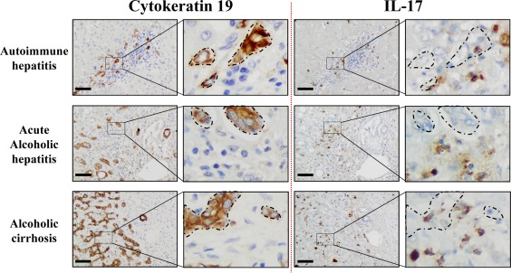
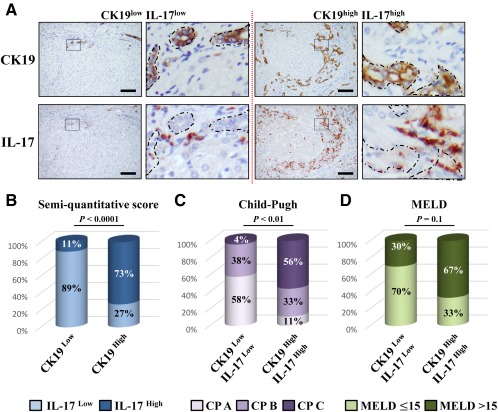
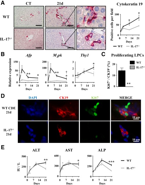
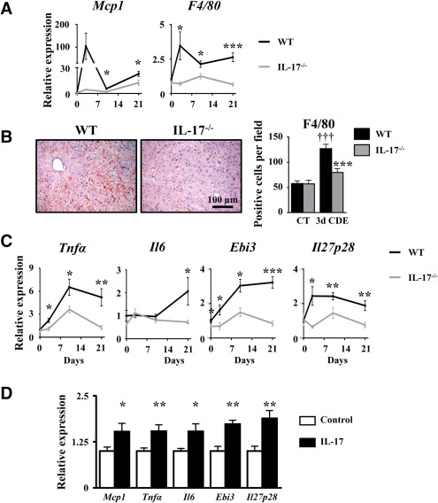
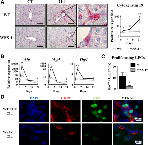
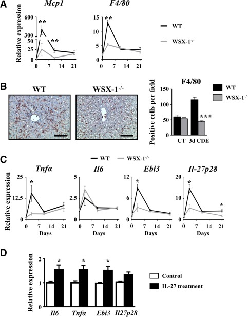
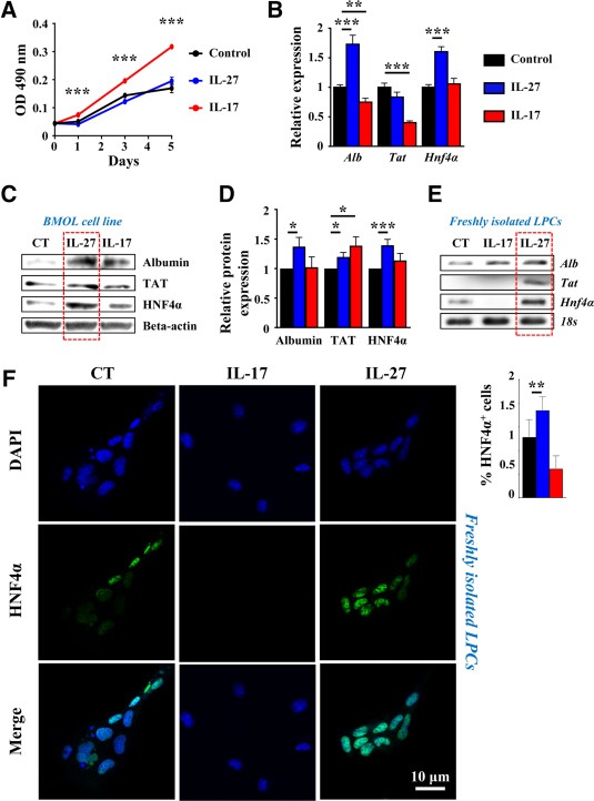
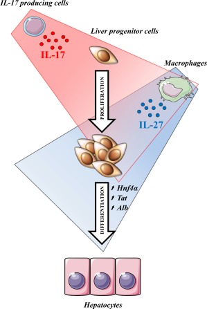
Liver progenitor cells (LPCs)/ductular reactions (DRs) are associated with inflammation and implicated in the pathogenesis of chronic liver diseases. However, how inflammation regulates LPCs/DRs remains largely unknown. Identification of inflammatory processes that involve LPC activation and expansion represent a key step in understanding the pathogenesis of liver diseases. In the current study, we found that diverse types of chronic liver diseases are associated with elevation of infiltrated interleukin (IL)-17-positive (+) cells and cytokeratin 19 (CK19)+ LPCs, and both cell types colocalized and their numbers positively correlated with each other. The role of IL-17 in the induction of LPCs was examined in a mouse model fed a choline-deficient and ethionine-supplemented (CDE) diet. Feeding of wild-type mice with the CDE diet markedly elevated CK19+Ki67+ proliferating LPCs and hepatic inflammation. Disruption of the IL-17 gene or IL-27 receptor, alpha subunit (WSX-1) gene abolished CDE diet-induced LPC expansion and inflammation. In vitro treatment with IL-17 promoted proliferation of bipotential murine oval liver cells (a liver progenitor cell line) and markedly up-regulated IL-27 expression in macrophages. Treatment with IL-27 favored the differentiation of bipotential murine oval liver cells and freshly isolated LPCs into hepatocytes. Conclusion: The current data provide evidence for a collaborative role between IL-17 and IL-27 in promoting LPC expansion and differentiation, respectively, thereby contributing to liver regeneration. (Hepatology Communications 2018;2:329-343).


Figures

Figure 1
Figure 1
IL‐17‐expressing cells and CK19+ cells are localized in similar areas in diseased livers. Representative CK19 and IL‐17 (brown color) immunostaining on human serial liver sections from diverse etiologies with an enlargement magnification field. Areas where CK19+ cells accumulate are delimited with dotted lines on serial sections to highlight their proximity with IL‐17+‐infiltrating cells. Scale bar, 100 µm.
Figure 2
Figure 2
Ductular reaction correlates with IL‐17‐producing cell infiltration in human diseased livers. (A) Representative liver sections from patients with CK19low IL‐17low (left) and CK19high IL‐17high (right) immunolabeling. Scale bar, 100 µm. (B) Relative CK19 and IL‐17 quantification was realized. (C) Child‐Pugh score class A (5 to 6), B (7 to 9), and C (10 to 15) in CK19low IL‐17low and CK19high IL‐17high patients. (D) Percentage of patients with a MELD score ≤15 or >15 in both groups. Abbreviation: CP, Child‐Pugh.
Figure 3
Figure 3
Disruption of the IL‐17 gene impairs liver progenitor cell activation in regenerating liver. Wild‐type and IL‐17−/− mice were fed a CDE diet and killed at the indicated time points. (A) Liver tissue sections were stained for CK19, and positive cell number quantification was realized. (B) Hepatic mRNA expression of LPC‐associated genes Afp, M2pk, and Thy1 was analyzed by qRT‐PCR and expressed as fold change over control diet‐fed WT mice. (C,D) Liver tissue sections were immunolabeled with antibodies directed against CK19 (red) and Ki67 (green), and the percentage of proliferating CK19+ cells was determined. (E) Serum ALT, AST, and ALP activities were measured. *P < 0.05, **P < 0.01, ***P < 0.005, WT versus IL‐17−/− mice; each group n = 4‐7 animals. Data represent mean ± SEM. Abbreviations: ALP, alkaline phosphatase; ALT, alanine aminotransferase; AST, aspartate aminotransferase; CT, control; d, day; DAPI, 4′,6‐diamidino‐2‐phenylindole; M2pk, type 2 muscle pyruvate kinase; qRT‐PCR, quantitative reverse‐transcription polymerase chain reaction.
Figure 4
Figure 4
IL‐17 deficiency represses liver inflammation, including IL‐27 production. (A) mRNA expression of MCP1 and F4/80 were quantified by qRT‐PCR. (B) Liver macrophage infiltration was analyzed by F4/80 immunostaining and counting. (C) TNF‐α, IL‐6, and IL‐27 (Ebi3 and IL‐27p28 subunits) mRNA expressions were quantified by qRT‐PCR in WT and IL‐17−/− mice after 3, 10, or 21 days of the CDE diet. *P < 0.05, **P < 0.01, ***P < 0.005, WT versus IL‐17−/− mice. (D) Inflammatory gene expressions were quantified by qRT‐PCR in the IL‐17‐treated RAW264.7 macrophage cell line. Data represent mean ± SEM. Abbreviations: CT, control; d, day; qRT‐PCR, quantitative reverse‐transcription polymerase chain reaction.
Figure 5
Figure 5
WSX‐1‐deficiency represses LPC‐driven liver regeneration. Wild‐type and WSX‐1−/− mice were fed a CDE diet, and samples were collected at the indicated time points. (A) CK19+ cells were stained and counted. Scale bar, 100 µm. (B) Hepatic mRNA expressions of LPC‐associated genes were measured by qRT‐PCR. (C,D) CK19 (red) and Ki67 (green) staining and counting after 21 days of the CDE diet. *P < 0.05, **P < 0.01, WT versus WSX‐1−/− mice; each group n = 4‐7 animals. Data represent mean ± SEM. Abbreviations: CT, control; d, day; DAPI, 4′,6‐diamidino‐2‐phenylindole; M2pk, type 2 muscle pyruvate kinase; qRT‐PCR, quantitative reverse‐transcription polymerase chain reaction.
Figure 6
Figure 6
CDE diet‐induced liver inflammation is reduced in WSX‐1−/−. Wild‐type and WSX‐1−/− mice were subjected to the CDE model. (A) Hepatic mRNA expression of macrophage‐related genes was assessed by qRT‐PCR. (B) Immunostaining of F4/80 was performed on WT and WSX‐1−/− mice, and positive cells were counted after 3 days of the CDE diet. Scale bar, 100 µm. (C) Hepatic mRNA expressions of inflammation‐related genes were quantified by qRT‐PCR; each group n = 4‐7 animals. *P < 0.05, **P < 0.01, ***P < 0.005, WT versus WSX‐1−/− mice. (D) RAW264.7 cells were cultured in the presence of 50 ng/mL IL‐27, and mRNA expressions of IL‐6, TNF‐α, and IL‐27 subunits were analyzed by qRT‐PCR. *P < 0.05, control versus IL‐27‐treated cells. Data represent mean ± SEM. Abbreviations: CT, control; d, day; qRT‐PCR, quantitative reverse‐transcription polymerase chain reaction.
Figure 7
Figure 7
IL‐17 favors LPC proliferation whereas IL‐27 induces their differentiation. BMOL cells were treated with 50 ng/mL recombinant IL‐27 or IL‐17, and (A) proliferation was assessed by optical density using an MTS assay. Hepatocyte differentiation marker expression was analyzed by (B) qRT‐PCR after 6 hours or (C) by western blot after 24 hours of treatment. (D) Western blot quantification (n = 6‐8 independent experiments). Results are expressed as fold change over untreated cells. *P < 0.05, **P < 0.01, ***P < 0.005. (D,E) Primary LPCs were cultured in the presence of 50 ng/mL recombinant IL‐17 or IL‐27 for 24 hours. Hepatocyte differentiation marker expressions were analyzed by (E) qRT‐PCR and (F) immunocytochemistry using an anti‐HNF4α antibody. HNF4α+ cells were counted in each condition. Data represent mean ± SEM. Abbreviations: CT, control; DAPI, 4′,6‐diamidino‐2‐phenylindole; MTS, 3‐(4,5‐dimethylthiazol‐2‐yl)‐5‐(3‐carboxymethoxyphenyl)‐2‐(4‐sulfophenyl)‐2H‐tetrazolium; OD, optical density; qRT‐PCR, quantitative reverse‐transcription polymerase chain reaction.
Figure 8
Figure 8
IL‐17 induces liver progenitor cell proliferation while IL‐27 favors their differentiation toward a hepatocytic phenotype. Taken together, these data provide evidence of a collaborative role of IL‐17 and IL‐27 in promoting liver regeneration. IL‐17 directly acts on LPCs to favor their proliferation. IL‐17 also induces macrophage IL‐27 production, which enhances LPC differentiation toward hepatocytes.

References

    1. Williams MJ, Clouston AD, Forbes SJ. Links between hepatic fibrosis, ductular reaction, and progenitor cell expansion. Gastroenterology 2014;146:349‐356. - PubMed
    1. Desmet VJ. Ductal plates in hepatic ductular reactions. Hypothesis and implications. I. Types of ductular reaction reconsidered. Virchows Arch 2011;458:251‐259. - PubMed
    1. Gouw AS, Clouston AD, Theise ND. Ductular reactions in human liver: diversity at the interface. Hepatology 2011;54:1853‐1863. - PubMed
    1. Roskams TA, Libbrecht L, Desmet VJ. Progenitor cells in diseased human liver. Semin Liver Dis 2003;23:385‐396. - PubMed
    1. Clouston AD, Powell EE, Walsh MJ, Richardson MM, Demetris AJ, Jonsson JR. Fibrosis correlates with a ductular reaction in hepatitis C: roles of impaired replication, progenitor cells and steatosis. Hepatology 2005;41:809‐818. - PubMed