. 2018 Mar 5;8(1):20.
doi: 10.1186/s13550-018-0374-8.

I-124 codrituzumab imaging and biodistribution in patients with hepatocellular carcinoma

Affiliations

I-124 codrituzumab imaging and biodistribution in patients with hepatocellular carcinoma

Jorge A Carrasquillo et al. EJNMMI Res. .

Abstract

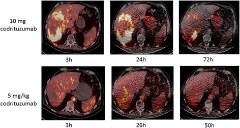
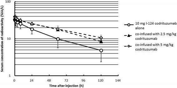
Background: I-124 codrituzumab (aka GC33), an antibody directed at Glypican 3, was evaluated in patients with hepatocellular carcinoma (HCC). Fourteen patients with HCC underwent baseline imaging with I-124 codrituzumab (~ 185 MBq, 10 mg). Seven of these patients undergoing sorafenib/immunotherapy with 2.5 or 5 mg/kg of cold codrituzumab had repeat imaging, with co-infusion of I-124 codrituzumab, as part of their immunotherapy treatment. Three patients who progressed while on sorafenib/immunotherapy were re-imaged after a 4-week washout period to assess for the presence of antigen. Serial positron emission tomography (PET) imaging and pharmacokinetics were performed following I-124 codrituzumab. An ELISA assay was used to determine "cold" codrituzumab serum pharmacokinetics and compare it to that of I-124 codrituzumab. Correlation of imaging results was performed with IHC. Short-term safety assessment was also evaluated.

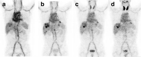
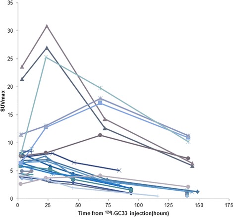
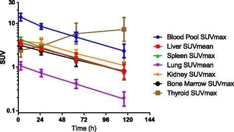
Results: Thirteen patients had tumor localization on baseline I-124 codrituzumab; heterogeneity in tumor uptake was noted. In three patients undergoing repeat imaging while on immunotherapy/sorafenib, evidence of decreased I-124 codrituzumab uptake was noted. All three patients who underwent imaging after progression while on immunotherapy continued to have I-124 codrituzumab tumor uptake. Pharmacokinetics of I-124 codrituzumab was similar to that of other intact IgG. No significant adverse events were observed related to the I-124 codrituzumab.

Conclusions: I-124 codrituzumab detected tumor localization in most patients with HCC. Pharmacokinetics was similar to that of other intact iodinated humanized IgG. No visible cross-reactivity with normal organs was observed.

Keywords: Antibody; Codrituzumab; Glypican; Hepatocellular; I-124.


Conflict of interest statement

Ethics approval and consent to participate

This study was reviewed and approved by MSK’s IRB, and all patients provided verbal and written informed consent.

Consent for publication

N/A.

Competing interests

Two authors, Norihisa Ohishi and Toshihiko Ohtomo, are members of Chugai Pharmaceutical Co., Ltd. The rights to the antibody are held by Hoffmann-La Roche, Inc., which funded the trial. The authors declare that they have no competing interests.

Publisher’s Note

Springer Nature remains neutral with regard to jurisdictional claims in published maps and institutional affiliations.

Figures

Fig. 1
Fig. 1
Patient 10 showed definite tumor accumulation in liver lesions at ~ 24 h and significant blood pool accumulation that decreased over time: 3 h (a), 22 h (b), 46 h (c), and 118 h (d). The highest SUVmax in the liver was 15.3 at 46 h. Normal organ distribution including the blood pool, liver, spleen, and kidney decreased over time. In contrast, free iodine accumulation in the thyroid increased over time
Fig. 2
Fig. 2
Each line represents a tumor in a single patient imaged over time; the tumor selected had the most intense uptake. Typically, maximal tumor uptake occurred within 24 h of tracer administration
Fig. 3
Fig. 3
Summary data from the baseline study in all patients receiving I-124 codrituzumab. VOIs were performed over the various organs and tissues. The amount of uptake was expressed in SUVmax or mean as indicated in the figure legend. The curves show that with the exception of the thyroid, which accumulates free I-124 over time, all other organs had a gradual decrease that was very similar to the clearance rate from the blood
Fig. 4
Fig. 4
Patient 7, serial-fused I-124 codrituzumab-PET/CT baseline images following injection of 190 MBq of I-124 codrituzumab (10 mg of codrituzumab) (upper panel). Upper panel images show prominent heterogeneous uptake in a large right liver tumor lesion, with more uptakes in the periphery than those in the central region. The lower panel shows serial-fused I-124 codrituzumab-PET/CT images obtained after treatment with sorafenib and codrituzumab (5 mg/kg) for 4 weeks and after injection of 183 MBq I-124 codrituzumab (co-infused with 5 mg/kg of codrituzumab). All images are displayed at the same SUV and show marked decrease in intensity of uptake in the periphery. For example, at 24 to 26 h, the SUVmax decreased in the cold co-infused images (lower panel) from 20 to 40% of the 10 mg injected mass, and in the periphery, there was less of a drop in SUVmax to 80–90% of the 10 mg injected mass, probably because of central necrosis
Fig. 5
Fig. 5
%IA/L of serum was plotted following injection of 10 mg I-124 codrituzumab alone (circle) or co-infused with 2.5 mg/kg (triangles) or 5 mg/kg (squares). The larger mass amount of antibody resulted in significant mass-dependent changes with longer half-life and greater AUC than the 10 mg injection (Table 2). There was an overlap of the clearance curves for the 2.5 and 5 mg/kg co-infused mass

Similar articles

Cited by

References

    1. Ayyar BV, Arora S, O’Kennedy R. Coming-of-age of antibodies in cancer therapeutics. Trends Pharmacol Sci. 2016;37:1009–1028. doi: 10.1016/j.tips.2016.09.005. - DOI - PubMed
    1. Smith-Jones PM, Solit D, Afroze F, Rosen N, Larson SM. Early tumor response to Hsp90 therapy using HER2 PET: comparison with F-18-FDG PET. J Nucl Med. 2006;47:793–796. - PMC - PubMed
    1. Smith-Jones PM, Solit DB, Akhurst T, Afroze F, Rosen N, Larson SM. Imaging the pharmacodynamics of HER2 degradation in response to Hsp90 inhibitors. Nat Biotechnol. 2004;22(6):701. doi: 10.1038/nbt968. - DOI - PubMed
    1. Gebhart G, Lamberts LE, Wimana Z, Garcia C, Emonts P, Ameye L, et al. Molecular imaging as a tool to investigate heterogeneity of advanced HER2-positive breast cancer and to predict patient outcome under trastuzumab emtansine (T-DM1): the ZEPHIR trial. Ann Oncol. 2016;27:619–624. doi: 10.1093/annonc/mdv577. - DOI - PubMed
    1. Carrasquillo JA, Pandit-Taskar N, O'Donoghue JA, Humm JL, Zanzonico P, Smith-Jones PM, et al. (124)I-huA33 antibody PET of colorectal cancer. J Nucl Med. 2011;52:1173–1180. doi: 10.2967/jnumed.110.086165. - DOI - PMC - PubMed

LinkOut - more resources