. 2018 Apr;50(4):647-656.
doi: 10.1007/s11255-018-1831-z. Epub 2018 Mar 5.

Epidermal growth factor receptor function in the human urothelium

Affiliations

Epidermal growth factor receptor function in the human urothelium

C Wasén et al. Int Urol Nephrol. 2018 Apr.

Abstract

Purpose: Epidermal growth factor receptor (EGFr)-targeted therapy may be used in subgroups of patients with urinary bladder cancer. Here we assessed the role of EGFr in urothelial proliferation and migration in a two- and three-dimensional cell culture system.

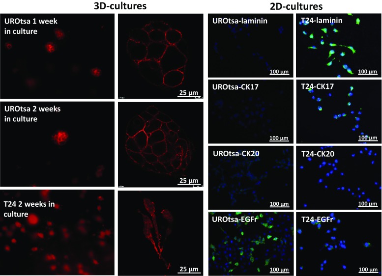
Methods: UROtsa cells derived from normal urothelium and malignant T24 cells were cultured in a Type I collagen gel. Proliferation and migration of urothelial cells, in the absence and presence of the EGFr inhibitor cetuximab, were assessed with a proliferation test (ATCC) and with the Axioplan 2 imaging microscope with a motorized stage (Carl Zeiss), respectively. The expressions of cytokeratin (CK) 17, CK20, EGFr, pEGFr, laminin, occludin and zonula occludens 1 (ZO-1) were assessed with immunohistochemistry and/or western blot.

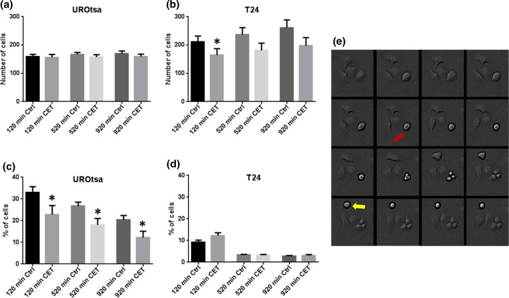
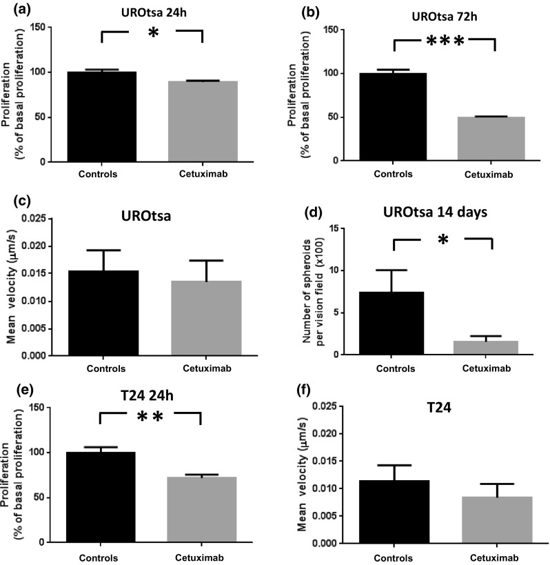
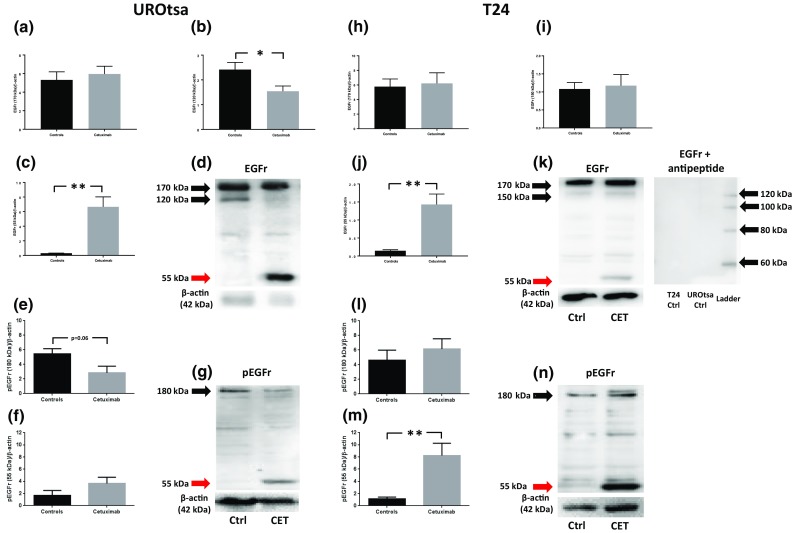
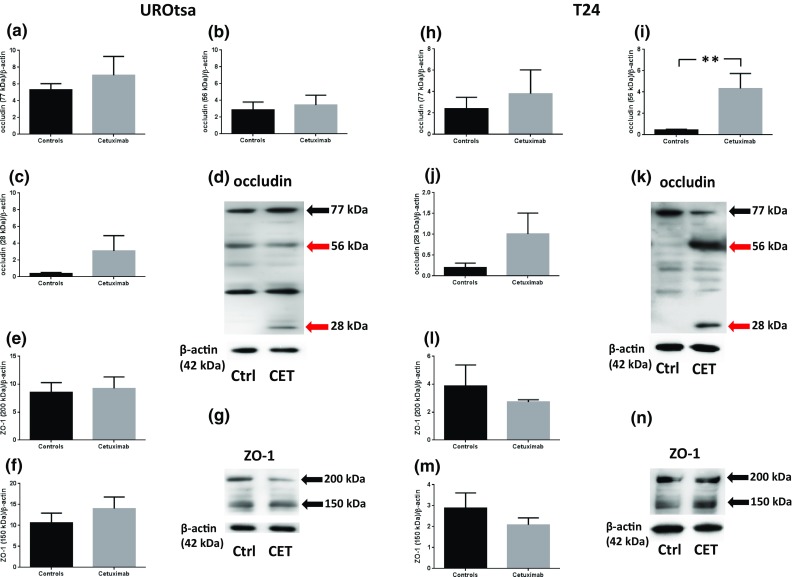
Results: UROtsa spheroids were formed after 7 days in culture, while T24 cells did not form spheroids. UROtsa expressed CK20 but not laminin or CK17 and consequently resembled umbrella cells. In UROtsa and T24, cetuximab inhibited urothelial proliferation, induced cleavage of EGFr and/or pEGFR but did not affect urothelial migration. The tight junction protein occludin was cleaved, and the formation of cellular spheroids was inhibited in UROtsa by the presence of cetuximab.

Conclusions: EGFr modulates urothelial proliferation and the formation of the three-dimensional structure of the urothelium possibly by interfering with occludin. The present data also show a cell culture technique enabling phenotypically normal urothelial cells to form epithelial structures in contrast to malignant urothelial cells.

Keywords: Epidermal growth factor receptor; Occludin; Proliferation; Three-dimensional cell culture; Urothelium.


Conflict of interest statement

Conflict of interest

The authors declare no conflict of interest.

Human and animal rights

This article does not contain any studies with human participants or animals performed by any of the authors.

Figures

Fig. 1
Fig. 1
First (fluorescence microscopy) and second (confocal microscopy) columns represent UROtsa grown for 1 and 2 weeks and T24 grown for 2 weeks. Cells were stained with phalloidin. Representative microphotographs of the expressions of laminin, CK17, CK20 and EGFr (green) with DAPI-stained (blue) nuclei in UROtsa (third column) and T24 (fourth column)
Fig. 2
Fig. 2
Total number of cells per vision field in a UROtsa and b T24 and percentages of cells with round shapes out of the total number of cells per vision field in c UROtsa and d T24 in the absence and presence of cetuximab (1.5 μM) after 120, 520 and 920 min after cell culture and e representative time-lapse series (320 s between frames) of T24 cells undergoing tripolar mitosis (red arrow) and regular dipolar mitosis (yellow arrow). *p < 0.05. Vertical bars represent the SEM
Fig. 3
Fig. 3
Urothelial proliferation (% of basal responses) in 24-h (a) and 72-h (b) UROtsa cultures, migration velocity in UROtsa cultures (c), number of round cells in 14-day UROtsa cultures (d), urothelial proliferation in 24-h T24 cell cultures and migration velocity in T24 cultures in the absence and presence of cetuximab (1.5 μM). *p < 0.05, **p < 0.01 and ***p < 0.001. Vertical bars represent the SEM
Fig. 4
Fig. 4
Western blot analyses of EGFr expressions in UROtsa (ac) and in T24 (hj); representative graphs in d, k, respectively, and pEGFr in UROtsa (e, f) and in T24 (lm); representative graphs in g, n, respectively. Black arrows indicate EGFr and pEGFr, respectively, while red arrows indicate fragments of protein at 55 kDa. Membrane incubated with anti-EGFr in the presence of EGFr peptide is displayed in 4 k. *p < 0.05 and **p < 0.01. Vertical bars represent the SEM
Fig. 5
Fig. 5
Western blot analyses of occludin expressions (77, 56 and 28 kDa in UROtsa (ac) and in T24 (hj); representative graphs in d, k, respectively) and ZO-1 (200 and 150 kDa in UROtsa (ef) and in T24 (lm); representative graphs in g, n, respectively). Black arrows indicate occludin and ZO-1, respectively, while red arrows indicate fragments of occludin. **p < 0.01. Vertical bars represent the SEM

Similar articles

Cited by

References

    1. Jaal J, Dorr W. Radiation effects on cellularity, proliferation and EGFR expression in mouse bladder urothelium. Radiat Res. 2010;173(4):479–485. doi: 10.1667/RR1759.1. - DOI - PubMed
    1. Daher A, de Boer WI, El-Marjou A, van der Kwast T, Abbou CC, Thiery JP, Radvanyi F, Chopin DK. Epidermal growth factor receptor regulates normal urothelial regeneration. Lab Invest. 2003;83(9):1333–1341. doi: 10.1097/01.LAB.0000086380.23263.52. - DOI - PubMed
    1. Varley C, Hill G, Pellegrin S, Shaw NJ, Selby PJ, Trejdosiewicz LK, Southgate J. Autocrine regulation of human urothelial cell proliferation and migration during regenerative responses in vitro. Exp Cell Res. 2005;306(1):216–229. doi: 10.1016/j.yexcr.2005.02.004. - DOI - PubMed
    1. Romih R, Koprivec D, Martincic DS, Jezernik K. Restoration of the rat urothelium after cyclophosphamide treatment. Cell Biol Int. 2001;25(6):531–537. doi: 10.1006/cbir.2000.0658. - DOI - PubMed
    1. Kramer C, Klasmeyer K, Bojar H, Schulz WA, Ackermann R, Grimm MO. Heparin-binding epidermal growth factor-like growth factor isoforms and epidermal growth factor receptor/ErbB1 expression in bladder cancer and their relation to clinical outcome. Cancer. 2007;109(10):2016–2024. doi: 10.1002/cncr.22627. - DOI - PubMed

MeSH terms