

Atlas of the Striatum and Globus Pallidus in the Tree Shrew: Comparison with Rat and Mouse
- PMID: 29508249
- PMCID: PMC5960448
- DOI: 10.1007/s12264-018-0212-z
Atlas of the Striatum and Globus Pallidus in the Tree Shrew: Comparison with Rat and Mouse
Abstract
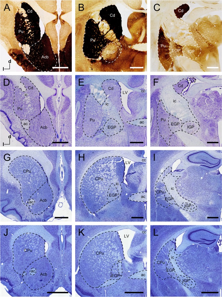
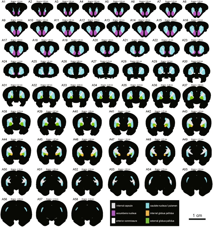
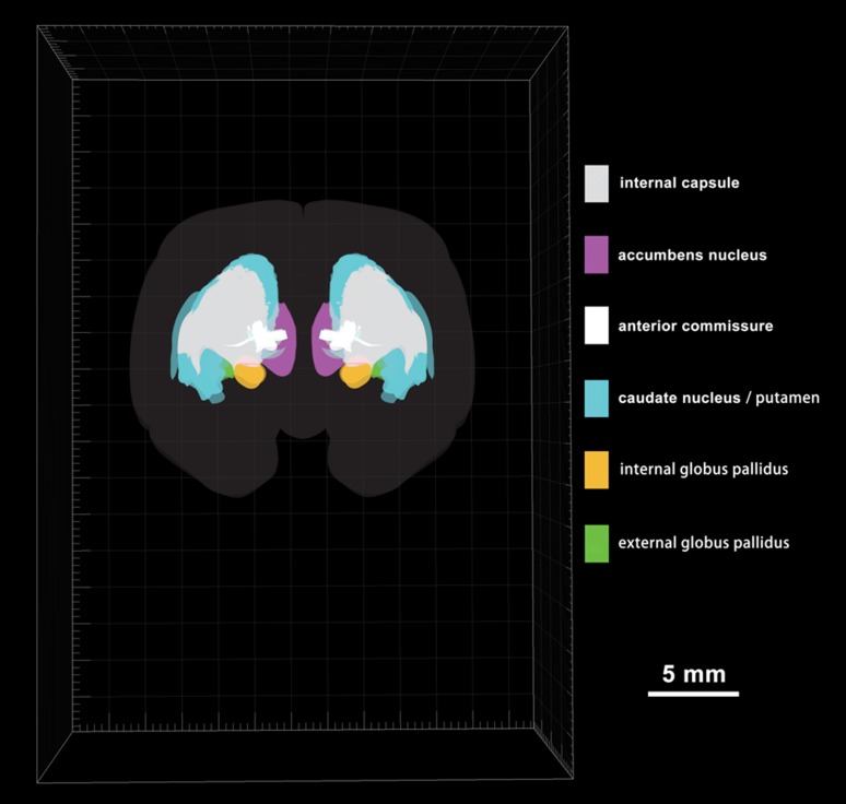
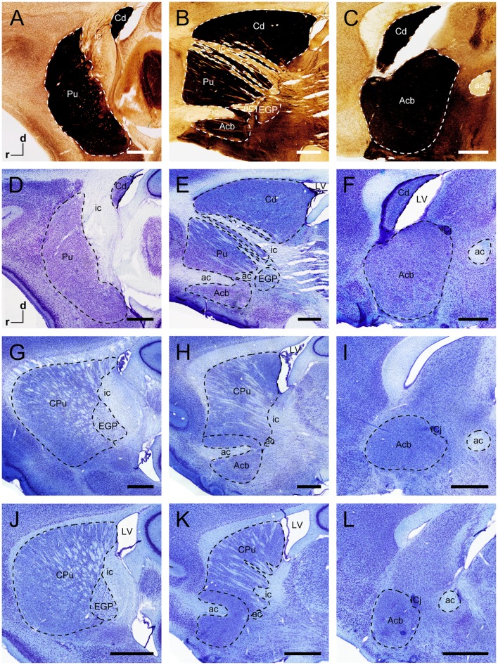
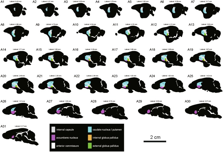
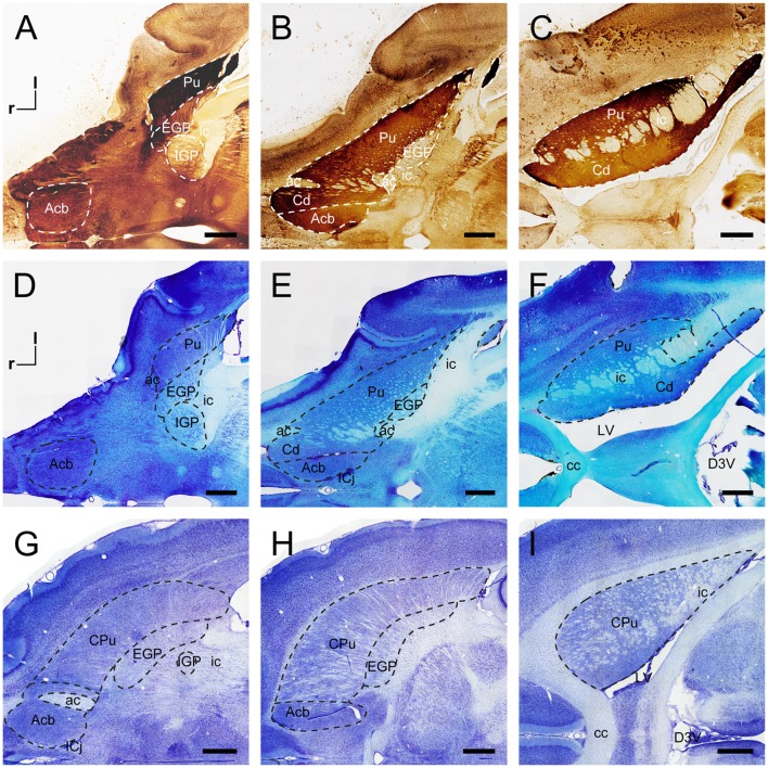
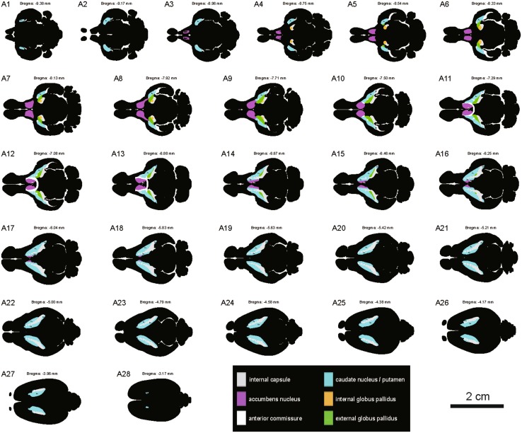
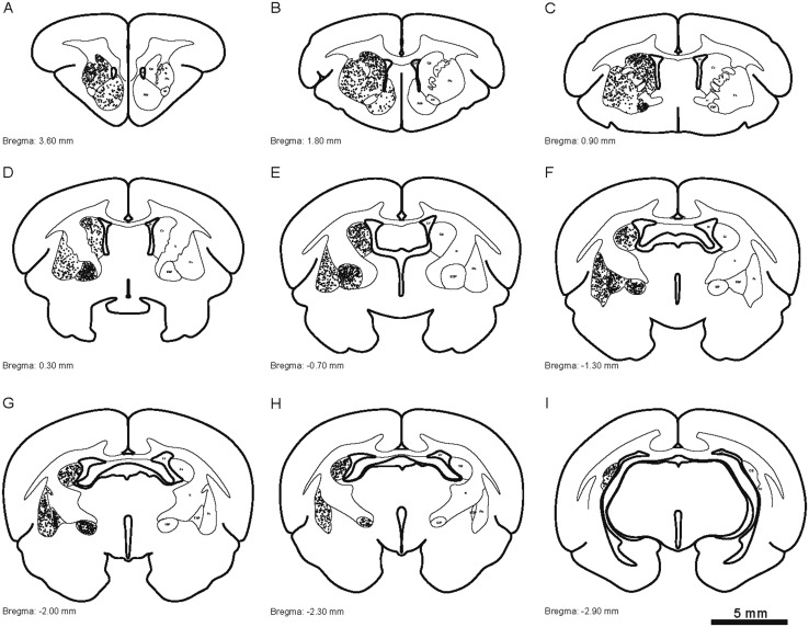
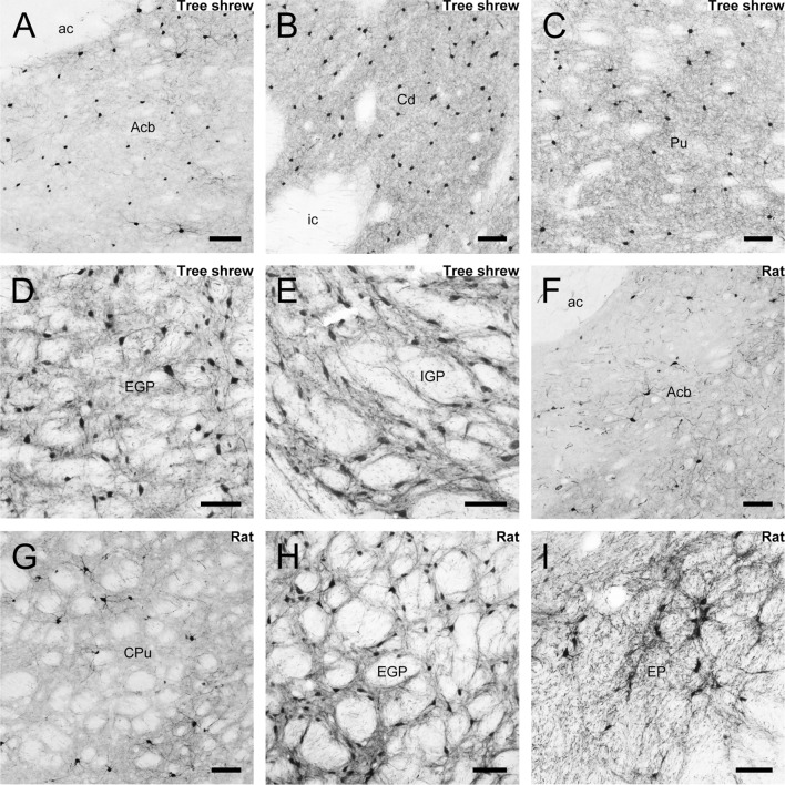
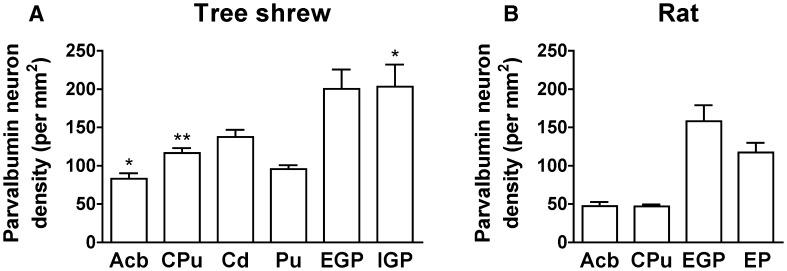
The striatum and globus pallidus are principal nuclei of the basal ganglia. Nissl- and acetylcholinesterase-stained sections of the tree shrew brain showed the neuroanatomical features of the caudate nucleus (Cd), internal capsule (ic), putamen (Pu), accumbens, internal globus pallidus, and external globus pallidus. The ic separated the dorsal striatum into the Cd and Pu in the tree shrew, but not in rats and mice. In addition, computer-based 3D images allowed a better understanding of the position and orientation of these structures. These data provided a large-scale atlas of the striatum and globus pallidus in the coronal, sagittal, and horizontal planes, the first detailed distribution of parvalbumin-immunoreactive cells in the tree shrew, and the differences in morphological characteristics and density of parvalbumin-immunoreactive neurons between tree shrew and rat. Our findings support the tree shrew as a potential model for human striatal disorders.
Keywords: Basal ganglia; Globus pallidus; Parvalbumin; Reconstruction; Rodent; Striatum.
Conflict of interest statement
All authors claim that there are no conflicts of interest.
Figures
Similar articles
-
Structural organization, GABAergic and tyrosine hydroxylase expression in the striatum and globus pallidus of the South American plains vizcacha, Lagostomus maximus (Rodentia, Caviomorpha).J Mol Histol. 2019 Dec;50(6):515-531. doi: 10.1007/s10735-019-09845-9. Epub 2019 Sep 12. J Mol Histol. 2019. PMID: 31515635
-
Heterogeneous and compartmental distribution of zinc in the striatum and globus pallidus of the rat.Neuroscience. 1995 Jun;66(3):523-37. doi: 10.1016/0306-4522(94)00592-s. Neuroscience. 1995. PMID: 7644017
-
Comparison of the rat dorsal and ventral striatopallidal system. A study using the GABA(A)-receptor alpha1-subunit and parvalbumin immunolabeling.Exp Brain Res. 1998 Jul;121(2):215-21. doi: 10.1007/s002210050454. Exp Brain Res. 1998. PMID: 9696391
-
[Paradigm of negative feedback via long-loop in the striatal dopamine release modulation in the rat].Rev Neurol. 2011 Mar 16;52(6):371-7. Rev Neurol. 2011. PMID: 21387254 Review. Spanish.
-
Striatal and subthalamic afferents to the primate pallidum: interactions between two opposite chemospecific neuronal systems.Prog Brain Res. 1993;99:89-104. doi: 10.1016/s0079-6123(08)61340-0. Prog Brain Res. 1993. PMID: 7509082 Review. No abstract available.
Cited by
-
Animal models of Alzheimer's disease: Current strategies and new directions.Zool Res. 2024 Nov 18;45(6):1385-1407. doi: 10.24272/j.issn.2095-8137.2024.274. Zool Res. 2024. PMID: 39572020 Free PMC article. Review.
-
Technical advance: The use of tree shrews as a model of pulmonary fibrosis.PLoS One. 2020 Nov 3;15(11):e0241323. doi: 10.1371/journal.pone.0241323. eCollection 2020. PLoS One. 2020. PMID: 33141839 Free PMC article.
-
Disrupting the α7nAChR-NR2A protein complex exerts antidepressant-like effects.Mol Brain. 2021 Jul 5;14(1):107. doi: 10.1186/s13041-021-00817-3. Mol Brain. 2021. PMID: 34225758 Free PMC article.
-
Study of tree shrew biology and models: A booming and prosperous field for biomedical research.Zool Res. 2024 Jul 18;45(4):877-909. doi: 10.24272/j.issn.2095-8137.2024.199. Zool Res. 2024. PMID: 39004865 Free PMC article. Review.
-
Homeobox Gene Six3 is Required for the Differentiation of D2-Type Medium Spiny Neurons.Neurosci Bull. 2021 Jul;37(7):985-998. doi: 10.1007/s12264-021-00698-5. Epub 2021 May 20. Neurosci Bull. 2021. PMID: 34014554 Free PMC article.
References
-
- Fujiyama F. Anatomical connections of the basal ganglia. Brain Nerve. 2009;61:341–349. - PubMed
MeSH terms
Substances
LinkOut - more resources
Full Text Sources
Other Literature Sources
