

Atlas of the Striatum and Globus Pallidus in the Tree Shrew: Comparison with Rat and Mouse
- PMID: 29508249
- PMCID: PMC5960448
- DOI: 10.1007/s12264-018-0212-z
Atlas of the Striatum and Globus Pallidus in the Tree Shrew: Comparison with Rat and Mouse
Abstract
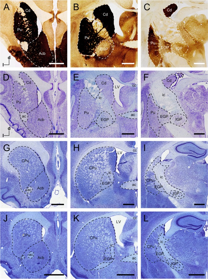
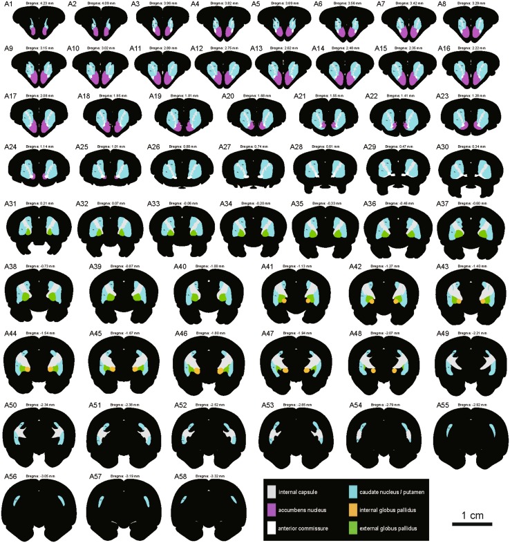
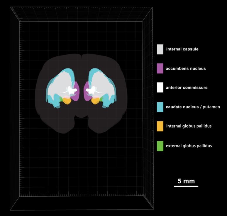
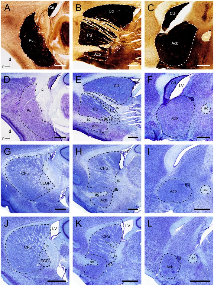
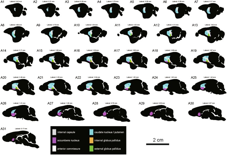
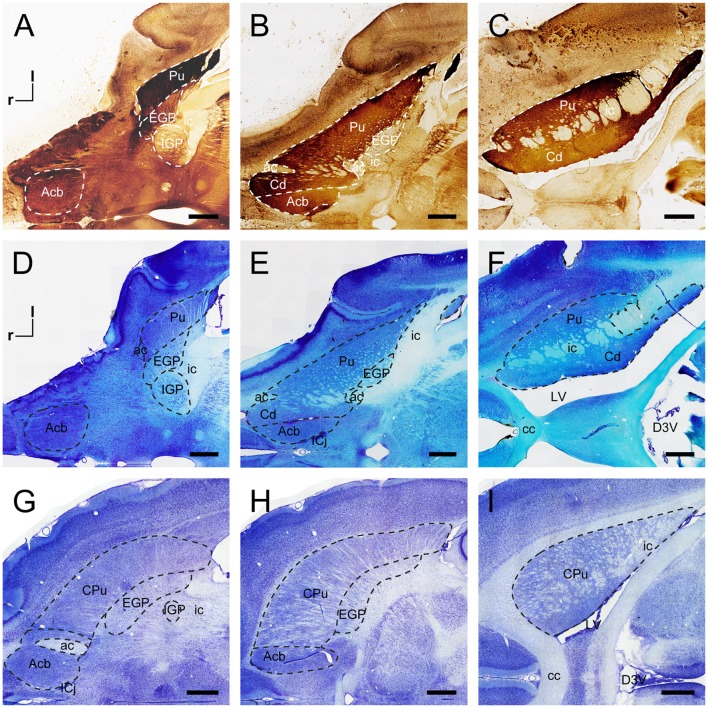
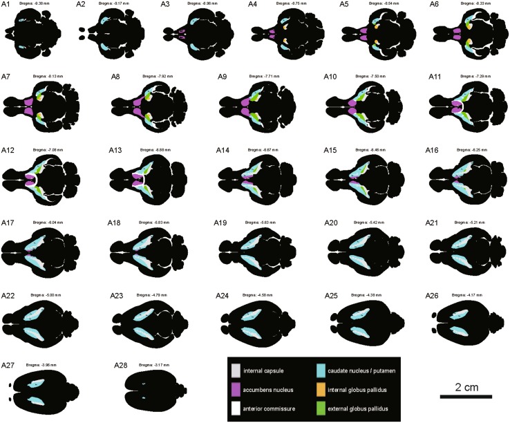
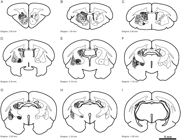
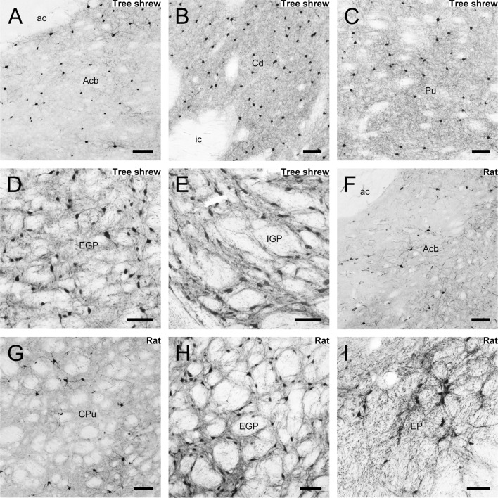
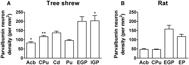
The striatum and globus pallidus are principal nuclei of the basal ganglia. Nissl- and acetylcholinesterase-stained sections of the tree shrew brain showed the neuroanatomical features of the caudate nucleus (Cd), internal capsule (ic), putamen (Pu), accumbens, internal globus pallidus, and external globus pallidus. The ic separated the dorsal striatum into the Cd and Pu in the tree shrew, but not in rats and mice. In addition, computer-based 3D images allowed a better understanding of the position and orientation of these structures. These data provided a large-scale atlas of the striatum and globus pallidus in the coronal, sagittal, and horizontal planes, the first detailed distribution of parvalbumin-immunoreactive cells in the tree shrew, and the differences in morphological characteristics and density of parvalbumin-immunoreactive neurons between tree shrew and rat. Our findings support the tree shrew as a potential model for human striatal disorders.
Keywords: Basal ganglia; Globus pallidus; Parvalbumin; Reconstruction; Rodent; Striatum.
Conflict of interest statement
All authors claim that there are no conflicts of interest.
Figures
References
-
- Fujiyama F. Anatomical connections of the basal ganglia. Brain Nerve. 2009;61:341–349. - PubMed
MeSH terms
Substances
LinkOut - more resources
Full Text Sources
Other Literature Sources
