

Elucidating the Structures of Amyloid Oligomers with Macrocyclic β-Hairpin Peptides: Insights into Alzheimer's Disease and Other Amyloid Diseases
- PMID: 29508987
- PMCID: PMC5911177
- DOI: 10.1021/acs.accounts.7b00554
Elucidating the Structures of Amyloid Oligomers with Macrocyclic β-Hairpin Peptides: Insights into Alzheimer's Disease and Other Amyloid Diseases
Abstract
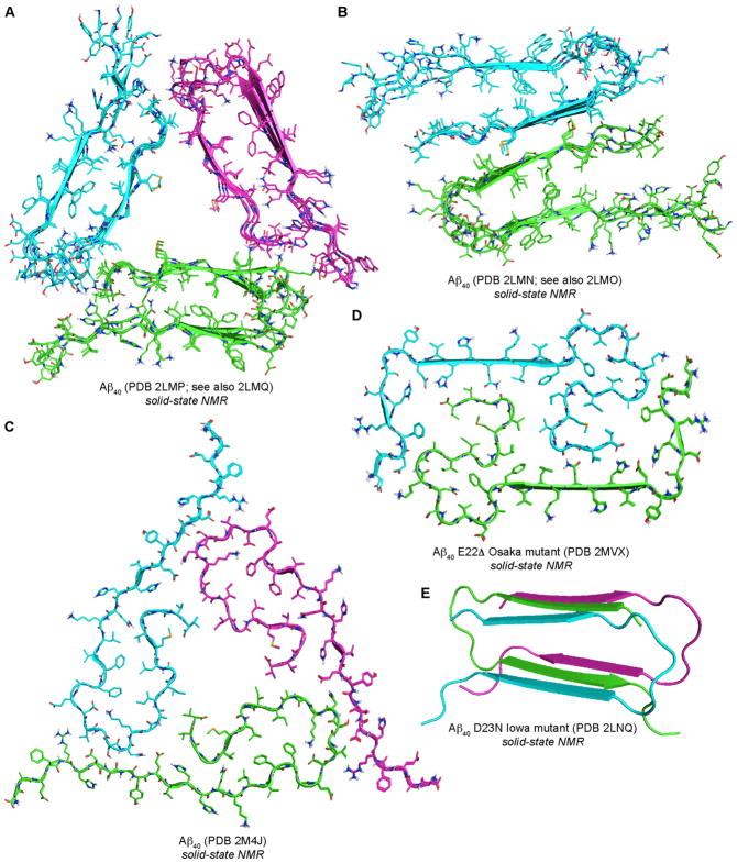
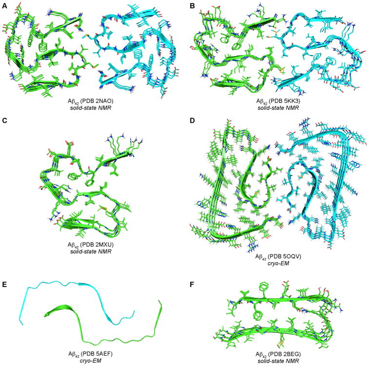
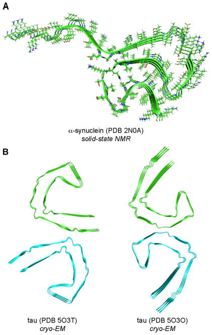
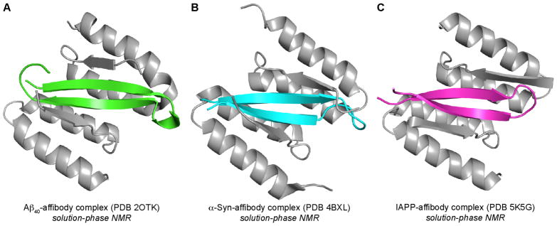
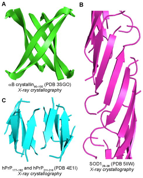
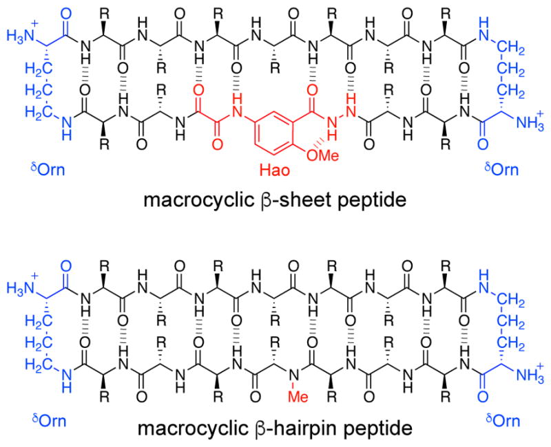
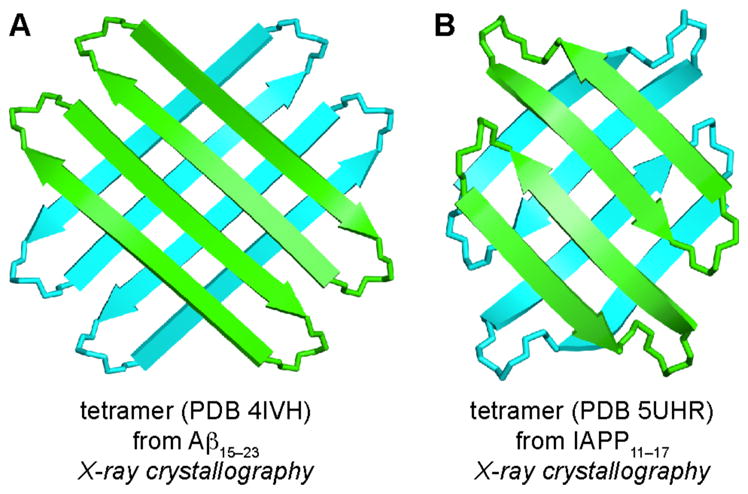
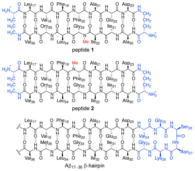
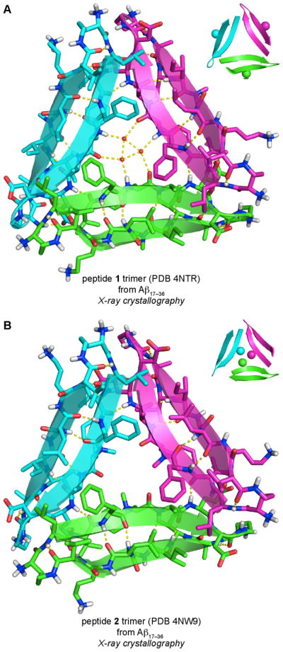
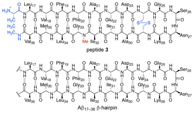
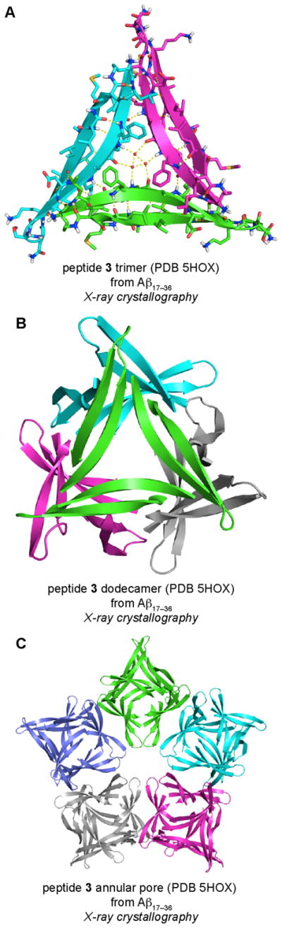
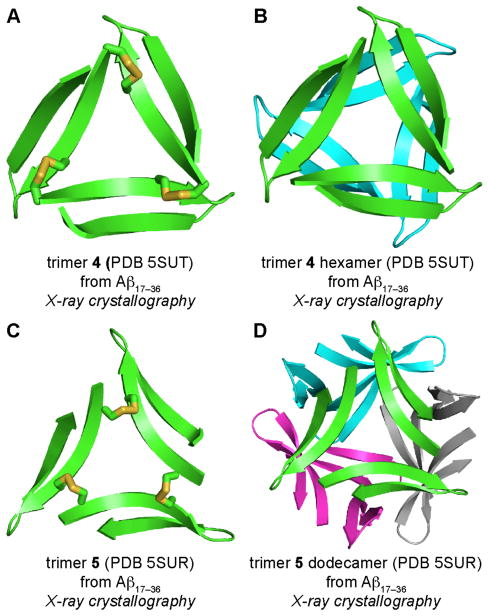
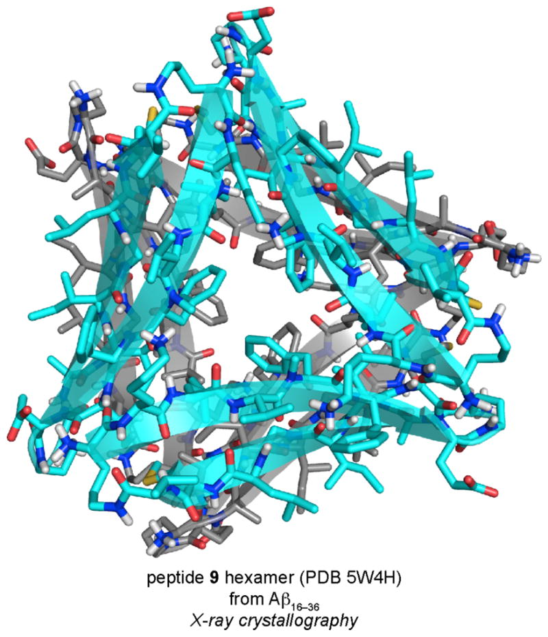
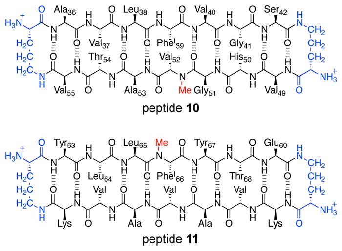
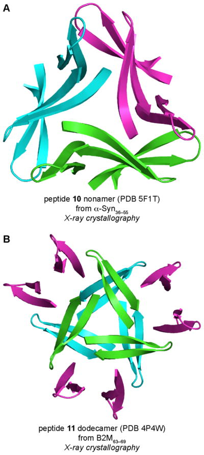
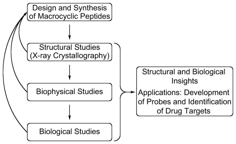
In the more than a century since its identification, Alzheimer's disease has become the archetype of amyloid diseases. The first glimpses of the chemical basis of Alzheimer's disease began with the identification of "amyloid" plaques in the brain in 1892 and extended to the identification of proteinaceous fibrils with "cross-β" structure in 1968. Further efforts led to the discovery of the β-amyloid peptide, Aβ, as a 40- or 42-amino acid peptide that is responsible for the plaques and fibrils. At this point, a three-decade-long marathon began to elucidate the structure of the fibrils and identify the molecular basis of Alzheimer's disease. Along the way, an alternative model began to emerge in which small aggregates of Aβ, called "oligomers", rather than fibrils, are the culprits that lead to neurodegeneration in Alzheimer's disease. This Account describes what is known about the structures of the fibrils and details our research group's efforts to understand the structural, biophysical, and biological properties of the oligomers in amyloid diseases. β-Sheets are the building blocks of amyloid fibrils and oligomers. Amyloid fibrils generally consist of extended networks of parallel β-sheets. Amyloid oligomers appear to be more compact enclosed structures, some of which are thought to be composed of antiparallel β-sheets comprising β-hairpins. β-Hairpins are special because their twisted shape, hydrophobic surfaces, and exposed hydrogen-bonding edges impart a unique propensity to form compact assemblies. Our laboratory has developed macrocyclic β-sheets that are designed to mimic β-hairpins formed by amyloidogenic peptides and proteins. The β-hairpin mimics contain two β-strand peptide fragments linked together at their N- and C-termini by two δ-linked ornithine turn mimics to create a macrocycle. An N-methyl group is installed on one of the β-strands to prevent uncontrolled aggregation. These design features facilitate crystallization of the β-hairpin mimics and determination of the X-ray crystallographic structures of the oligomers that they form. During the past few years, our laboratory has elucidated the X-ray crystallographic structures of oligomers formed by β-hairpin mimics derived from Aβ, α-synuclein, and β2-microglobulin. Out of these three amyloidogenic peptides and proteins, the Aβ β-hairpin mimics have provided the most insight into amyloid oligomers. Our studies have revealed a previously undiscovered mode of self-assembly, whereby three Aβ β-hairpin mimics assemble to form a triangular trimer. The triangular trimers are remarkable, because they contain two largely hydrophobic surfaces that pack together with other triangular trimers to form higher-order oligomers, such as hexamers and dodecamers. Some of the dodecamers pack in the crystal lattice to form annular porelike assemblies. Some of the β-hairpin mimics and triangular trimers assemble in solution to form oligomers that recapitulate the crystallographically observed oligomers. These oligomers exhibit toxicity toward neuronally derived cells, recapitulating the toxicity of the oligomers formed by full-length amyloidogenic peptides and proteins. These findings are significant, because they address a gap in understanding the molecular basis of amyloid diseases. We anticipate that these studies will pave the way for developing diagnostics and therapeutics to combat Alzheimer's disease, Parkinson's disease, and other amyloid diseases.
Conflict of interest statement
The authors declare no competing financial interest.
Figures
References
-
- Benilova I, Karran E, De Strooper B. The Toxic Aβ Oligomer and Alzheimer's Disease: An Emperor in Need of Clothes. Nat Neurosci. 2012;15:349–357. - PubMed
-
-
For leading references, see the Supporting Information.
-
Publication types
MeSH terms
Substances
Grants and funding
LinkOut - more resources
Full Text Sources
Other Literature Sources
Medical
Research Materials
Miscellaneous
