

Epicutaneous administration of the pattern recognition receptor agonist polyinosinic-polycytidylic acid activates the MDA5/MAVS pathway in Langerhans cells
- PMID: 29509510
- PMCID: PMC6053315
- DOI: 10.1096/fj.201701090R
Epicutaneous administration of the pattern recognition receptor agonist polyinosinic-polycytidylic acid activates the MDA5/MAVS pathway in Langerhans cells
Abstract
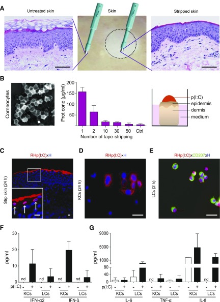
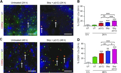
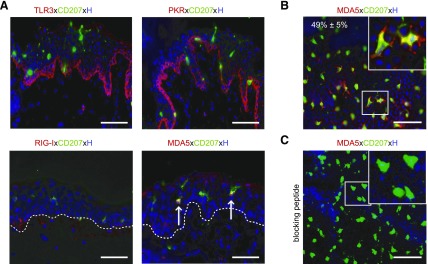
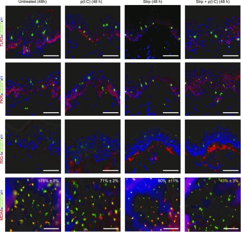
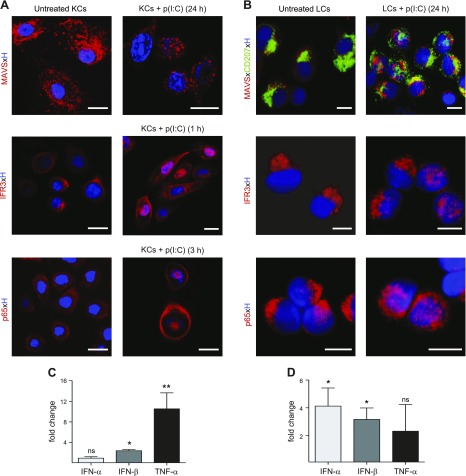
Together with keratinocytes (KCs) and the dense network of Langerhans cells (LCs), the epidermis is an ideal portal for vaccine delivery. Pattern recognition receptor agonists, in particular polyinosinic-polycytidylic acid [p(I:C)], are promising adjuvant candidates for therapeutic vaccination to generate protective T-cell immunity. Here we established an ex vivo skin explant model to study the expression and activation of double-stranded RNA (dsRNA)-sensing pattern recognition receptors in LCs and KCs in human skin. Whereas KCs expressed all known dsRNA sensing receptors at a constitutive and inducible level, LCs exclusively expressed melanoma differentiation-associated protein 5 (MDA5) in untreated skin and freshly isolated cells. Comparative assessments of downstream signaling pathways induced by p(I:C) revealed distinct mitochondrial antiviral-signaling protein, IFN-regulatory factor 3, and NF-κB activation in LCs and KCs. Consequently, p(I:C) treatment of LCs significantly induced IFN-α and IFN-β mRNA expression, while in KCs an up-regulation of IFN-β and TNF-α mRNA was detectable. Stimulation of LCs with specific ligands revealed that not the TLR3- but only the MDA5-specific ligand induced IFN-α2, IFN-β, and TNF-α cytokines, but no IL-6 and -8. In KCs, both ligands induced production of high IL-6 and IL-8 levels, and low IFN-α2 and IFN-β levels, indicating that different dsRNA-sensing receptors and/or downstream signaling pathways are activated in both cell types. Our data suggest that MDA5 may be an attractive adjuvant target for epicutaneous delivery of therapeutic vaccines with the goal to target LCs.-Tajpara, P., Schuster, C., Schön, E., Kienzl, P., Vierhapper, M., Mildner, M., Elbe-Bürger, A. Epicutaneous administration of the pattern recognition receptor agonist polyinosinic-polycytidylic acid activates the MDA5/MAVS pathway in Langerhans cells.
Keywords: downstream signaling; nuclear translocation; skin; tape stripping; therapeutic vaccination.
Conflict of interest statement
This work was supported by the Austrian Science Fund (FWF) DK W1248-B30. The authors thank W. Bauer, B. Reininger, J. Strobl and M. Buchberger (Department of Dermatology, Medical University of Vienna) for excellent help with the sorting and analysis of epidermal cells. The authors are grateful for helpful discussions with J. Stöckl (Institute of Immunology, Medical University of Vienna) and R. de Martin (Department of Vascular Biology and Thrombosis Research, Medical University of Vienna). The authors declare no conflicts of interest.
Figures
Similar articles
-
DNAJB1/HSP40 Suppresses Melanoma Differentiation-Associated Gene 5-Mitochondrial Antiviral Signaling Protein Function in Conjunction with HSP70.J Innate Immun. 2018;10(1):44-55. doi: 10.1159/000480740. Epub 2017 Oct 26. J Innate Immun. 2018. PMID: 29069650 Free PMC article.
-
Double-stranded RNA induces an antiviral defense status in epidermal keratinocytes through TLR3-, PKR-, and MDA5/RIG-I-mediated differential signaling.J Immunol. 2008 Aug 15;181(4):2694-704. doi: 10.4049/jimmunol.181.4.2694. J Immunol. 2008. PMID: 18684960
-
Role of double-stranded RNA pattern recognition receptors in rhinovirus-induced airway epithelial cell responses.J Immunol. 2009 Dec 1;183(11):6989-97. doi: 10.4049/jimmunol.0901386. Epub 2009 Nov 4. J Immunol. 2009. PMID: 19890046 Free PMC article.
-
Particulate formulations for the delivery of poly(I:C) as vaccine adjuvant.Adv Drug Deliv Rev. 2013 Oct;65(10):1386-99. doi: 10.1016/j.addr.2013.05.013. Epub 2013 Jun 7. Adv Drug Deliv Rev. 2013. PMID: 23751781 Review.
-
MDA5 Is a Major Determinant of Developing Symptoms in Critically Ill COVID-19 Patients.Clin Rev Allergy Immunol. 2024 Dec;67(1-3):58-72. doi: 10.1007/s12016-024-09008-z. Epub 2024 Oct 26. Clin Rev Allergy Immunol. 2024. PMID: 39460899 Review.
Cited by
-
Octenidine-based hydrogel shows anti-inflammatory and protease-inhibitory capacities in wounded human skin.Sci Rep. 2021 Jan 8;11(1):32. doi: 10.1038/s41598-020-79378-9. Sci Rep. 2021. PMID: 33420112 Free PMC article.
-
Significance and Role of Pattern Recognition Receptors in Malignancy.Arch Immunol Ther Exp (Warsz). 2019 Jun;67(3):133-141. doi: 10.1007/s00005-019-00540-x. Epub 2019 Apr 11. Arch Immunol Ther Exp (Warsz). 2019. PMID: 30976817 Free PMC article. Review.
-
A Preclinical Model for Studying Herpes Simplex Virus Infection.J Invest Dermatol. 2019 Mar;139(3):673-682. doi: 10.1016/j.jid.2018.08.034. Epub 2018 Nov 8. J Invest Dermatol. 2019. PMID: 30414908 Free PMC article.
-
Comparative assessment of commercially available wound gels in ex vivo human skin reveals major differences in immune response-modulatory effects.Sci Rep. 2022 Oct 19;12(1):17481. doi: 10.1038/s41598-022-20997-9. Sci Rep. 2022. PMID: 36261541 Free PMC article.
-
Interoperability of RTN1A in dendrite dynamics and immune functions in human Langerhans cells.Elife. 2022 Oct 12;11:e80578. doi: 10.7554/eLife.80578. Elife. 2022. PMID: 36223176 Free PMC article.
References
-
- Brewer J. M., Conacher M., Satoskar A., Bluethmann H., Alexander J. (1996) In interleukin-4–deficient mice, alum not only generates T helper 1 responses equivalent to Freund’s complete adjuvant, but continues to induce T helper 2 cytokine production. Eur. J. Immunol. 26, 2062–2066 10.1002/eji.1830260915 - DOI - PubMed
-
- Zhu Q., Egelston C., Vivekanandhan A., Uematsu S., Akira S., Klinman D. M., Belyakov I. M., Berzofsky J. A. (2008) Toll-like receptor ligands synergize through distinct dendritic cell pathways to induce T cell responses: implications for vaccines. Proc. Natl. Acad. Sci. USA 105, 16260–16265 10.1073/pnas.0805325105 - DOI - PMC - PubMed
Publication types
MeSH terms
Substances
Grants and funding
LinkOut - more resources
Full Text Sources
Other Literature Sources
Research Materials
Miscellaneous
