

Maternal Diets Deficient in Vitamin D Increase the Risk of Kyphosis in Offspring: A Novel Kyphotic Porcine Model
- PMID: 29509618
- PMCID: PMC6818982
- DOI: 10.2106/JBJS.17.00182
Maternal Diets Deficient in Vitamin D Increase the Risk of Kyphosis in Offspring: A Novel Kyphotic Porcine Model
Abstract
Background: The purpose of this study was to explore the role of perinatal vitamin-D intake on the development and characterization of hyperkyphosis in a porcine model.
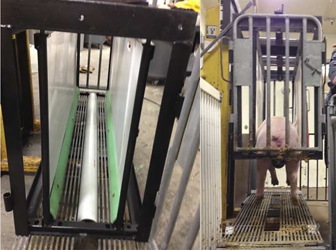
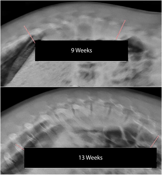
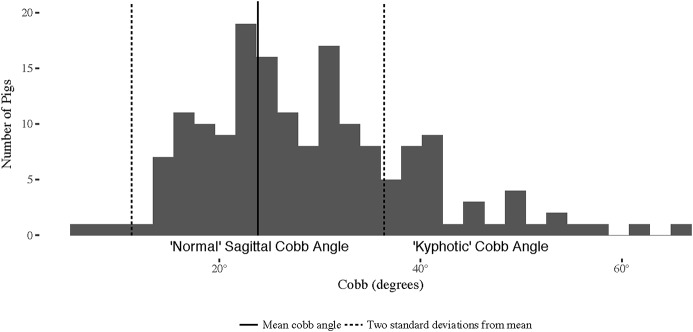
Methods: The spines of 16 pigs were assessed at 9, 13, and 17 weeks of age with radiography and at 17 weeks with computed tomography (CT), magnetic resonance imaging (MRI), histology, and bone-density testing. An additional 169 pigs exposed to 1 of 3 maternal dietary vitamin-D levels from conception through the entire lactation period were fed 1 of 4 nursery diets supplying different levels of vitamin D, calcium, and phosphorus. When the animals were 13 weeks of age, upright lateral spinal radiography was performed with use of a custom porcine lift and sagittal Cobb angles were measured in triplicate to determine the degree of kyphosis in each pig.
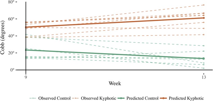
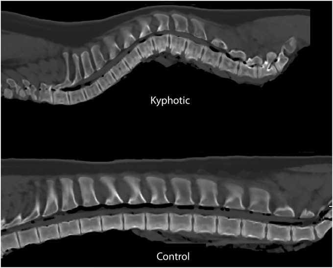
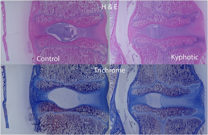
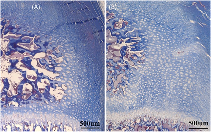
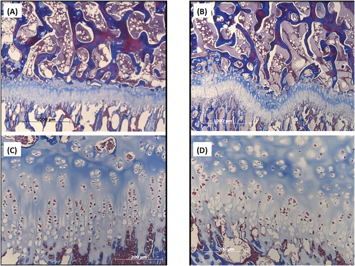
Results: The experimental animals had significantly greater kyphotic sagittal Cobb angles at all time points when compared with the control animals. These hyperkyphotic deformities demonstrated no significant differences in Hounsfield units, contained a slightly lower ash content (46.7% ± 1.1% compared with 50.9% ± 1.6%; p < 0.001), and demonstrated more physeal irregularities. Linear mixed model analysis of the measured kyphosis demonstrated that maternal diet had a greater effect on sagittal Cobb angle than did nursery diet and that postnatal supplementation did not completely eliminate the risk of hyperkyphosis.
Conclusions: Maternal diets deficient in vitamin D increased the development of hyperkyphosis in offspring in this model.
Clinical relevance: This study demonstrates that decreased maternal dietary vitamin-D intake during pregnancy increases the risk of spinal deformity in offspring. In addition, these data show the feasibility of generating a large-animal spinal-deformity model through dietary manipulation alone.
Figures
Comment in
-
Spinal Deformity in Vitamin D-Deprived Pigs: Why and What Next?: Commentary on an article by Matthew A. Halanski, MD, et al.: "Maternal Diets Deficient in Vitamin D Increase the Risk of Kyphosis in Offspring. A Novel Kyphotic Porcine Model".J Bone Joint Surg Am. 2018 Mar 7;100(5):e33. doi: 10.2106/JBJS.17.01345. J Bone Joint Surg Am. 2018. PMID: 29509629 No abstract available.
Similar articles
-
Serum and tissue 25-OH vitamin D3 concentrations do not predict bone abnormalities and molecular markers of vitamin D metabolism in the hypovitaminosis D kyphotic pig model.Br J Nutr. 2017 Jul;118(1):30-40. doi: 10.1017/S0007114517001751. Epub 2017 Jul 26. Br J Nutr. 2017. PMID: 28745259
-
Expression of kyphosis in young pigs is induced by a reduction of supplemental vitamin D in maternal diets and vitamin D, Ca, and P concentrations in nursery diets.J Anim Sci. 2012 Dec;90(13):4905-15. doi: 10.2527/jas.2012-5173. Epub 2012 Oct 16. J Anim Sci. 2012. PMID: 23100590
-
Maternal dietary vitamin D carry-over alters offspring growth, skeletal mineralisation and tissue mRNA expressions of genes related to vitamin D, calcium and phosphorus homoeostasis in swine.Br J Nutr. 2016 Sep;116(5):774-87. doi: 10.1017/S0007114516002658. Epub 2016 Aug 2. Br J Nutr. 2016. PMID: 27480125 Clinical Trial.
-
Spinal Deformity in Vitamin D-Deprived Pigs: Why and What Next?: Commentary on an article by Matthew A. Halanski, MD, et al.: "Maternal Diets Deficient in Vitamin D Increase the Risk of Kyphosis in Offspring. A Novel Kyphotic Porcine Model".J Bone Joint Surg Am. 2018 Mar 7;100(5):e33. doi: 10.2106/JBJS.17.01345. J Bone Joint Surg Am. 2018. PMID: 29509629 No abstract available.
-
The rehabilitation of hyperkyphotic posture in the elderly.Eur J Phys Rehabil Med. 2009 Dec;45(4):583-93. Eur J Phys Rehabil Med. 2009. PMID: 20032918 Review.
Cited by
-
Impact of dietary vitamin D3 supplements in nursery diets on subsequent growth and bone responses of pigs during an immune challenge.J Anim Sci. 2019 Dec 17;97(12):4895-4903. doi: 10.1093/jas/skz347. J Anim Sci. 2019. PMID: 31701141 Free PMC article.
-
Alleged predisposing dietary factors fail to increase the incidence of osteochondrosis-like lesions in growing pigs at 14 and 24 wk of age.J Anim Sci. 2020 Apr 1;98(4):skaa103. doi: 10.1093/jas/skaa103. J Anim Sci. 2020. PMID: 32249288 Free PMC article.
-
Osteochondrosis and other lesions in all intervertebral, articular process and rib joints from occiput to sacrum in pigs with poor back conformation, and relationship to juvenile kyphosis.BMC Vet Res. 2022 Jan 18;18(1):44. doi: 10.1186/s12917-021-03091-6. BMC Vet Res. 2022. PMID: 35042517 Free PMC article.
-
Biomechanical study of spinal cord and nerve root in idiopathic scoliosis: based on finite element analysis.BMC Musculoskelet Disord. 2024 Sep 6;25(1):717. doi: 10.1186/s12891-024-07832-0. BMC Musculoskelet Disord. 2024. PMID: 39243084 Free PMC article.
-
Understanding the etiopathogenesis of lumbar intervertebral disc herniation: From clinical evidence to basic scientific research.JOR Spine. 2023 Oct 18;7(1):e1289. doi: 10.1002/jsp2.1289. eCollection 2024 Mar. JOR Spine. 2023. PMID: 38222810 Free PMC article. Review.
References
-
- Damborg F, Engell V, Andersen M, Kyvik KO, Thomsen K. Prevalence, concordance, and heritability of Scheuermann kyphosis based on a study of twins. J Bone Joint Surg Am. 2006. October;88(10):2133-6. - PubMed
-
- Halal F, Gledhill RB, Fraser C. Dominant inheritance of Scheuermann’s juvenile kyphosis. Am J Dis Child. 1978. November;132(11):1105-7. - PubMed
MeSH terms
Substances
Grants and funding
LinkOut - more resources
Full Text Sources
Other Literature Sources
Medical
Research Materials
Miscellaneous
