

Collagens of Poriferan Origin
- PMID: 29510493
- PMCID: PMC5867623
- DOI: 10.3390/md16030079
Collagens of Poriferan Origin
Abstract
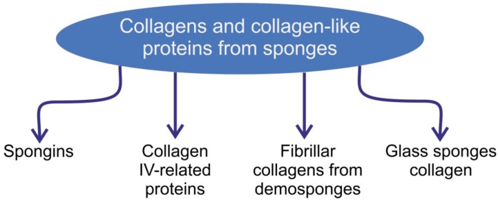
The biosynthesis, structural diversity, and functionality of collagens of sponge origin are still paradigms and causes of scientific controversy. This review has the ambitious goal of providing thorough and comprehensive coverage of poriferan collagens as a multifaceted topic with intriguing hypotheses and numerous challenging open questions. The structural diversity, chemistry, and biochemistry of collagens in sponges are analyzed and discussed here. Special attention is paid to spongins, collagen IV-related proteins, fibrillar collagens from demosponges, and collagens from glass sponge skeletal structures. The review also focuses on prospects and trends in applications of sponge collagens for technology, materials science and biomedicine.
Keywords: biomaterials; collagen; collagen-related proteins; scaffolds; sponges; spongin.
Conflict of interest statement
The authors declare no conflict of interest.
Figures
Similar articles
-
Marine Spongin: Naturally Prefabricated 3D Scaffold-Based Biomaterial.Mar Drugs. 2018 Mar 9;16(3):88. doi: 10.3390/md16030088. Mar Drugs. 2018. PMID: 29522478 Free PMC article. Review.
-
Discovery of mammalian collagens I and III within ancient poriferan biopolymer spongin.Nat Commun. 2025 Mar 13;16(1):2515. doi: 10.1038/s41467-025-57460-y. Nat Commun. 2025. PMID: 40082406 Free PMC article.
-
Insights into early extracellular matrix evolution: spongin short chain collagen-related proteins are homologous to basement membrane type IV collagens and form a novel family widely distributed in invertebrates.Mol Biol Evol. 2006 Dec;23(12):2288-302. doi: 10.1093/molbev/msl100. Epub 2006 Aug 31. Mol Biol Evol. 2006. PMID: 16945979
-
First Report on Chitin in a Non-Verongiid Marine Demosponge: The Mycale euplectellioides Case.Mar Drugs. 2018 Feb 20;16(2):68. doi: 10.3390/md16020068. Mar Drugs. 2018. PMID: 29461501 Free PMC article.
-
Secondary Metabolites from the Marine Sponge Genus Phyllospongia.Mar Drugs. 2017 Jan 6;15(1):12. doi: 10.3390/md15010012. Mar Drugs. 2017. PMID: 28067826 Free PMC article. Review.
Cited by
-
Spider Chitin: An Ultrafast Microwave-Assisted Method for Chitin Isolation from Caribena versicolor Spider Molt Cuticle.Molecules. 2019 Oct 16;24(20):3736. doi: 10.3390/molecules24203736. Molecules. 2019. PMID: 31623238 Free PMC article.
-
Discovery of chitin in skeletons of non-verongiid Red Sea demosponges.PLoS One. 2018 May 15;13(5):e0195803. doi: 10.1371/journal.pone.0195803. eCollection 2018. PLoS One. 2018. PMID: 29763421 Free PMC article.
-
Biocompatibility of a Marine Collagen-Based Scaffold In Vitro and In Vivo.Mar Drugs. 2020 Aug 11;18(8):420. doi: 10.3390/md18080420. Mar Drugs. 2020. PMID: 32796603 Free PMC article.
-
Spongin as a Unique 3D Template for the Development of Functional Iron-Based Composites Using Biomimetic Approach In Vitro.Mar Drugs. 2023 Aug 22;21(9):460. doi: 10.3390/md21090460. Mar Drugs. 2023. PMID: 37755073 Free PMC article.
-
On the Path to Thermo-Stable Collagen: Culturing the Versatile Sponge Chondrosia reniformis.Mar Drugs. 2021 Nov 26;19(12):669. doi: 10.3390/md19120669. Mar Drugs. 2021. PMID: 34940668 Free PMC article.
References
-
- Tzaphlidou M., Berillis P. Structural alterations caused by lithium in skin and liver collagen using an image processing method. J. Trace Microprobe Tech. 2002;20:493–504. doi: 10.1081/TMA-120015611. - DOI
-
- Ehrlich H., Deutzmann R., Brunner E., Cappellini E., Koon H., Solazzo C., Yang Y., Ashford D., Thomas-Oates J., Lubeck M., et al. Mineralization of the metre-long biosilica structures of glass sponges is templated on hydroxylated collagen. Nat. Chem. 2010;2:1084–1088. doi: 10.1038/nchem.899. - DOI - PubMed
Publication types
MeSH terms
Substances
LinkOut - more resources
Full Text Sources
Other Literature Sources
