

Biomechanical efficacy of AP, PA lag screws and posterior plating for fixation of posterior malleolar fractures: a three dimensional finite element study
- PMID: 29510693
- PMCID: PMC5840778
- DOI: 10.1186/s12891-018-1989-7
Biomechanical efficacy of AP, PA lag screws and posterior plating for fixation of posterior malleolar fractures: a three dimensional finite element study
Abstract
Background: Clinically there are different fixation methods used for fixation of the posterior malleolar fractures (PMF), but the best treatment modality is still not clear. Few studies have concentrated on this issue, least of all using a biomechanical comparison. The purpose of this study was to carry out a computational comparative biomechanics of three different commonly used fixation constructs for the fixation of PMF by finite element analysis (FEA).
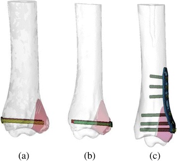
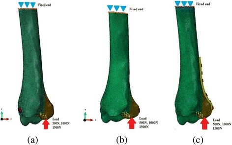
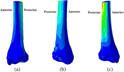
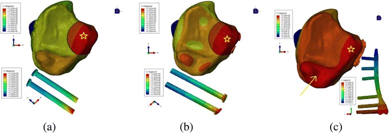
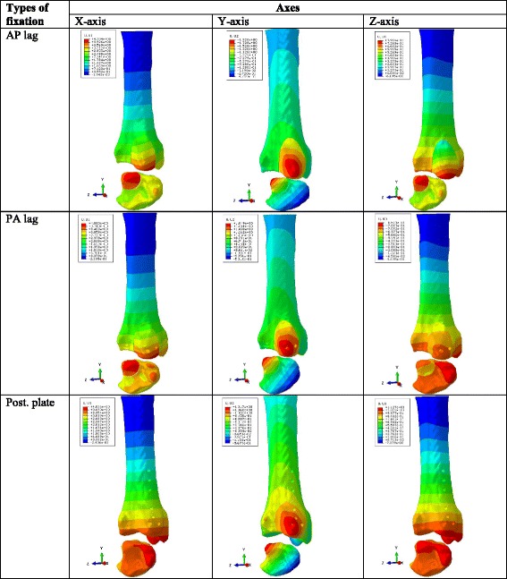
Methods: Computed tomography (CT) images were used to reconstruct three dimensional (3D) model of the tibia. Computer aided design (CAD) software was used to design 3D models of PMF. Finally, 3D models of PMF fixed with two antero-posterior (AP) lag screws, two postero-anterior (PA) lag screws and posterior plate were simulated through computational processing. Simulated loads of 500 N, 1000 N and 1500 N were applied to the PMF and proximal ends of the models were fixed in all degrees of freedom. Output results representing the model von Mises stress, relative fracture micro-motion and vertical displacement of the fracture fragment were analyzed.
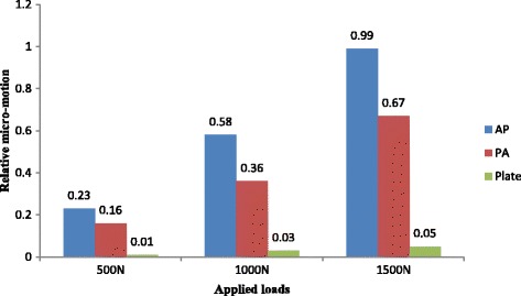
Results: The mean vertical displacement value in the posterior plate group (0.52 mm) was lower than AP (0.68 mm) and PA (0.69 mm) lag groups. Statistically significant low amount of the relative micro-motion (P < 0.05) was observed in the posterior plate group.
Conclusions: It was concluded that the posterior plate is biomechanically the most stable fixation method for fixation of PMF.
Keywords: Biomechanical; Finite element analysis; Fixation; Posterior malleolar fracture; Three dimensional.
Conflict of interest statement
Consent for publication
Not applicable.
Competing interests
The authors declare that they have no competing interest.
Publisher’s Note
Springer Nature remains neutral with regard to jurisdictional claims in published maps and institutional affiliations.
Figures


References
MeSH terms
LinkOut - more resources
Full Text Sources
Other Literature Sources
Medical
Miscellaneous
