. 2018 Mar 6;20(1):16.
doi: 10.1186/s13058-018-0944-8.

The hypoxic tumor microenvironment in vivo selects the cancer stem cell fate of breast cancer cells

Affiliations

The hypoxic tumor microenvironment in vivo selects the cancer stem cell fate of breast cancer cells

Hoon Kim et al. Breast Cancer Res. .

Abstract

Background: Tumor hypoxia is an independent prognostic factor associated with poor patient survival. Emerging evidence suggests that hypoxia can potentially maintain or enhance the stem cell phenotype of both normal stem cells and cancer cells. However, it remains to be determined whether cell fate is regulated in vivo by the hypoxic tumor microenvironment (TME).

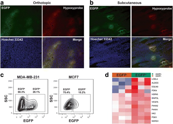
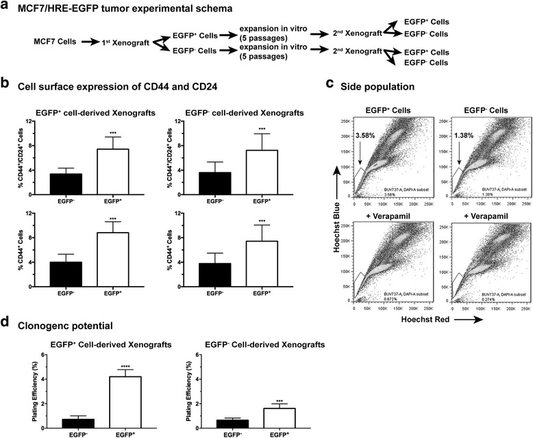
Methods: We established a hypoxia-sensing xenograft model to identify hypoxic tumor cell in vivo primarily using human breast cancer cell lines MDA-MB-231 and MCF7. Hypoxic tumor cells were identified in situ by fluorescence of green fluorescence protein. They were further isolated from xenografts, purified and sorted by flow cytometry for detailed analysis of their stem cell characteristics.

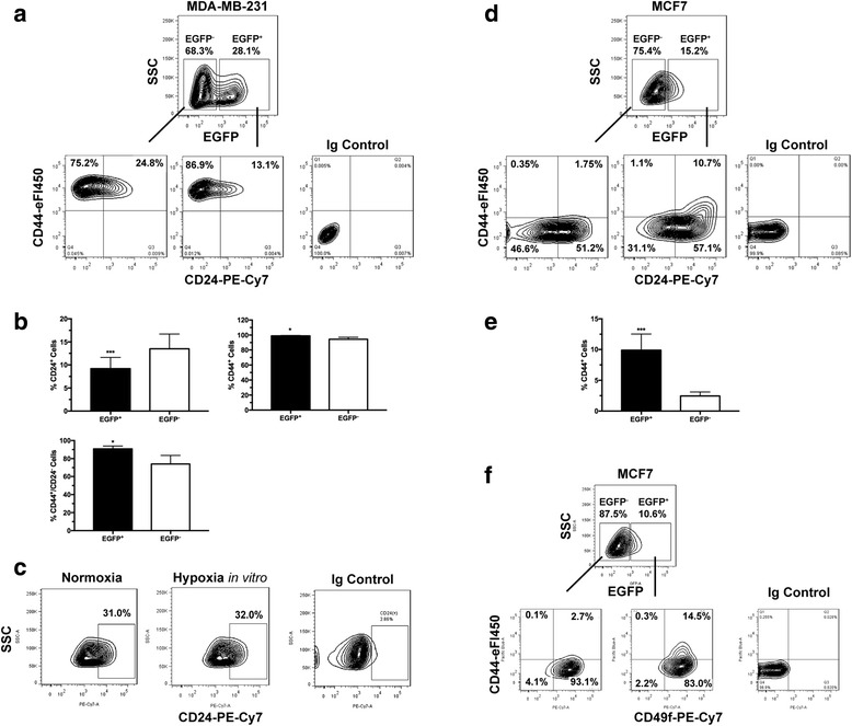
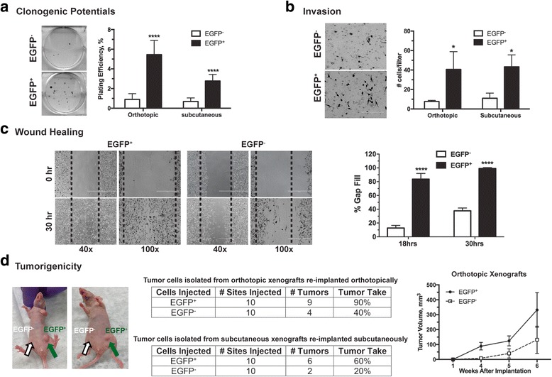
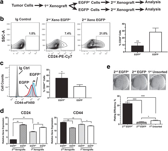
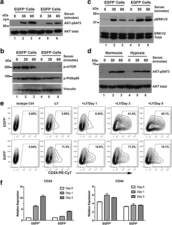
Results: We have found that hypoxic tumor cells freshly isolated from xenografts contain increased subpopulations of tumor cells with cancer stem cell (CSC)-like characteristics. The CSC characteristics of the hypoxic tumor cells are further enhanced upon re-implantation in vivo, whereas secondary xenografts derived from the non-hypoxic tumor cells remain similar to the primary xenografts. Interestingly, the phenotypes exhibited by the hypoxic tumor cells are stable and remain distinctively different from those of the non-hypoxic tumor cells isolated from the same tumor mass even when they are maintained under the same ambient culture conditions. Mechanistically, the PI3K/AKT pathway is strongly potentiated in the hypoxic tumor cells and is required to maintain the CSC-like phenotype. Importantly, the differential cell fates between hypoxic and non-hypoxic tumor cells are only found in tumor cells isolated from the hypoxic TME in vivo and are not seen in tumor cells treated by hypoxia in vitro alone.

Conclusions: These previously unknown observations suggest that the hypoxic TME may promote malignant progression and therapy resistance by coordinating induction, selection and/or preferential maintenance of the CSC-like phenotype in tumor cells.

Keywords: AKT; Breast cancer cell; Cancer stem cell; Cell fate; Hypoxia; PI3K; Tumor microenvironment; Xenograft.


Conflict of interest statement

Ethics approval and consent to participate

All animal procedures were reviewed and approved by the Institutional Animal Care & Use Committee (IACUC) of Yale University.

Consent for publication

Not applicable.

Competing interests

The authors declare that they have no competing interests.

Publisher’s Note

Springer Nature remains neutral with regard to jurisdictional claims in published maps and institutional affiliations.

Figures

Fig. 1
Fig. 1
The hypoxia-sensing human breast cancer xenograft model. a, b MDA-MB-231 cells stably expressing the HRE-EGPF reporter gene are implanted either orthotopically in mammary fat pads (a) or ectopically in the hind back (b) of female athymic mice. Hypoxic regions are visualized by immunostaining of the Hypoxyprobe (red). Expression of the hypoxia reporter gene is shown by fluorescence of enhanced green fluorescent protein (EGFP). Nuclei are counterstained with Hoechst 33342. c The hypoxic populations from the MDA-MB-231/HRE-EGFP and MCF7/HRE-EGFP xenografts, respectively, are analyzed by fluorescence-activated cell sorting. d Microarray analysis shows that expression of a panel of commonly observed hypoxia-induced genes is significantly upregulated in the EGFP+ cells freshly isolated from the MDA-MB-231/HRE-EGFP xenografts, compared to the EGFP cells from the same xenografts (n = 3; analysis of variance, p < 0.05). SSC, side scatter
Fig. 2
Fig. 2
Cancer stem cell (CSC)-like cells are enriched in the hypoxic populations freshly isolated from xenografts. Tumor cells are enzymatically dissociated and isolated from either the MDA-MB-231/HRE-EGFP (a-c) or MCF7/HRE-EGFP (d-f) xenografts. Stem cell characteristics are evaluated by fluorescence-activated cell sorting (FACS) for the expression of CSC-associated surface markers CD24, CD44 and CD49f. Representative FACS plots are shown in a, c, d and f. Quantitative population analyses are shown in b (n = 4–5; *p < 0.05, ***p < 0.001, Student’s t test) and e (n = 4; ***p < 0.001, Student’s t test). These results are confirmed by three or more independent experiments. EGFP, enhanced green fluorescent protein; SSC, side scatter
Fig. 3
Fig. 3
The ex vivo hypoxic tumor cells possess properties functionally associated with self-renewal and tumorigenic potentials. Tumor cells are enzymatically dissociated and isolated from the MDA-MB-231/HRE-EGFP xenografts. After sorting into the enhanced green fluorescent protein (EGFP)+ and EGFP populations, tumor cells were plated for in vitro assays (a-c) or directly re-implanted in athymic mice (d). Detailed experimental conditions are described in “Methods”. a Clonogenic potential (n = 6, ***p < 0.001, Student’s t test). b Tumor cell invasion (n = 3, *p < 0.05, Student’s t test). c Wound healing potentials (n = 5, *p < 0.001, Student’s t test). d Tumorigenic potentials in vivo are primarily reflected by percent tumor take (the ability of implanted cells to produce a tumor)
Fig. 4
Fig. 4
The cancer stem cell (CSC)-like population is further enriched in secondary xenografts derived from the enhanced green fluorescent protein (EGFP)+ MDA-MB-231 cells. a Generation of the secondary xenografts. b, c Surface levels of CD24 and CD44 are analyzed by fluorescence-activated cell sorting. b Average CD24+ populations from six individual tumors (***p < 0.001, Student’s t test). c Average CD44++ (right-pointing arrow) populations from three individual tumors (*p < 0.05, Student’s t test). d Quantitative RT-PCR analysis of expression of CD24 and CD44 genes in the EGFP+ and EGFP cells freshly isolated from either the 2nd or 1st xenografts (n = 3; *p < 0.05, **p < 0.01, Student’s t test). e Clonogenic growth of sorted EGFP+ and EGFP cells freshly isolated from the 2nd xenografts in comparison to the unsorted tumor cells from the 1st xenografts (n = 3; *p < 0.05, ***p < 0.0001, Student’s t test)
Fig. 5
Fig. 5
The cancer stem cell (CSC)-like characteristics of tumor cells isolated from the secondary enhanced green fluorescent protein (EGFP)+ MCF7/HRE-EGFP xenografts. a Generation of secondary MCF7/HRE-EGFP xenografts by re-implantation of sorted EGFP+ and EGFP cells isolated from the primary MCF7/HRE-EGFP xenografts. Tumor cells freshly isolated from the secondary xenografts were sorted into EGFP+ and EGFP populations for (b) fluorescence-activated cell sorting (FACS) analysis of the CD44+/CD24+ and CD44+ populations (n = 5 for EGFP+ cells, n = 4 for EGFP cells; ***p < 0.001, Student’s t test). c Side population (SP) of the secondary xenograft-derived tumor cells. MCF7 tumor cells were isolated from the secondary xenografts derived from EGFP+ and EGFP- tumor cells, respectively, and expanded in vitro for three passages. Cells were stained with Hoechst 33342 for side population analysis by FACS. Verapamil (50 μM) was used to block nuclear export of Hoechst 33342. These results were validated in two independent experiments. d Clonogenic potential of the freshly sorted EGFP+ and EGFP populations from the secondary xenografts (n = 6; ****p < 0.0001, ***p < 0.001, Student’s t test)
Fig. 6
Fig. 6
The PI3K/AKT pathway is required for maintenance of the CD44+/CD24 cancer stem cell (CSC) phenotype. The enhanced green fluorescent protein (EGFP)+ and EGFP cells sorted from the 1st MDA-MB-231 xenografts underwent serum starvation overnight. After serum stimulation, phosphorylation of AKT (a), PI3Kp85 (b), mTOR (b), and ERK1/2 (c) was analyzed using Western blots. d Serum-stimulated AKT phosphorylation in the parental MDA-MB-231/HRE-EGFP cells under normoxia and hypoxia (1% O2). e Increase in the CD24+ population induced by the PI3K inhibitor LY294002 (20 μM). f Quantitative RT-PCR analysis of expression of CD24 and CD44 genes in response to LY294002. These observations are confirmed by independent experiments

References

    1. Dhani N, Fyles A, Hedley D, Milosevic M. The clinical significance of hypoxia in human cancers. Semin Nucl Med. 2015;45(2):110–121. doi: 10.1053/j.semnuclmed.2014.11.002. - DOI - PubMed
    1. Vaupel P, Mayer A. Hypoxia in cancer: significance and impact on clinical outcome. Cancer Metastasis Rev. 2007;26(2):225–239. doi: 10.1007/s10555-007-9055-1. - DOI - PubMed
    1. Hockel M, Schlenger K, Aral B, Mitze M, Schaffer U, Vaupel P. Association between tumor hypoxia and malignant progression in advanced cancer of the uterine cervix. Cancer Res. 1996;56(19):4509–4515. - PubMed
    1. Lin Q, Yun Z. Impact of the hypoxic tumor microenvironment on the regulation of cancer stem cell characteristics. Cancer Biol Ther. 2010;9(12):949–956. doi: 10.4161/cbt.9.12.12347. - DOI - PMC - PubMed
    1. Simon MC, Keith B. The role of oxygen availability in embryonic development and stem cell function. Nat Rev Mol Cell Biol. 2008;9(4):285–296. doi: 10.1038/nrm2354. - DOI - PMC - PubMed

Publication types