

The role of the peritrophic matrix and red blood cell concentration in Plasmodium vivax infection of Anopheles aquasalis
- PMID: 29510729
- PMCID: PMC5840820
- DOI: 10.1186/s13071-018-2752-5
The role of the peritrophic matrix and red blood cell concentration in Plasmodium vivax infection of Anopheles aquasalis
Abstract
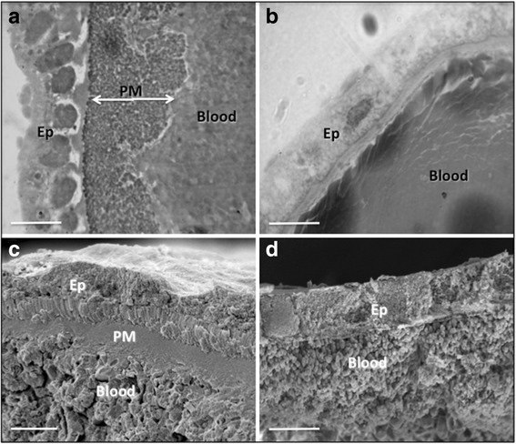
Background: Plasmodium vivax is predominant in the Amazon region, and enhanced knowledge of its development inside a natural vector, Anopheles aquasalis, is critical for future strategies aimed at blocking parasite development. The peritrophic matrix (PM), a chitinous layer produced by the mosquito midgut in response to blood ingestion, is a protective barrier against pathogens. Plasmodium can only complete its life-cycle, and consequently be transmitted to a new host, after successfully passing this barrier. Interestingly, fully engorged mosquitoes that had a complete blood meal form a thicker, well-developed PM than ones that feed in small amounts. The amount of red blood cells (RBC) in the blood meal directly influences the production of digestive enzymes and can protect parasites from being killed during the meal digestion. A specific study interrupting the development of the PM associated with the proteolytic activity inhibition, and distinct RBC concentrations, during the P. vivax infection of the New World malaria vector An. aquasalis is expected to clarify whether these factors affect the parasite development.
Results: Absence of PM in the vector caused a significant reduction in P. vivax infection. However, the association of chitinase with trypsin inhibitor restored infection rates to those of mosquitoes with a structured PM. Also, only the ingestion of trypsin inhibitor by non-chitinase treated mosquitoes increased the infection intensity. Moreover, the RBC concentration in the infected P. vivax blood meal directly influenced the infection rate and its intensity. A straight correlation was observed between RBC concentrations and infection intensity.
Conclusions: This study established that there is a balance between the PM role, RBC concentration and digestive enzyme activity influencing the establishment and development of P. vivax infection inside An. aquasalis. Our results indicate that the absence of PM in the midgut facilitates digestive enzyme dispersion throughout the blood meal, causing direct damage to P. vivax. On the other hand, high RBC concentrations support a better and thick, well-developed PM and protect P. vivax from being killed. Further studies of this complex system may provide insights into other details of the malaria vector response to P. vivax infection.
Keywords: Chitinase; Hematocrit; Malaria; Peritrophic matrix; Plasmodium vivax; Trypsin.
Conflict of interest statement
Authors’ information
ASO, RNP, OVL and DCBS received PhD scholarships from one of the following Brazilian agencies: FAPEAM, FAPESP, FIOCRUZ, CNPq and CAPES. DCBS is a PhD student from the Graduation Program in Tropical Medicine from the State University of Amazonas, Brazil. PFPP, FTMC, NFCS and MVGL are senior fellows supported by CNPq.
Consent for publication
Not applicable.
Competing interests
The authors declare that they have no competing interests.
Publisher’s Note
Springer Nature remains neutral with regard to jurisdictional claims in published maps and institutional affiliations.
Figures


Similar articles
-
Interactions of human malaria parasites, Plasmodium vivax and P.falciparum, with the midgut of Anopheles mosquitoes.Med Vet Entomol. 1997 Jul;11(3):290-6. doi: 10.1111/j.1365-2915.1997.tb00409.x. Med Vet Entomol. 1997. PMID: 9330262 Review.
-
Anopheles aquasalis transcriptome reveals autophagic responses to Plasmodium vivax midgut invasion.Parasit Vectors. 2019 May 24;12(1):261. doi: 10.1186/s13071-019-3506-8. Parasit Vectors. 2019. PMID: 31126324 Free PMC article.
-
Long-lasting infectivity of Plasmodium vivax present in malarial patient blood to Anopheles aquasalis.Exp Parasitol. 2021 Mar;222:108064. doi: 10.1016/j.exppara.2021.108064. Epub 2021 Jan 6. Exp Parasitol. 2021. PMID: 33421382
-
Promising approach to reducing Malaria transmission by ivermectin: Sporontocidal effect against Plasmodium vivax in the South American vectors Anopheles aquasalis and Anopheles darlingi.PLoS Negl Trop Dis. 2018 Feb 14;12(2):e0006221. doi: 10.1371/journal.pntd.0006221. eCollection 2018 Feb. PLoS Negl Trop Dis. 2018. PMID: 29444080 Free PMC article.
-
Plasmodium ookinete development in the mosquito midgut: a case of reciprocal manipulation.Parasitology. 1998;116 Suppl:S83-93. doi: 10.1017/s0031182000084973. Parasitology. 1998. PMID: 9695113 Review.
Cited by
-
Rosette formation by Plasmodium vivax gametocytes favors the infection in Anopheles aquasalis.Front Cell Infect Microbiol. 2023 Feb 15;13:1108348. doi: 10.3389/fcimb.2023.1108348. eCollection 2023. Front Cell Infect Microbiol. 2023. PMID: 36875524 Free PMC article.
-
Effects of Polystyrene Diet on the Growth and Development of Tenebrio molitor.Toxics. 2022 Oct 13;10(10):608. doi: 10.3390/toxics10100608. Toxics. 2022. PMID: 36287887 Free PMC article.
-
Viability and Infectivity of Plasmodium vivax Gametocytes in Short-Term Culture.Front Cell Infect Microbiol. 2021 Jun 1;11:676276. doi: 10.3389/fcimb.2021.676276. eCollection 2021. Front Cell Infect Microbiol. 2021. PMID: 34141630 Free PMC article.
-
Gut microbiota is essential in PGRP-LA regulated immune protection against Plasmodium berghei infection.Parasit Vectors. 2020 Jan 6;13(1):3. doi: 10.1186/s13071-019-3876-y. Parasit Vectors. 2020. PMID: 31907025 Free PMC article.
-
Sexual forms obtained in a continuous in vitro cultured Colombian strain of Plasmodium falciparum (FCB2).Malar J. 2020 Feb 3;19(1):57. doi: 10.1186/s12936-020-3142-y. Malar J. 2020. PMID: 32014000 Free PMC article.
References
-
- WHO . World malaria report. Geneva: World Health Organization; 2016.
-
- Deane LM. Malaria vectors in Brazil. Mem Inst Oswaldo Cruz. 1986;81(Suppl. 2):5–14. doi: 10.1590/S0074-02761986000600002. - DOI
Publication types
MeSH terms
Substances
Grants and funding
LinkOut - more resources
Full Text Sources
Other Literature Sources
Medical
