. 2018 Mar 6;37(1):49.
doi: 10.1186/s13046-018-0717-3.

MicroRNA-1468 promotes tumor progression by activating PPAR-γ-mediated AKT signaling in human hepatocellular carcinoma

Affiliations

MicroRNA-1468 promotes tumor progression by activating PPAR-γ-mediated AKT signaling in human hepatocellular carcinoma

Zhikui Liu et al. J Exp Clin Cancer Res. .

Retraction in

Abstract

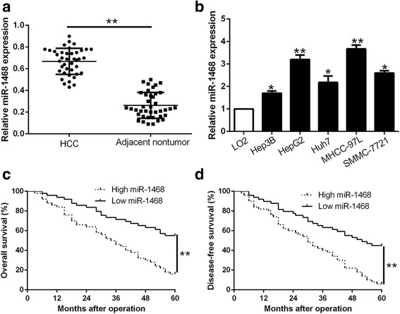
Background: Accumulating evidence confirm that aberrant microRNAs (miRNAs) expression contributes to hepatocellular carcinoma (HCC) development and progression. Previous study reported that miR-1468 showed an up-regulated tendency and might be a potential prognostic biomarker in HCC samples derived from TCGA database. However, the role of miR-1468 and its underlying mechanisms involved in the growth and metastasis of HCC remain poorly investigated.

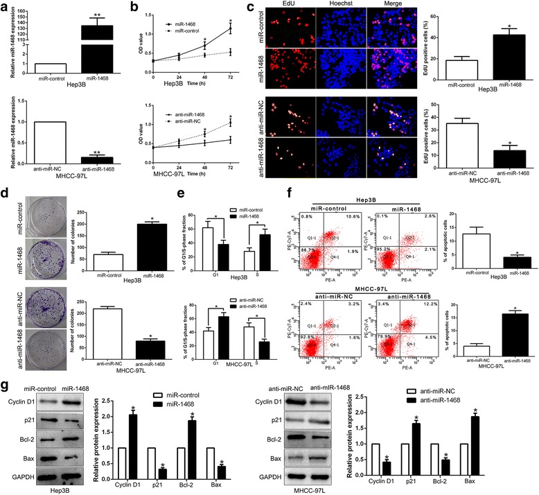
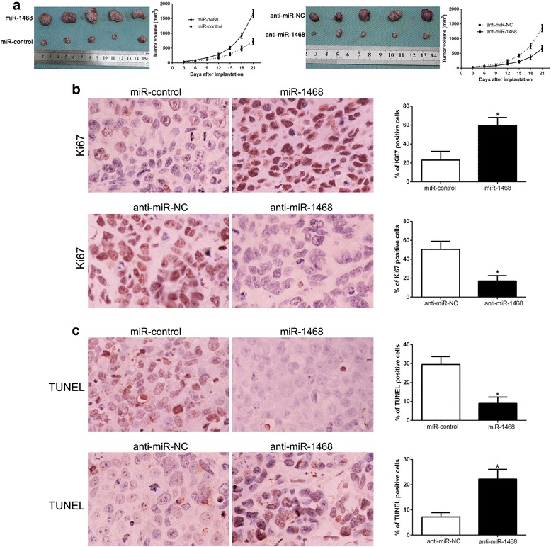
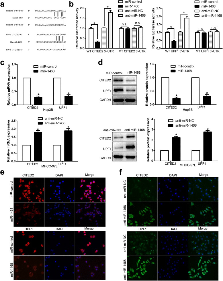
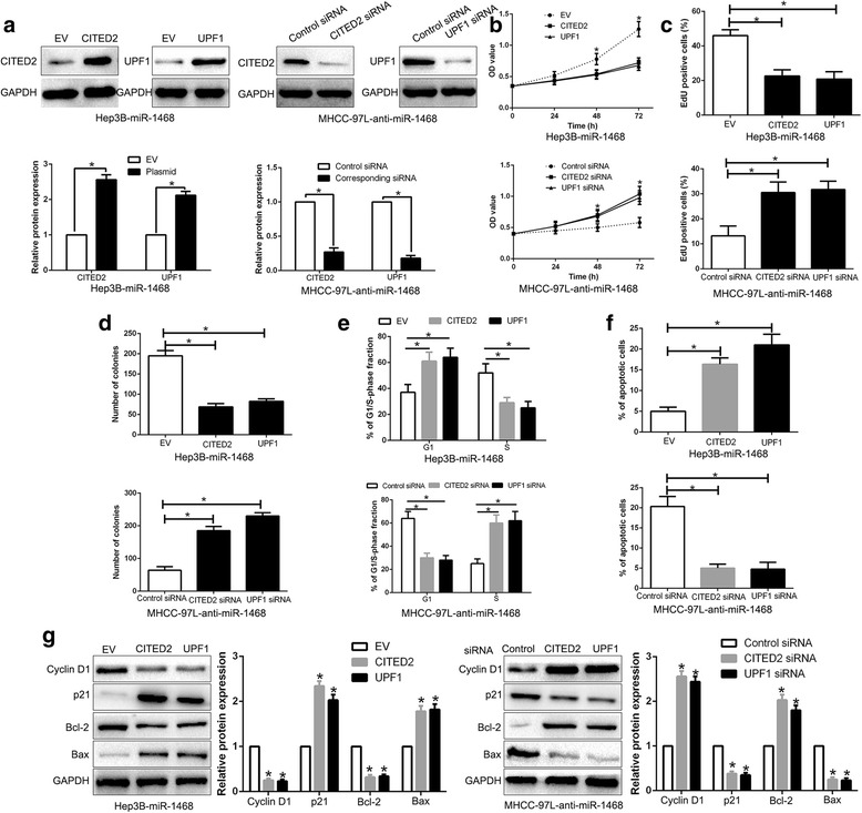
Methods: CCK-8, EdU, colony formation and flow cytometry were used to determine proliferation, cell cycle progression and apoptosis of HCC cells in vitro. The subcutaneous tumor model in nude mice was established to detect tumor growth of HCC in vivo. The direct binding of miR-1468 to 3'UTR of Cbp/p300 interacting transactivator with Glu/Asp rich carboxy-terminal domain 2 (CITED2) and Up-frameshift protein 1 (UPF1) was confirmed by luciferase reporter assay.

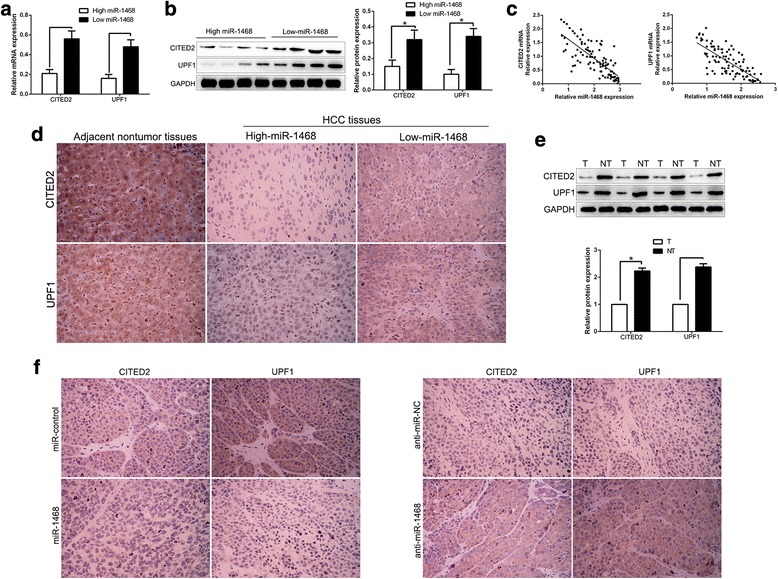
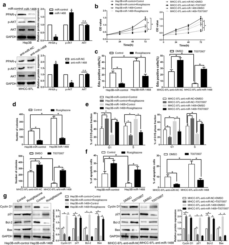
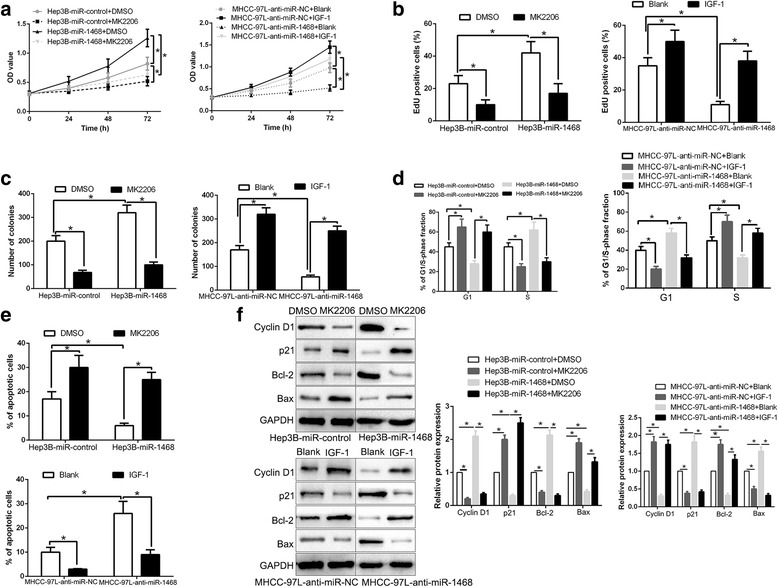
Results: Here, we demonstrated that miR-1468 expression was up-regulated in HCC tissues and cell lines. Clinical analysis revealed that increased miR-1468 level was significantly correlated with malignant prognostic features and shorter survival. Gain- and loss-of-function experiments indicated that miR-1468 promoted cell proliferation, colony formation, cell cycle progression and induced apoptosis of HCC cells in vitro and in vivo. Moreover, CITED2 and UPF1 were identified as direct downstream targets of miR-1468 in HCC cells, and mediated the functional effects of miR-1468 in HCC, resulting in peroxisome proliferator-activated receptor-γ (PPAR-γ)/AKT signaling activation. In clinical samples of HCC, miR-1468 inversely correlated with the levels of CITED2 and UPF1, which were confirmed to be down-regulated in HCC. Restoration of CITED2 or UPF1 expression at least partially abolished the biological effects of miR-1468 on HCC cells. Moreover, alteration of PPAR-γ or AKT phosphorylation could reverse the function of miR-1468 in HCC.

Conclusions: Taken together, this research supports the first evidence that miR-1468 plays an oncogenic role in HCC via activating PPAR-γ/AKT pathway by targeting CITED2 and UPF1, and represents a promising therapeutic strategy for HCC patients.

Keywords: CITED2; Hepatocellular carcinoma; PPAR-γ; Tumor growth; UPF2; miR-1468.


Conflict of interest statement

Ethics approval and consent to participate

All procedures performed in studies involving human participants were in accordance with the ethical standards of the Research Ethics Committee of The First Affiliated Hospital of Xi’an Jiaotong University and with the 1964 Helsinki declaration and its later amendments. ALL written informed consent to participate in the study was obtained from HCC patients for samples to be collected from them.

Consent for publication

Not applicable

Competing interests

The authors declare that they have no competing interests.

Publisher’s Note

Springer Nature remains neutral with regard to jurisdictional claims in published maps and institutional affiliations.

Figures

Fig. 1
Fig. 1
MiR-1468 is down-regulated in HCC and correlates with HCC progression and survival. Comparing differences in the expression of miR-1468 between (a) HCC and matched tumor-adjacent tissues and (b) HCC cell lines and the immortalized hepatic cell line LO2. HCC patients with higher expression of miR-1468 had worse overall survival (c) and disease-free survival (d). *P < 0.05, **P < 0.01
Fig. 2
Fig. 2
MiR-1468 promotes cell proliferation, cell cycle progression, colony formation and inhibits apoptosis in HCC cells in vitro. a Hep3B and MHCC-97 L cells that were transfected with corresponding miRNA vectors were subjected to qRT-PCR for miR-1468 expression. Overexpression of miR-1468 promoted cell proliferation (b, c), colony formation (d), cell cycle progression (e) and inhibited apoptosis (f) in Hep3B cells, while down-regulation of miR-1468 inhibited cell proliferation (b, c), colony formation (d), cell cycle progression (e) and promoted apoptosis (f) in MHCC-97 L cells. g Western blot analysis of cycle regulator Cyclin D1 and p21, apoptosis-related protein Bcl2/Bax expression in the presence and absence of miR-1468. n = six independent experiments. *P < 0.05, **P < 0.01
Fig. 3
Fig. 3
MiR-1468 promotes tumor growth and inhibits apoptosis in vivo. a Representative pictures of HCC xenografts from both Hep3B-miR-1468 (left panel) and MHCC-97 L-anti-miR-1468 cells (right panel). Tumor growth curve revealed that miR-1468 overexpression significantly promoted, while miR-1468 knockdown inhibited tumor growth in vivo. Tumor nodules were subjected to immunohistochemical staining for Ki-67 (b) and TUNEL (c) assays and quantitative analysis. Representative immunostaining and TUNEL assays revealed that miR-1468 overexpression significantly increased the number of Ki-67 positive cells and inhibited the number of apoptotic cells. However, the percentage of Ki-67 positive cells in tumors arising from the miR-1468 knockdown group was significantly lower and the percentage of apoptotic cells was significantly higher than that in the negative control (NC) group. *P < 0.05
Fig. 4
Fig. 4
CITED2 and UPF1 are direct targets of miR-1468 in HCC cells. a miR-1468 and its putative binding sequences in the 3’-UTR of CITED2 and UPF1. The mutant binding site was generated in the complementary site for the seed region of miR-1468. b miR-1468 overexpression significantly suppressed, while miR-1468 loss increased the luciferase activity that carried wild-type (wt) but not mutant (mt) 3’-UTR of CITED2 or UPF1. c Hep3B and MHCC-97 L cells that were transfected with precursor miR-1468 and miR-1468 inhibitors (anti-miR-1468), respectively, were subjected to qRT-PCR for CITED2 and UPF1 mRNA expression. d miR-1468 overexpression reduced the expression of CITED2 and UPF1 protein in Hep3B cells and miR-1468 knockdown increased the level of CITED2 and UPF1 protein in MHCC-97 L cells. e, f Immunofluorescence staining of CITED2 and UPF1 after transduction of miR-1468. n.s, no significance, *P < 0.05
Fig. 5
Fig. 5
An inverse correlation between miR-1468 and CITED2, UPF1 expression is observed in HCC tissues. a and b The expression of CITED2 and UPF1 in miR-1468 high-expressing tumors was significantly lower than that in miR-1468 low-expressing tumors, as determined by qRT-PCR and immunoblotting. c An inverse correlation between the levels of miR-1468 and CITED2, UPF1 mRNA was observed in HCC tissues. d Representative immunohistochemical staining showed a weak staining of CITED2, UPF1 in miR-1468 high-expressing HCC tissue and strong staining of CITED2, UPF1 in the miR-1468 low-expressing tumor. The adjacent nontumor tissues show a stronger staining of CITED2 and UPF1 compared to HCC tissues. e The expression of CITED2 and UPF1 were down-regulated in HCC tissues compared with matched non-tumor tissues. f Xenografts tissues immunohistochemical staining of CITED2 and UPF1 in miR-1468 overexpression or knockdown subcutaneous tumor nodules. *P < 0.05
Fig. 6
Fig. 6
Modulation of CITED2 or UPF1 partially abolishes miR-1468-mediated cellular processes in HCC. a miR-1468-overexpressing Hep3B cells that were transfected with empty vector (EV) or CITED2, UPF1 overexpression plasmid were subjected to western blot for CITED2 and UPF1. miR-1468-suppressive MHCC-97 L cells that were transfected with scrambled siRNA or CITED2, UPF1 siRNA were subjected to western blot for CITED2, UPF1. CITED2 or UPF1 restoration abrogated the effects of miR-1468 overexpression on cell proliferation (b, c), colony formation (d), cell cycle progression (e) and apoptosis (f) of Hep3B cells. CITED2 or UPF1 knockdown reversed the suppressive effects of miR-1468 knockdown in MHCC-97 L cells (b-f). g Alteration of CITED2 or UPF1 abolished the effects of miR-1468 on cell cycle and apoptosis associated factors. *P < 0.05
Fig. 7
Fig. 7
PPAR-γ/AKT signaling is essential for the biological function of miR-1468 in HCC. a Hep3B and MHCC-97 L cells that were transfected with corresponding miRNA vectors were subjected to immunoblotting for phosphorylated AKT and AKT. PPAR-γ agonist rosiglitazone treatment inhibited the promotive effects on cell proliferation (b, c), colony formation (d), cell cycle progression (e) and apoptosis (f) of miR-1468 overexpressing Hep3B cells. PPAR-γ antagonist, T0070907, reversed the suppressive effects of miR-1468 knockdown in MHCC-97 L cells. g Western blot analysis indicated that modulating PPAR-γ activation reversed the effects of miR-1468 alteration on cell cycle and apoptosis associated factors of HCC cells. *P < 0.05
Fig. 8
Fig. 8
AKT phosphorylation is essential for the biological function of miR-1468 in HCC. AKT inhibitor MK2206, or AKT phosphorylation activator IGF-1, abolished the cell proliferation (a, b), colony formation (c), cell cycle progression (d) and apoptosis (e) of HCC cells which were transduced of miR-1468 vectors. f Western blot analysis indicated that modulating AKT phosphorylation reversed the effects of miR-1468 alteration on cell cycle and apoptosis associated factors of HCC cells. *P < 0.05

References

    1. Torre LA, Bray F, Siegel RL, Ferlay J, Lortet-Tieulent J, Jemal A. Global cancer statistics, 2012. CA Cancer J Clin. 2015;65(2):87–108. doi: 10.3322/caac.21262. - DOI - PubMed
    1. Forner A, Llovet JM, Bruix J. Hepatocellular carcinoma. Lancet. 2012;379(9822):1245–1255. doi: 10.1016/S0140-6736(11)61347-0. - DOI - PubMed
    1. El-Serag HB, Rudolph KL. Hepatocellular carcinoma: epidemiology and molecular carcinogenesis. Gastroenterology. 2007;132(7):2557–2576. doi: 10.1053/j.gastro.2007.04.061. - DOI - PubMed
    1. Maluccio M, Covey A. Recent progress in understanding, diagnosing, and treating hepatocellular carcinoma. CA Cancer J Clin. 2012;62(6):394–399. doi: 10.3322/caac.21161. - DOI - PubMed
    1. Garzon R, Calin GA, Croce CM. MicroRNAs in cancer. Annu Rev Med. 2009;60:167–179. doi: 10.1146/annurev.med.59.053006.104707. - DOI - PubMed

Publication types

MeSH terms