. 2018 Mar 20;115(12):E2849-E2858.
doi: 10.1073/pnas.1722344115. Epub 2018 Mar 6.

Murine knockin model for progranulin-deficient frontotemporal dementia with nonsense-mediated mRNA decay

Affiliations

Murine knockin model for progranulin-deficient frontotemporal dementia with nonsense-mediated mRNA decay

Andrew D Nguyen et al. Proc Natl Acad Sci U S A. .

Abstract

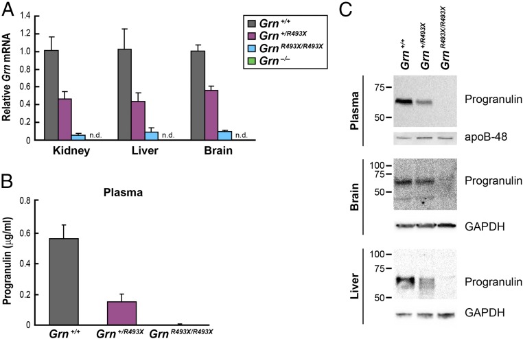
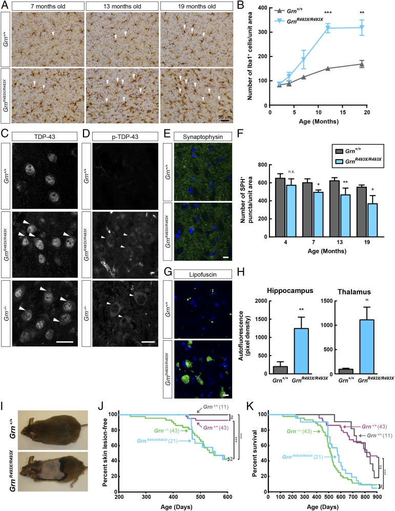
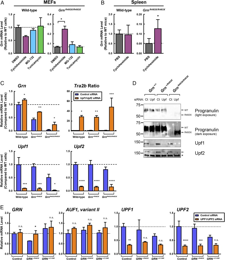
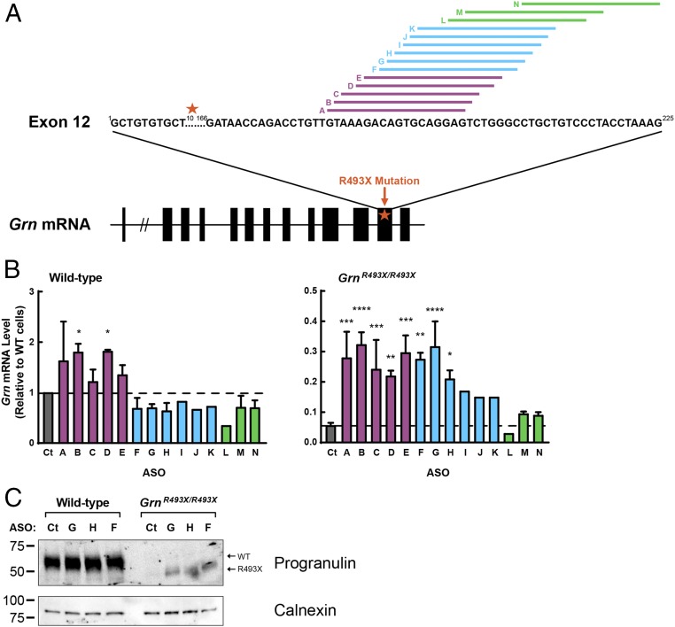
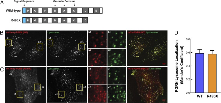
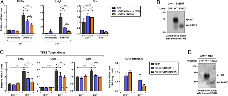
Frontotemporal dementia (FTD) is the most common neurodegenerative disorder in individuals under age 60 and has no treatment or cure. Because many cases of FTD result from GRN nonsense mutations, an animal model for this type of mutation is highly desirable for understanding pathogenesis and testing therapies. Here, we generated and characterized GrnR493X knockin mice, which model the most common human GRN mutation, a premature stop codon at arginine 493 (R493X). Homozygous GrnR493X mice have markedly reduced Grn mRNA levels, lack detectable progranulin protein, and phenocopy Grn knockout mice, with CNS microgliosis, cytoplasmic TDP-43 accumulation, reduced synaptic density, lipofuscinosis, hyperinflammatory macrophages, excessive grooming behavior, and reduced survival. Inhibition of nonsense-mediated mRNA decay (NMD) by genetic, pharmacological, or antisense oligonucleotide-based approaches showed that NMD contributes to the reduced mRNA levels in GrnR493X mice and cell lines and in fibroblasts from patients containing the GRNR493X mutation. Moreover, the expressed truncated R493X mutant protein was functional in several assays in progranulin-deficient cells. Together, these findings establish a murine model for in vivo testing of NMD inhibition or other therapies as potential approaches for treating progranulin deficiency caused by the R493X mutation.

Keywords: frontotemporal dementia; lysosome; neurodegeneration; nonsense-mediated mRNA decay; progranulin.


Conflict of interest statement

The authors declare no conflict of interest.

Figures

Fig. 1.
Fig. 1.
Homozygous GrnR493X targeted mice have markedly reduced Grn mRNA levels and lack progranulin protein. (A) Grn mRNA levels in tissues were determined by qPCR. (B) ELISA of progranulin in mouse plasma. Data are presented as mean ± SD. (C) Immunoblot analysis of progranulin in mouse tissues. apo, apolipoprotein; n.d., not detected.
Fig. 2.
Fig. 2.
Phenotype of GrnR493X/R493X mice recapitulates features of global knockout mice. (A and B) GrnR493X/R493X mice exhibit age-dependent microgliosis. (A) Immunostaining of Iba1 in the thalamus. Arrowheads indicate selected Iba1+ cells. (B) Quantification of Iba1+ microglial density in 12-mo-old mice, n = 3–4 mice per genotype. (C and D) Increased levels of total (C) and phosphorylated (D) TDP-43 in the cytoplasm of thalamic neurons of 12-mo-old GrnR493X/R493X and Grn−/− mice. Arrowheads indicate cytoplasmic accumulation of TDP-43. (Scale bars, 20 μm.) (E and F) GrnR493X/R493X mice exhibit age-dependent reduction of synaptic density. (E) Immunostaining of synaptophysin in the thalamus of 13-mo-old mice. (Scale bar, 10 μm.) (F) Quantification of synaptophysin density, n = 3–4 mice per genotype at each age. (G and H) GrnR493X/R493X mice have increased lipofuscin in the brain. (G) Images of autofluorescent lipofuscin (green channel) in the thalamus of 19-mo-old mice. (Scale bar, 10 μm.) (H) Quantification of autofluorescence. (IK) GrnR493X/R493X mice have increased skin lesions resulting in decreased survival. (I) Skin lesions in 16-mo old mice. (I and J) Kaplan–Meier curves for skin lesion onset (J) and survival (K) in Grn+/+ (gray curves) and GrnR493X/R493X (blue curves) littermate mice. For comparison, curves for Grn+/+ (purple curves) and Grn−/− (green curves) littermate mice are also shown. Data are presented as mean ± SD; *P < 0.05, **P < 0.01, ***P < 0.001, as determined by Student’s t test (B, F, and H), Long-rank (Mantel–Cox) test (J and K). n.s., not significant; SPH, synaptophysin.
Fig. 3.
Fig. 3.
Blocking NMD increases Grn mRNA in GrnR493X mouse model and GRN+/R493X patient fibroblasts. (A) Wild-type and GrnR493X/R493X MEFs were treated for 5 h with 50 μM cycloheximide, 10 μM MG-132, or 10 μg/mL tunicamycin. Gene expression was determined by qPCR. (B) Wild-type and GrnR493X/R493X mice (n = 4–7 per group) were acutely injected with 20 mg/kg body weight cycloheximide dissolved in PBS. After 4 h, gene expression was determined by qPCR. (C and D) Upf1 and Upf2 were knocked down in wild-type, Grn+/R493X, and GrnR493X/R493X MEFs using DharmaFECT1. After 3 d, gene expression was determined by qPCR (C), and protein levels were determined by Western blot analyses (D). (E) UPF1 and UPF2 were knocked down in control and GRN+/R493X HDFs using DharmaFECT1. After 3 d, gene expression was determined by qPCR. All data are presented as mean ± SD; *P < 0.05, **P < 0.01, ***P < 0.001, ****P < 0.0001, as determined by one-way ANOVA followed by Tukey’s post hoc test (A), or two-way ANOVA followed by Bonferroni’s post hoc test (B, C, and E). Asterisk in D indicates a nonspecific band. n.s., not significant.
Fig. 4.
Fig. 4.
ASOs designed to inhibit NMD of GrnR493X increase Grn mRNA in GrnR493X cells. (A) Schematic of ASO targets in exon 12 of the Grn mRNA. Black boxes, coding exons; star, R493X mutation. (B and C) MEFs were transfected with ASOs using Lipofectamine 2000. After 24 h, gene expression was determined by qPCR (B); after 48 h, protein levels in cell lysates were determined by immunoblot analysis (C). Data are presented as mean ± SD; *P < 0.05, **P < 0.01, ***P < 0.001, ****P < 0.0001, as determined by one-way ANOVA followed by Dunnett post hoc test. Ct, control.
Fig. 5.
Fig. 5.
Progranulin R493X mutant is delivered to lysosomes. (A) Schematic representation of the wild-type progranulin and the R493X truncated mutant proteins. Blue boxes, signal sequence; gray boxes, granulin domains. (B and C) Images show the colocalization of mCherry-PGRN wild-type (B) and mCherry-PGRN R493X (C) with Alexa Fluor 647-dextran–labeled lysosomes in progranulin-deficient HeLa cells. Inset boxes (b1, b2, c1, and c2) obtained from the highlighted regions show the robust overlap of mCherry-PGRN puncta with lysosomes. (Scale bars, 10 μm.) (Magnification: B and C, Insets, 3×.) (D) Quantitative colocalization analysis shows similar lysosome localization for both wild-type and R493X forms of progranulin. Colocalization coefficient was quantified on a per cell basis, and 22–28 transfected cells were measured for each plasmid. Data are presented as mean ± SEM from four independent experiments. P = 0.897 (nonsignificant), as determined by unpaired t test.
Fig. 6.
Fig. 6.
Progranulin R493X mutant is functional in cell-based assays. (A) Overexpression of wild-type progranulin and the R493X mutant in Grn−/− BMDMs suppresses the proinflammatory response. Macrophages were infected with the indicated lentivirus for 24 h, and subsequently treated with 10 ng/mL LPS and 10 ng/mL IFN-γ for an additional 24 h. Gene expression was determined by qPCR. (B) Immunoblot analysis of progranulin in conditioned media from Grn−/− macrophages. (C) Overexpression of wild-type progranulin and the R493X mutant in Grn−/− MEFs restores normal expression of TFEB target genes. (D) Immunoblot analysis of progranulin in conditioned media from Grn−/− MEFs. All data are presented as mean ± SD; *P < 0.05, **P < 0.01, ***P < 0.001, ****P < 0.0001, as determined by two-way ANOVA followed by Bonferroni’s post hoc test. n.s., not significant.

References

    1. Baker M, et al. Mutations in progranulin cause tau-negative frontotemporal dementia linked to chromosome 17. Nature. 2006;442:916–919. - PubMed
    1. Cruts M, et al. Null mutations in progranulin cause ubiquitin-positive frontotemporal dementia linked to chromosome 17q21. Nature. 2006;442:920–924. - PubMed
    1. Gass J, et al. Mutations in progranulin are a major cause of ubiquitin-positive frontotemporal lobar degeneration. Hum Mol Genet. 2006;15:2988–3001. - PubMed
    1. Cruts M. 2012 The AD & FTLD mutation database. Available at www.molgen.vib-ua.be/FTDMutations. Accessed November 8, 2016.
    1. Cruts M, Theuns J, Van Broeckhoven C. Locus-specific mutation databases for neurodegenerative brain diseases. Hum Mutat. 2012;33:1340–1344. - PMC - PubMed

Publication types