. 2018 Mar 6;11(520):eaan6480.
doi: 10.1126/scisignal.aan6480.

A role for corticotropin-releasing factor signaling in the lateral habenula and its modulation by early-life stress

Affiliations

A role for corticotropin-releasing factor signaling in the lateral habenula and its modulation by early-life stress

Michael E Authement et al. Sci Signal. .

Abstract

Centrally released corticotropin-releasing factor or hormone (extrahypothalamic CRF or CRH) in the brain is involved in the behavioral and emotional responses to stress. The lateral habenula (LHb) is an epithalamic brain region involved in value-based decision-making and stress evasion. Through its inhibition of dopamine-mediated reward circuitry, the increased activity of the LHb is associated with addiction, depression, schizophrenia, and behavioral disorders. We found that extrahypothalamic CRF neurotransmission increased neuronal excitability in the LHb. Through its receptor CRFR1 and subsequently protein kinase A (PKA), CRF application increased the intrinsic excitability of LHb neurons by affecting changes in small-conductance SK-type and large-conductance BK-type K+ channels. CRF also reduced inhibitory γ-aminobutyric acid-containing (GABAergic) synaptic transmission onto LHb neurons through endocannabinoid-mediated retrograde signaling. Maternal deprivation is a severe early-life stress that alters CRF neural circuitry and is likewise associated with abnormal mental health later in life. LHb neurons from pups deprived of maternal care exhibited increased intrinsic excitability, reduced GABAergic transmission, decreased abundance of SK2 channel protein, and increased activity of PKA, without any substantial changes in Crh or Crhr1 expression. Furthermore, maternal deprivation blunted the response of LHb neurons to subsequent, acute CRF exposure. Activating SK channels or inhibiting postsynaptic PKA activity prevented the effects of both CRF and maternal deprivation on LHb intrinsic excitability, thus identifying potential pharmacological targets to reverse central CRF circuit dysregulation in patients with associated disorders.


Conflict of interest statement

Competing interests: The authors declare that they have no conflicts of interest.

Figures

Figure 1
Figure 1
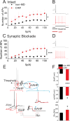
CRF increased the excitability of LHb neurons. A: Whole cell patch clamp recording of action potentials (APs) in LHb neurons from non-MD rats in response to depolarizing current injections (I) and CRF (250nM) bath application in slices, with synaptic transmission intact (n=7 cells from 7 rats; F(1,6)=12.3). B: Sample AP recordings of LHb neurons in response to a 50pA depolarizing current step before (baseline, black) and after CRF (red) application (calibration bar: 20mV/1s) from data in (A). C: Whole cell patch clamp recordings of APs as described in (A), with fast synaptic transmission blocked. (n=10 cells from 10 rats; F(1,9)=13.901). D: Sample AP recordings as described in (B) with synaptic blockade. E: Schematic of the method to measure AP threshold, mAHP and fAHP in sample AP recordings in response to the lowest depolarizing current step that generated the first AP/s before (black) and after CRF (red) application (calibration bar: 20mV/1s). Input resistance (Rin) was calculated from the steady-state voltage deflections generated in response to a 50pA hyperpolarizing current (calibration bar: 10mV/1s). F: Average amplitude of fAHP, mAHP and Rin derived from recordings in (C). Data are means ± SEM from cells for each condition (one cell from each rat); *P<0.05, ***P<0.001, by two-way RM-ANOVA (A and C) or paired Student's t-test (F).
Figure 2
Figure 2
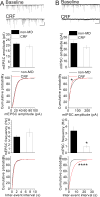
CRF depressed GABAergic transmission without affecting glutamatergic synaptic transmission. A: Top: Representative AMPAR-mediated mEPSC traces recorded from LHb neurons from non-MD rats before (baseline) and after CRF (250 nM) application (calibration bars: 30 pA/5s). Graphs below: Average mEPSC amplitude and frequency, and the cumulative probability plots of amplitude and frequency (inter-event interval) for all mEPSCs before and after CRF application. (n=10 cells from 10 non-MD rats). B: As described in (A) for GABAAR-mediated mIPSCs before and after CRF application (calibration bars: 50 pA/5s). (n=6 cells from 6 non-MD rats). Data are means ± SEM from cells for each condition (one cell from each rat). *P<0.05, ****P<0.0001 by paired Student's t-tests, or KS tests for cumulative distribution curves.
Figure 3
Figure 3
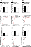
CRF suppressed GABA release from GABAergic terminals through retrograde eCB signaling. (A–C): Average mIPSC amplitude and frequency, and cumulative probability plots of amplitude and frequency (inter-event interval) for all mIPSCs before and after the application of (A) CRF (250 nM) with GDPβS (300 µM) present in the patch pipette (n=10 cells from 10 non-MD rats), (B) WIN55,212-2 (2 µM; n=8 cells from 8 non-MD rats), or (C) CRF with AM251 (10 µM) present in the perfusate (n=7 cells recorded from 7 non-MD rats). Data are means ± SEM from cells for each condition (one cell from each rat). *P< 0.05, ***P<0.001, ****P<0.001, by paired Student's t-tests, or KS tests for the cumulative distribution curves.
Figure 4
Figure 4
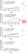
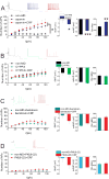
CRF increased LHb intrinsic excitability through G-protein-dependent CRFR1 signaling and requires intracellular Ca2+. (A to D) Whole cell patch clamp recordings of APs in LHb neurons from non-MD rats in response to depolarizing step currents (left), with representative AP traces (right) in response to a 100pA depolarizing current step before (baseline, black) and after application of CRF (250 nM, red), CRFR1 antagonist antalarmin (A; 1 µM; F(1,6)=0.222), CRFR2 antagonist antisauvagine (B; 25 nM; n=6 cells from 6 non-MD rats, F(1,5) = 5.057), G-protein inhibitor GDPβS (C; 300 µM; n=4 cells from 4 non-MD rats, F(1,3)=0.86), and calcium chelator BAPTA (D; 30 mM; n=6 cells from 6 non-MD rats, F(1,5) = 0.327) was included in the patch pipette or perfusate). All recordings performed with fast synaptic transmission blocked. Data are means ± SEM from cells for each condition (one cell from each rat); **P<0.01 by two-way RM-ANOVA.
Figure 5
Figure 5
Pharmacological modulation of SK and BK channels altered CRF-induced increases in LHb intrinsic excitability in a PKA-dependent manner. (A–D): AP recordings in response to depolarizing current steps with representative AP traces in LHb neurons from non-MD rats in response to a 100pA current step before (baseline, black) and after (A) SK channel blocker apamin (100nM, blue; n=11 cells from 11 non-MD rats, F(2,20)=9.127), (B) SK channel positive modulator CYPPA (10µM, green; n=9 cells from 9 non-MD rats, F(2,16)=2.058), (C) BK channel blocker iberiotoxin (100nM, teal blue; n=7 cells from 7 non-MD rats, F(1,6)=3.039), or (D) PKI(–22) (10 µM, sky blue; n=6 cells from 6 non-MD rats, F(1,5)=2.773) alone or with subsequent CRF (250 nM, red) bath application. Right: Amplitudes of fAHP, mAHP and Rin derived from the AP recordings. All recordings were performed with fast synaptic transmission blocked. Data are means ± SEM from cells for each condition (one cell from each rat). *P<0.05, ***P< 0.001, by two-way RM-ANOVA (left panels) or paired Student's t-tests (right panels).
Figure 6
Figure 6
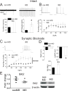
MD increased the excitability of LHb neurons. A. Top: Representative spontaneous AP recordings of LHb neurons from non-MD and MD rats using cell-attached voltage clamp recordings. Bottom: Percent of spontaneously active LHb neurons (left) and mean AP frequency (right) from each group (n= 33 cells from 18 rats or 29 cells from 19 rats, respectively). B. Representative traces (top) and number of APs (bottom) recorded from LHb neurons using whole cell patch clamp recording, with synaptic transmission intact, in slices from non-MD and MD rats in response to depolarizing current steps (calibration bar: 20mV/1s) [n= 24 cells from 11 rats (non-MD) or 24 cells from 9 rats (MD), F(1,460)=1.69]C. As described in (B), with fast synaptic transmission blocked [n= 25 cells from 17 rats (non-MD) or 31 cells from 18 rats (MD), F(1,600)=35.84]. D: Average amplitude of fAHP, mAHP and Rin derived from AP recordings in (C). E: Representative Western blots and quantitation of SK2 abundance (β-actin: loading control) in LHb tissue homogenates from non-MD and MD rats (n= 6 biological replicates). Data are means ± SEM. *P<0.05, **P<0.01 by unpaired Student's t-test; ****P<0.0001 by two-way ANOVA.
Figure 7
Figure 7
MD potentiated both glutamatergic and GABAergic synaptic transmission onto LHb neurons and increased E/I ratio. A: Representative AMPAR-mediated mEPSC traces from non-MD and MD rats (calibration bars: 30 pA/5s). Average mEPSC amplitude and frequency (left) and cumulative probability plots of amplitude and frequency (inter-event interval) (right) in non-MD and MD rats (n= 20 cells from 20 rats or 17 cells from 17 rats, respectively). B: As described in (A) for GABAAR-mediated mIPSC traces from non-MD and MD rats (calibration bars: 50 pA/5s; n=13 cells from 13 rats or 9 cells from 9 rats, respectively). C: Summary of E/I ratios obtained from non-MD and MD rats (n=16 cells from 8 rats or 12 cells from 9 rats, respectively), with representative traces of evoked EPSCs (black, recorded at −55 mV holding potential) and IPSCs (red, recorded at +10 mV holding potential) in response to the stria medullaris stimulation for LHb neurons. *P<0.05, **P<0.01, ****P< 0.0001 by unpaired Student's t-tests, or KS tests for the cumulative distribution curves.
Figure 8
Figure 8
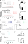
The effects of CRF and apamin on LHb intrinsic excitability were blunted following MD in a PKA-dependent manner. A: Whole cell patch clamp AP recordings in LHb neurons in slices from MD rats in response to depolarizing current steps with representative AP traces (in response to a 100pA current step) before (baseline, black) and after application of CRF (250 nM, red), with fast synaptic transmission intact (top) or blocked (bottom). n= 6 cells from 6 MD rats; F(1,5)=0.38 and F(1,5)=2.335, respectively. B: Relative expression of Crh and Crhr1 mRNA in the LHb from non-MD and MD rats (Crh: n = 8 and 10 biological replicates, respectively; Crhr1: n = 9 and 12 biological replicates, respectively). Data are normalized to expression in the non-MD group and are means ± SEM. C: AP recordings in LHb neurons from MD rats in response to depolarizing current steps with representative AP traces (in response to a 100pA current step) before (baseline, black) and after application of (C) apamin (100nM, blue) or (D) 1-EBIO (300 mM, green), with fast synaptic transmission blocked. n=12 cells from 12 rats (C; F(1,11)=1.816) or 7 cells from 7 rats (D; F(1,6)=5.677). E: AP recordings in response to depolarizing current steps with representative AP traces (in response to a 100pA current step) in LHb neurons from MD rats with patch pipettes filled with normal internal solution (black) or along with PKI(6–22) (10 µM; sky blue). Each condition n=8 cells from 8 rats; F(1,140)=45.02. Data are means ± SEM. *P<0.05 by two-way RM-ANOVA; ****P<0.0001 by two-way ANOVA. F: Representative Western blots and quantitative data of PKA (GAPDH: loading control) in LHb tissue homogenates from non-MD and MD rats (n= 11 biological replicates).

References

    1. Valentino RJ, Lucki I, Van Bockstaele E. Corticotropin-releasing factor in the dorsal raphe nucleus: Linking stress coping and addiction. Brain research. 2010;1314:29–37. - PMC - PubMed
    1. Koob GF. The role of CRF and CRF-related peptides in the dark side of addiction. Brain research. 2010;1314:3–14. - PMC - PubMed
    1. Silberman Y, Winder DG. Emerging role for corticotropin releasing factor signaling in the bed nucleus of the stria terminalis at the intersection of stress and reward. Front Psychiatry. 2013;4:42. - PMC - PubMed
    1. Sanders J, Nemeroff C. The CRF System as a Therapeutic Target for Neuropsychiatric Disorders. Trends Pharmacol Sci. 2016;37:1045–1054. - PMC - PubMed
    1. Silberman Y, Matthews RT, Winder DG. A corticotropin releasing factor pathway for ethanol regulation of the ventral tegmental area in the bed nucleus of the stria terminalis. J Neurosci. 2013;33:950–960. - PMC - PubMed

Publication types

MeSH terms

Substances

LinkOut - more resources