. 2018 Mar 6;9(3):370.
doi: 10.1038/s41419-018-0411-6.

Rb is required for retinal angiogenesis and lamination

Affiliations

Rb is required for retinal angiogenesis and lamination

Yi Zhou et al. Cell Death Dis. .

Abstract

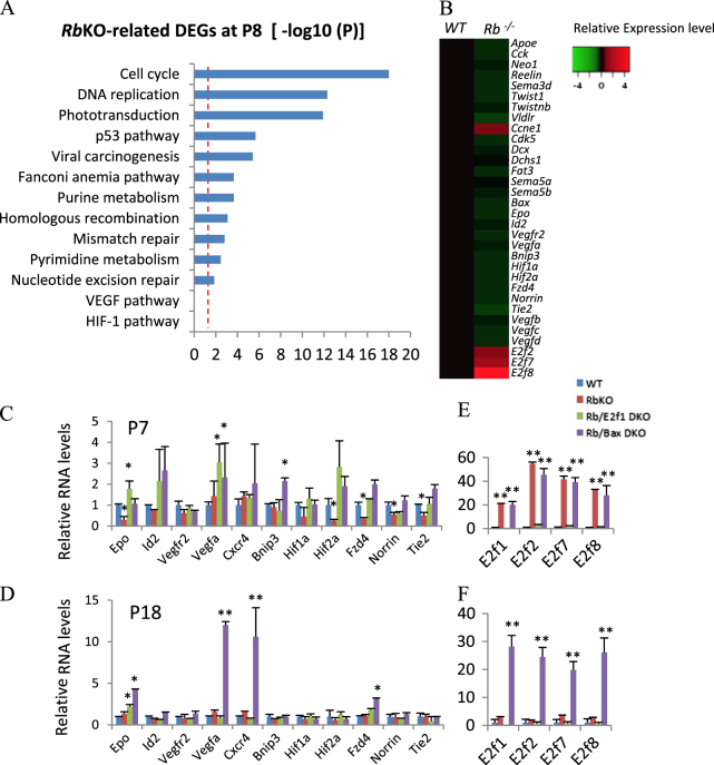
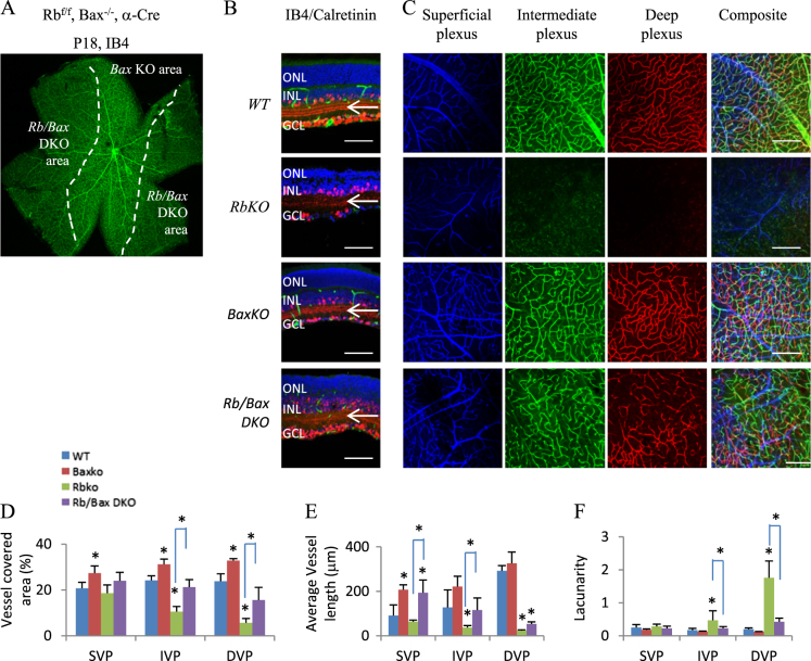
Retinoblastoma tumor suppressor (Rb) promotes cell cycle exit, survival, differentiation, and tumor suppression in the retina. Here, we show it is also essential for vascularization and lamination. Despite minimal effects on Hif1a target expression, intraretinal vascular plexi did not form in the Rb -/- murine retina. Deleting adenovirus E2 promoter binding factor 3 (E2f3), which rescues starburst amacrine cell differentiation, or E2f2, had no effect, but deleting E2f1, which promotes neuronal cell cycle exit and survival, restored retinal vasculature. We specifically linked cell loss to the defect because removing Bax rescued rod and bipolar neurons and the vasculature, but not cell cycle exit. Despite rescuing Rb -/- neurons, Bax deletion exacerbated a delay in outer retina lamination, and exposed a requirement for Rb in inner retina lamination. The latter resembled Sem5 or FAT atypical cadherin 3 (Fat3) mutants, but expression of Sem5/Fat3 pathway components, or that of Neogenin, which perturbs migration in the Rb -/- cortex, was unchanged. Instead, lamination defects correlated with ectopic division, and were E2f1-dependent, implicating the cell cycle machinery. These in vivo studies expose new developmental roles for Rb, pinpoint aberrant E2f1 and Bax activity in neuronal death and vascular loss, and further implicate E2f1 in defective lamination. Links between Rb, angiogenesis and lamination have implications for the treatment of neovascularization, neurodegeneration and cancer.


Conflict of interest statement

The authors declare that they have no conflict of interest.

Figures

Fig. 1
Fig. 1. Rb is required for the development of intraretinal vascular plexi and the OPL.
a Isolectin B4 (IB4) staining of P18 whole-mount retinas of Rbf/f (WT control) and Rbf/f;Z/Red;a-Cre (RbKO in red areas) mouse. Selected areas are blown up to show the vascular density. b Whole-mount retinas of the indicated ages and genotypes were stained for IB4. c Confocal images of IB4-stained SVP, IVP, and DVP of P18 whole-mount retinas of the indicated genotypes. Pseudo-colors were used to differentiate these three plexi. d IB4 (green) and DAPI (blue) staining of P18 retinal sections of WT or RbKO retina. e Horizontal sections of indicated ages and genotypes were stained for nuclear (DAPI, blue), mitosis (PH3, green at P0, red at P8). Arrows in d and e indicate the position of outer plexiform layer (OPL). The dotted lines in ac indicate the boundary between WT (in the center) and RbKO areas (in the periphery). NBL neuroblast layer, ONL outer nuclear layer, INL inner nuclear layer, IPL inner plexiform layer, GCL ganglion cell layer. Scale bar is 50 µm
Fig. 2
Fig. 2. Gene expression changes in the Rb-null retina and the effect of E2f1 or Bax loss.
a Gene list enrichment analysis using Kyoto Encyclopedia of Genes and Genomes (KEGG) 2016 datasets in Enrichr of RbKO-regulated retinal DEGs at P8 (−log 10(P)). Dotted line indicates adjusted p < 0.05. b Heatmap of relative expression level of selected genes of P8 RbKO and WT retina, based on the microarray analysis. cf Real-time RT-PCR analysis of angiogenesis genes (c, d) and the E2f family (e, f) at P7, P18 retinas of the indicated genotypes, respectively. Error bars represent SD of measurements from three animals, and asterisks indicate a significant difference between the WT and the indicated genotypes (*p < 0.05; **p < 0.01, one-way ANOVA followed by Bonferroni correction)
Fig. 3
Fig. 3. E2f1 mediates the RbKO-induced retinal angiogenesis and lamination defects.
a P18 retinal sections of the indicated genotypes were stained for nuclear (DAPI, blue), vascular endothelium cells (IB4, green), and amacrine cells (Calretinin, red). White arrows indicate Calretinin+ tracks in the IPL; loss of the outer two tracks reveals the starburst amacrine cell defect. b P18 whole-mount retinas of indicated genotypes were stained for IB4 to label vasculature. Dotted lines indicate the boundary between WT (center) and RbKO (peripheral) areas. c Confocal images of IB4-stained SVP, IVP, and DVP of P18 whole-mount retinas of indicated genotypes. Pseudo-colors were used to differentiate these three plexi. df Quantification of vessel coverage (d), average vessel length (e), and lacunarity (f) by the AngioTool software. Error bars represent SD of measurements from at least three animals and asterisks indicate significant differences between retinas of RbKO and the indicated genotypes (*p < 0.05, **p < 0.01, one-way ANOVA followed by Bonferroni correction). Scale bar is 50 μm. ONL outer nuclear layer, INL inner nuclear layer, GCL ganglion cell layer, ON optic head
Fig. 4
Fig. 4. Bax loss rescues RbKO neurons.
a Retinal sections of indicated ages and genotypes were stained for nuclei (DAPI, blue) and division (Ki67, green). b Quantification of data in a. c Sections were stained for nuclei (DAPI, blue) and apoptosis (Casp3, red). d Quantification of data in c. e Sections were stained for nuclei (DAPI, blue), ganglion cells (Brn3, red), rod bipolar cells (PKCα, green), or rod photoreceptors (Rho, green). f, g Quantification of the data in e. White and yellow arrowheads in a, c, e highlight the OPL and OMPL, respectively. Data in b, d, f, g are mean ± SD. Asterisks indicate significant difference between WT and other genotypes, or between RbKO and Rb/Bax DKO as indicated by square brackets (*p < 0.05, **p < 0.01, one-way ANOVA followed by Bonferroni correction). ONL outer nuclear layer, INL inner nuclear layer, GCL ganglion cell layer, OPL outer plexiform layer, OMPL outer misplaced plexiform layer. Scale bar is 50 µm
Fig. 5
Fig. 5. BaxKO ameliorates the vascular defect in the RbKO retina.
a Isolectin B4 (IB4) staining of P18 whole-mount retinas of Rbf/f;Bax−/−;a-Cre mouse. b P18 retinal sections of indicated genotypes were stained for nuclei (DAPI, blue), vascular endothelial cells (IB4, green), and amacrine cells (Calretinin, red). Arrows highlight the amacrine neurite tracks in the IPL. ONL outer nuclear layer, INL inner nuclear layer, GCL ganglion cell layer. c Confocal images of IB4-stained superficial, intermediate, and deep plexi of P18 whole-mount retinas of indicated genotypes. Pseudo-colors were used to differentiate the three plexi. df Quantification of vessel coverage (d), average vessel length (e), and lacunarity (f) by the AngioTool software. Error bars represent SD of measurements from at least three animals and asterisks indicate significant difference between WT and other genotypes, or between RbKO and Rb/Bax DKO as indicated by square brackets (*p < 0.05, one-way ANOVA followed by Bonferroni correction). Scale bar in b and c is 50 µm
Fig. 6
Fig. 6. Lamination defects in the Rb/Bax DKO retina.
a Retinal sections of indicated genotypes and ages were stained for nuclei (DAPI, blue), synaptic vesicles (SV2a, red; Syt1, green), cones (M opsin, red), and horizontal cells (OC2, blue). b RT-PCR analysis of the indicated genes and ages (n = 3). c Retinal sections of indicated genotypes and ages were stained for nuclei (DAPI, blue), division (Ki67, green), and synaptic vesicles (SV2a, red). Arrows indicate ectopically dividing cells disrupting the SV2+ OPL. d Retinal sections of indicated genotypes and ages were stained for nuclei (DAPI, blue), cyclin E (red), Cdk5 (red), and Dcx (red). e P18 Rb/Bax DKO retinal sections were stained for nuclei (DAPI, blue), cone (cone arrestin, red), horizontal (D28K, green), Müller (GS, green), or amacrine cells (Calretinin, red; Ap2a, red). Arrowheads highlight the OMPL separating the INL. f Retinal thickness for the indicated genotypes and ages. g Cell counts of cone (cone arrestin+), horizontal (D28K+), amacrine (Ap2a+), and Müller cells (GS+) of the indicated genotypes at P18. h P18 Rb/Bax DKO retinal sections were stained for nuclei (DAPI, blue), division (Ki67, green), and amacrine cells (Calretinin, red). Dendrites of Calretinin+ amacrine cell stratify in the IPL, as usual, but also the OMPL (arrow). Data are mean ± SD. Asterisks indicate significant difference between WT and other genotypes, or between RbKO and Rb/Bax DKO as indicated by square brackets (*p < 0.05, one-way ANOVA followed by Bonferroni correction). ONL outer nuclear layer, OMPL outer misplaced plexiform layer, OPL outer plexiform layer, INL inner nuclear layer, IPL inner plexiform layer, GCL ganglion cell layer. Scale bar is 50 µm

Similar articles

Cited by

References

    1. Schaal C, Pillai S, Chellappan SP. The Rb-E2F transcriptional regulatory pathway in tumor angiogenesis and metastasis. Adv. Cancer Res. 2014;121:147–182. doi: 10.1016/B978-0-12-800249-0.00004-4. - DOI - PubMed
    1. Fruttiger M. Development of the retinal vasculature. Angiogenesis. 2007;10:77–88. doi: 10.1007/s10456-007-9065-1. - DOI - PubMed
    1. Ye X, Wang Y, Nathans J. The Norrin/Frizzled4 signaling pathway in retinal vascular development and disease. Trends Mol. Med. 2010;16:417–425. doi: 10.1016/j.molmed.2010.07.003. - DOI - PMC - PubMed
    1. Usui Y, et al. Neurovascular crosstalk between interneurons and capillaries is required for vision. J. Clin. Invest. 2015;125:2335–2346. doi: 10.1172/JCI80297. - DOI - PMC - PubMed
    1. Junge HJ, et al. TSPAN12 regulates retinal vascular development by promoting Norrin- but not Wnt-induced FZD4/beta-catenin signaling. Cell. 2009;139:299–311. doi: 10.1016/j.cell.2009.07.048. - DOI - PubMed

Publication types

MeSH terms

Substances

Grants and funding

LinkOut - more resources