. 2018 Mar 6;9(3):369.
doi: 10.1038/s41419-018-0412-5.

Group 2 innate lymphoid cells protect lung endothelial cells from pyroptosis in sepsis

Affiliations

Group 2 innate lymphoid cells protect lung endothelial cells from pyroptosis in sepsis

Dengming Lai et al. Cell Death Dis. .

Abstract

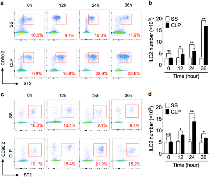
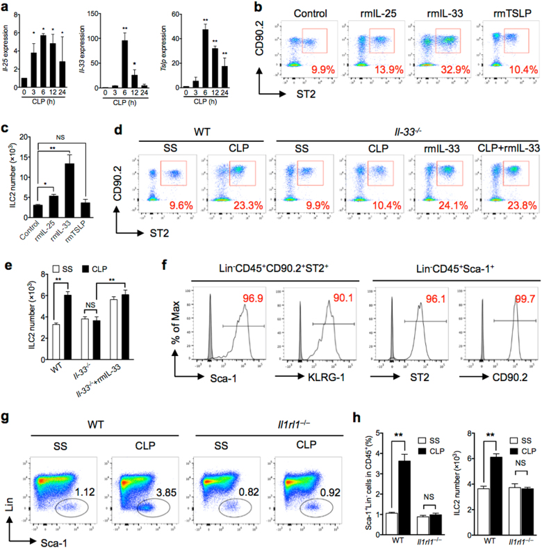
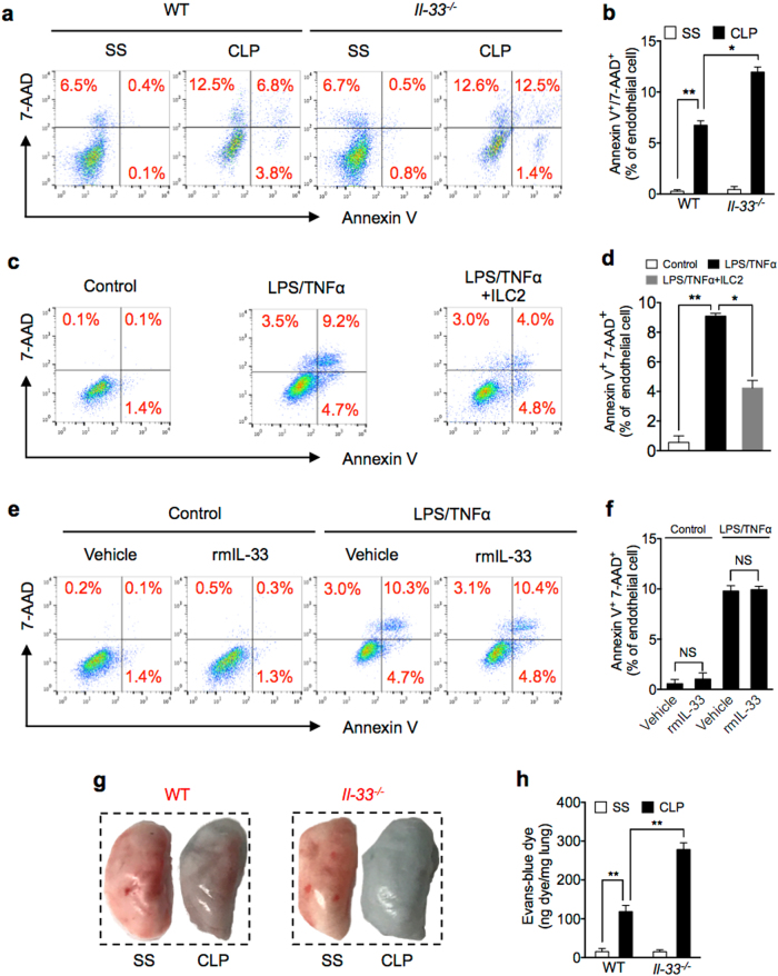
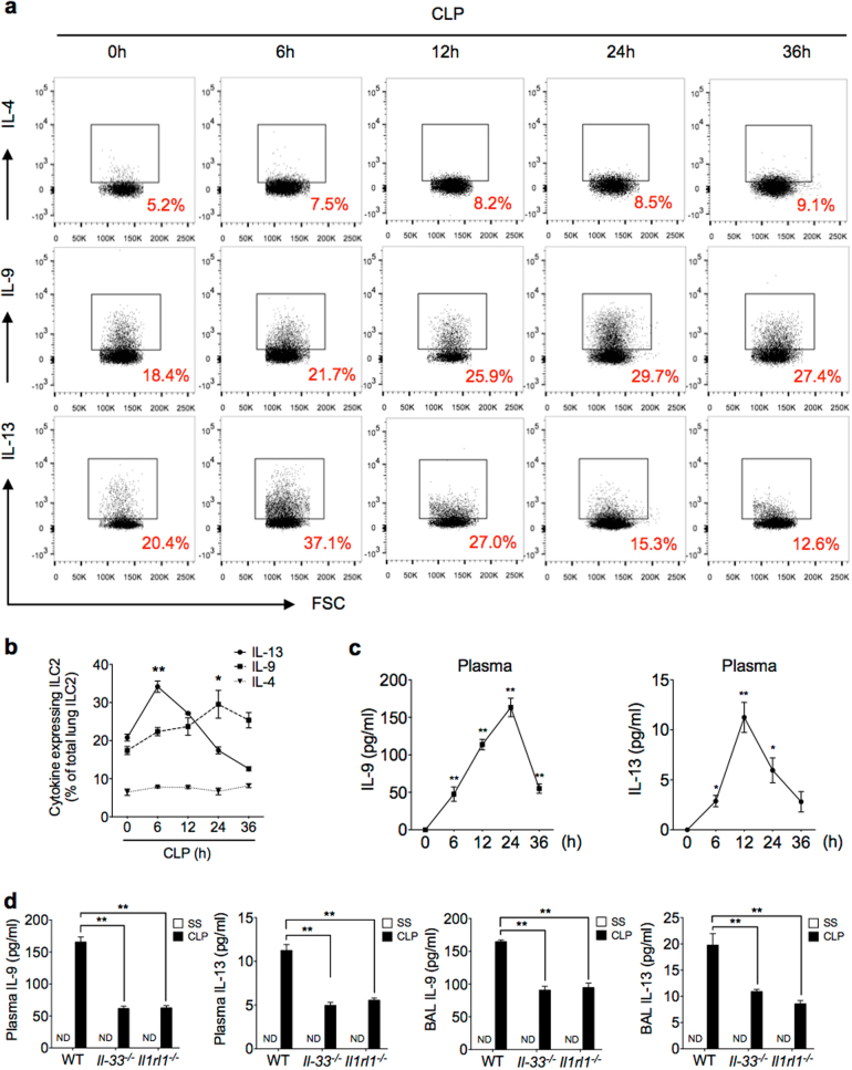
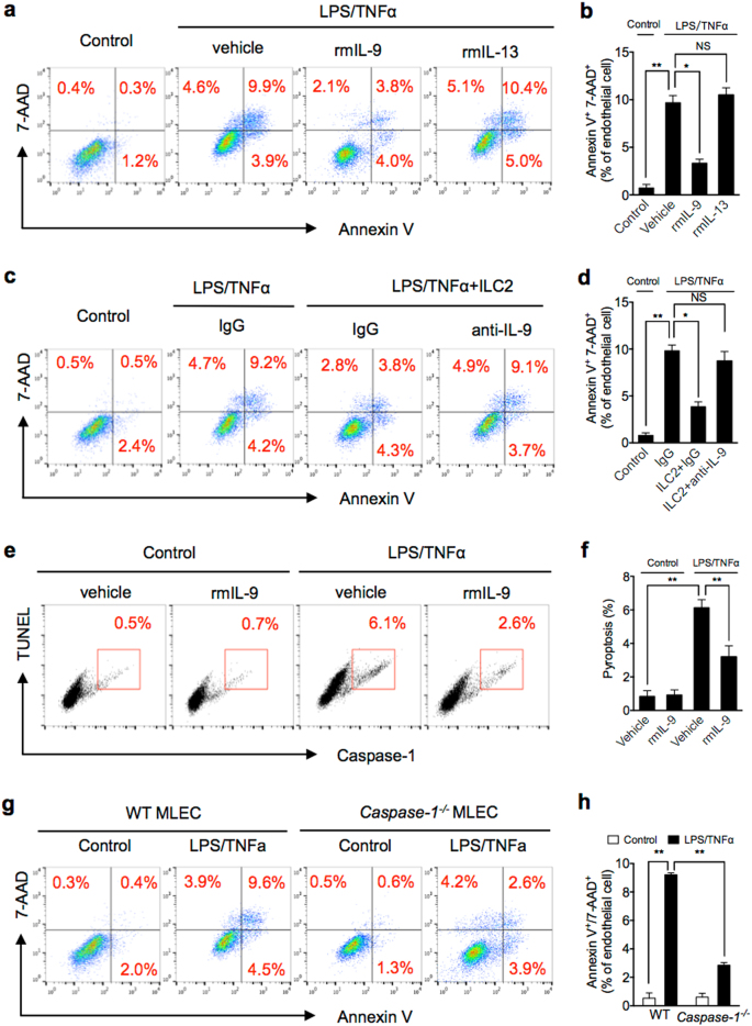
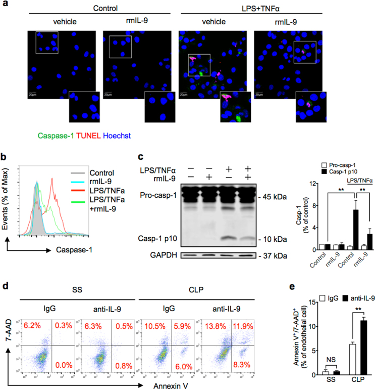
Group 2 innate lymphoid cells (ILC2) are one of three subgroups of innate lymphoid cells (ILC1, ILC2, and ILC3), and the major ILC population detected in the lungs. The function of ILC2 in the regulation of lung inflammation remains unclear. In the current study, we explored an important role of ILC2 in protecting lung endothelial cell (EC) from pyroptosis in sepsis-induced acute lung inflammation and the underlying mechanism. Using a cecal ligation and puncture (CLP) mouse sepsis model, we demonstrated that IL-33, which is released in response to sepsis, acting through its receptor ST2 mediates ILC2 expansion in the lungs. We further showed that the increased ILC2 in the lungs secrete IL-9, which in turn prevents lung EC from undergoing pyroptosis, a pro-inflammatory cell death form, by attenuating caspase-1 activation. These findings suggest a previously unidentified innate pathway that negatively regulates lung inflammation following sepsis.


Conflict of interest statement

The authors declare that they have no conflict of interest.

Figures

Fig. 1
Fig. 1. Sepsis induces ILC2 expansion in lungs and peritoneal cavity.
Representative flow cytometry plots showing percentages of LinCD45+CD90.2+ST2+ ILC2 in a lung and c peritoneal lavage fluid of WT mice at 0, 12, 24, and 36 h after CLP or sham surgery (SS). Bar graphs showing ILC2 absolute cell number in b lung and d peritoneal cavity in SS or CLP mice at time points up to 36 h. n = 6 mice/group. Data are shown as mean ± SEM. *P < 0.05, **P < 0.01, NS not significant
Fig. 2
Fig. 2. IL-33/ST2 signaling is required for sepsis-induced ILC2 recruitment.
a RT-qPCR expression of Il-25, Il-33, and Tslp mRNA in lung tissue, which are relative to 18 s, at time points up to 24 h after CLP compared to SS of each time points (n = 3–5 mice/group). b Representative FACS plots and c bar graph showing absolute number of ILC2 in lungs harvested from control (PBS), rmIL-25, rmIL-33, and rmTSLP-treated mice (1 μg in 50 μl PBS intratracheal (n = 5 mice/group). d Representative flow cytometry plots and e absolute number of lung ILC2 (LinCD45+CD90.2+ST2+) in WT and Il-33−/− mice with/without rmIL-33 treatment mice at 24 h after CLP or SS. Data are representative of three experiments, n = 3–6. f Expression of ILC2-identifying cell surface markers (Sca-1 and KLRG1) on LinCD45+CD90.2+ST2+ cells, and expression of ST2 and CD90.2 on LinCD45+Sca-1+ cells. g Bar graphs showing representative FACS plots and h absolute cell number of lung ILC2 in WT or Il1rl1−/− mice as above (n = 5 mice/group). The plots were gated at CD45+ cells. Data shown as mean ± SEM. *P < 0.05, **P < 0.01, NS not significant
Fig. 3
Fig. 3. ILC2 protect lung EC from death following sepsis.
a Representative flow cytometry plots of Annexin V/7-AAD staining of MLEC from WT and Il-33−/− mice at 24 h after SS or CLP. MLECs were identified as CD31+ and Annexin V/7-AAD double-stained cells were analyzed as dying (n = 5 mice/group). b Bar graph of Annexin V/7-AAD double-stained cells as a percentage of MLEC in WT and Il-33−/− mice at 24 h after SS or CLP. c Representative flow cytometry plots of Annexin V/7-AAD staining of in vitro primary isolated MLEC from WT mice cultured alone (control), with LPS (1 μg/ml) + TNFα (20 ng/ml), or LPS + TNFα + co-culture with ILC2 (1 × 104 cells/well) for 24 h. d Bar graph of Annexin V/7-AAD double-stained cells as a percentage of total MLEC. e Representative flow cytometry plots and f bar graph of Annexin V/7-AAD staining of purified MLEC from WT mice cultured with LPS (1 μg/ml) + TNFα (20 ng/ml) and with or without rmIL-33 (50 ng/ml) for 24 h. g Representative images of whole lungs stained with Evans blue dye from WT and Il-33−/− mice at 24 h following SS or CLP. h Colorimetric quantitative analysis of Evans blue dye extracted from stained lungs from WT and Il-33−/− mice at 24 h following SS or CLP (n = 5 mice/group). Data are representative of three independent in vitro experiments. Data shown are the mean ± SEM. *P < 0.05, **P < 0.01
Fig. 4
Fig. 4. Characterization of cytokine expression and secretion from lung ILC2 in sepsis.
a, b Representative flow cytometry plots and graph of quantitation of percentages of IL-4+, IL-9+, and IL-13+ lung ILC2 from WT mice collected at time points up to 36 h after CLP or SS and stimulated with PMA (50 ng/ml), ionomycin (500 ng/ml), and brefeldin A (1 μg/ml) for 4 h prior to intracellular cytokine determination by flow cytometry. n = 5–8 mice/group. c Plasma IL-9 and IL-13 concentration in WT mice measured by ELISA at time points up to 36 h after CLP or SS. d Plasma and bronchoalveolar lavage fluid (BALF) levels of IL-9 at 24 h and IL-13 at 12 h in WT, Il-33−/−, and Il1rl1−/− mice after SS or CLP determined by ELISA (n = 5 mice/group). Data shown are the mean ± SEM. *P < 0.05, **P < 0.01
Fig. 5
Fig. 5. ILC2-derived IL-9 protects MLEC from pyroptosis.
a, b Representative flow cytometry plots and quantitation of Annexin V/7-AAD staining of MLEC cultured with LPS/TNFα and with or without rmIL-9 or rmIL-13 for 24 h. c, d Representative flow cytometry plots and quantitation of Annexin V/7-AAD staining of MLEC co-cultured with ILC2 (1 × 104 cells/well) and LPS (1 μg/ml) + TNFα (20 ng/ml) and with IgG or anti-IL-9 for 24 h. e, f Representative flow cytometry plots and quantitation of MLEC pyroptosis (caspase-1/TUNEL double-positive cells) with rmIL-9 (50 ng/ml), LPS, and TNFα for 24 h. g, h Representative flow cytometry plots and quantitation of Annexin V/7-AAD staining of MLEC from WT or Caspase-1−/− mice with LPS/TNFα for 24 h. MLECs were identified as CD31+ cells. Data shown are the mean ± SEM, n = 3–6 mice/group. *P < 0.05, **P < 0.01
Fig. 6
Fig. 6. IL-9 decreases caspase-1 activation in lung EC.
a Confocal microscopy immunofluorescence of MLEC stained for activated caspase-1 (green), TUNEL (red), and Hoescht (blue) at 24 h cultured with rmIL-9 (50 ng/ml), LPS, and TNFα. b Number of MLEC expressing activated caspase-1 at 6 h after treatment with LPS and TNFα with or without rmIL-9 (50 ng/ml). c Western blots of pro-caspase-1 (Pro-casp-1) and activated/cleaved caspase-1 (Casp-1 p10) in whole-cell lysates of MLEC at 6 h after LPS, TNFα, and/or rmIL-9 (50 ng/ml). d Representative flow cytometry plots of MLEC from WT mice treated with nonspecific IgG or anti-IL-9 antibody (1 mg/kg) at 30 min before SS or CLP. MLECs were isolated at 24 h after CLP, and identified as CD31+, and Annexin V/7-AAD double-stained cells were analyzed as dying (n = 5 mice/group). e Bar graph of Annexin V/7-AAD double-stained cells as demonstrated in d. Data shown are the mean ± SEM, n = 3–6 mice/group in vivo. *P < 0.05, **P < 0.01

Similar articles

Cited by

References

    1. Singer M, et al. The Third International Consensus Definitions for Sepsis and Septic Shock (Sepsis-3) JAMA. 2016;31:801–810. doi: 10.1001/jama.2016.0287. - DOI - PMC - PubMed
    1. Kaukonen KM, Bailey M, Suzuki S, Pilcher D, Bellomo R. Mortality related to severe sepsis and septic shock among critically ill patients in Australia and New Zealand, 2000-2012. JAMA. 2014;311:1308–1316. doi: 10.1001/jama.2014.2637. - DOI - PubMed
    1. Ani C, Farshidpanah S, Bellinghausen Stewart A, Nguyen HB. Variations in organism-specific severe sepsis mortality in the United States: 1999-2008. Crit. Care Med. 2015;43:65–77. doi: 10.1097/CCM.0000000000000555. - DOI - PubMed
    1. Fleischmann C, et al. Assessment of global incidence and mortality of hospital-treated sepsis. Current estimates and limitations. Am. J. Respir. Crit. Care Med. 2016;193:259–272. doi: 10.1164/rccm.201504-0781OC. - DOI - PubMed
    1. Angus DC, van der Poll T. Severe sepsis and septic shock. N. Engl. J. Med. 2013;369:840–851. doi: 10.1056/NEJMra1208623. - DOI - PubMed

Publication types

MeSH terms