. 2018 Nov;25(10):1781-1795.
doi: 10.1038/s41418-018-0079-6. Epub 2018 Mar 6.

Post-translational modification of OCT4 in breast cancer tumorigenesis

Affiliations

Post-translational modification of OCT4 in breast cancer tumorigenesis

Yunhee Cho et al. Cell Death Differ. 2018 Nov.

Erratum in

Abstract

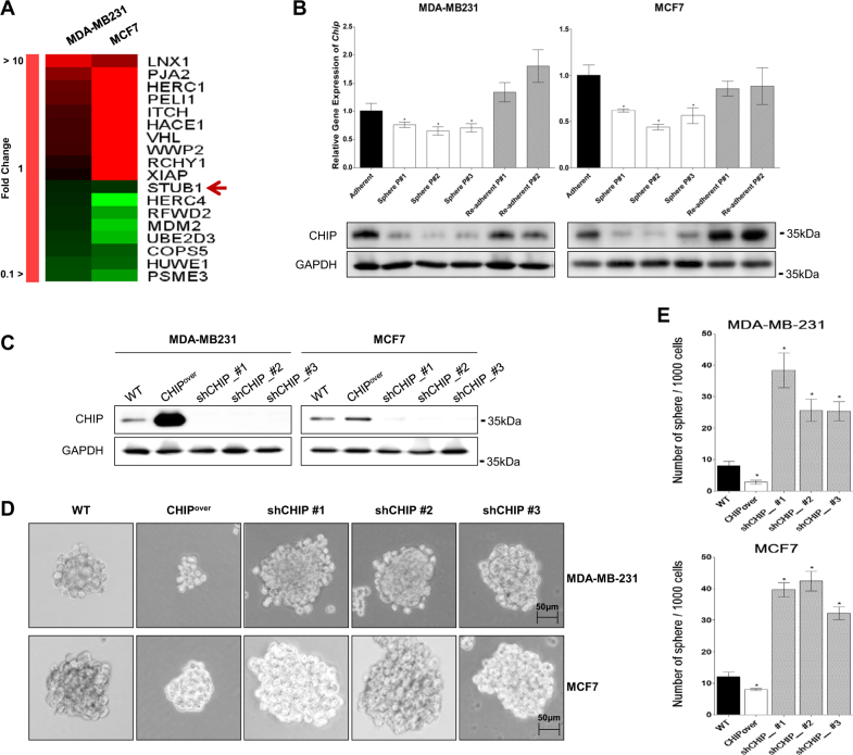
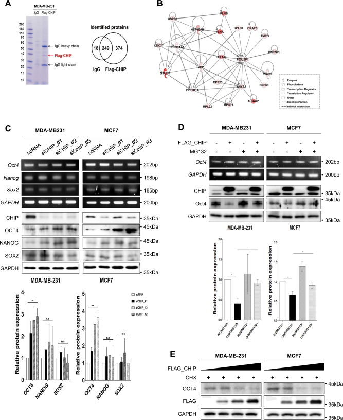
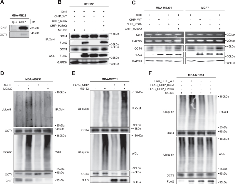
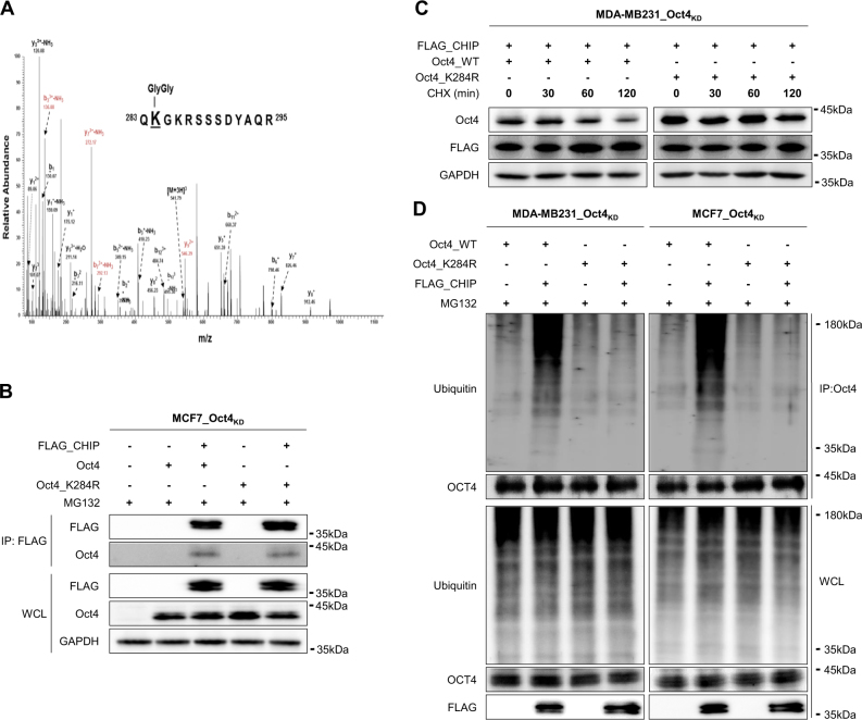
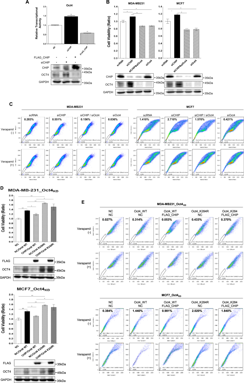
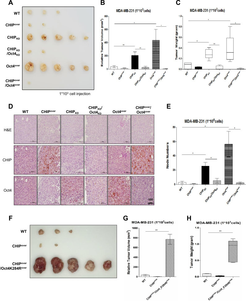
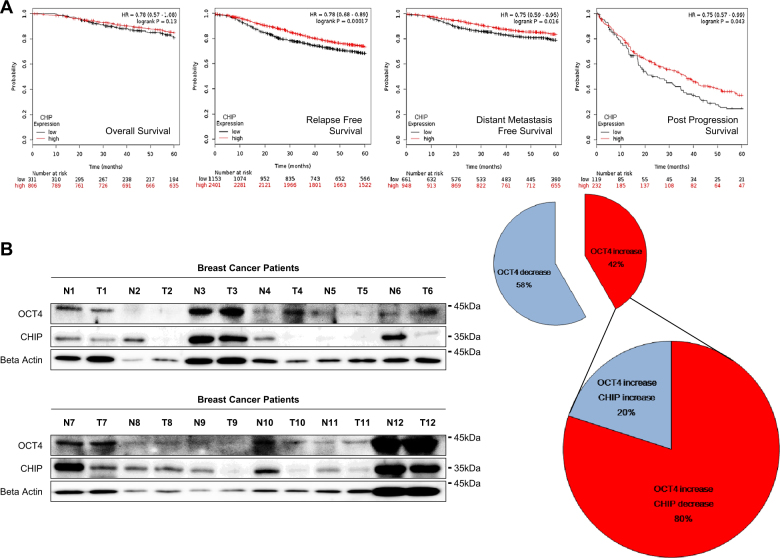
Recurrence and drug resistance of breast cancer are still the main reasons for breast cancer-associated deaths. Cancer stem cell (CSC) model has been proposed as a hypothesis for the lethality of breast cancer. Molecular mechanisms underlying CSC maintenance are still unclear. In this study, we generated mammospheres derived from breast cancer MDA-MB231 cells and MCF7 cells to enrich CSCs and performed DNA microarray analysis. We found that the expression of carboxy terminus of HSP70-interacting protein (CHIP) E3 ubiquitin ligase was significantly downregulated in breast CSCs. CHIP depletion increased mammosphere formation, whereas CHIP overexpression reversed this effect. We identified interactomes by mass spectrometry and detected CHIP directly interacted with OCT4, a stemness factor. CHIP overexpression decreased OCT4 stability through proteasomal degradation. CHIP induced OCT4 ubiquitination, whereas H260Q, a catalytic CHIP mutant, did not. Interestingly, we determined that OCT4 was ubiquitinated at lysine 284, and CHIP overexpression did not degrade K284R mutant OCT4. CHIP overexpression decreased the proliferation and side population of breast cancer cells, but these were not occurred in K284R mutant OCT4 overexpressed cells. Only 1000 cells showing CHIP depletion or OCT4 overexpression sufficiently generated breast tumors and lung metastases in xenografted mice. Ubiquitination-defective mutant of OCT4(K284R) overexpressed cells drastically generated tumor burdens in mice. Patients with breast cancer who showed low CHIP expression had poor survival probability. Taken together, we suggest that CHIP-induced OCT4 ubiquitination is important in breast CSCs. Regulation of CHIP expression and OCT4 protein stability is a considerable approach for breast cancer therapy.


Conflict of interest statement

:The authors declare that they have no conflict of interest.

Figures

Fig. 1
Fig. 1
CHIP expression affects mammosphere-forming ability. a Expression of different E3 ligases was compared by performing microarray analysis of MDA-MB-231 and MCF7 cells in mammosphere and normal cultures. b CHIP expression level was detected by performing qRT-PCR (upper) and western blotting (lower). GAPDH was used as a loading control. c The cells were infected with lentiviral vectors to generate the indicated stable cell lines. CHIP overexpression and depletion were confirmed by performing western blotting. d Mammosphere-forming ability of the stable cell lines was measured for 15 days under sphere-forming conditions, and (e) the number of spheres formed was quantified. Data are presented as mean ± SD (n = 3). Significant differences are indicated by an asterisk (*p < 0.05); p values were calculated using Student’s t test
Fig. 2
Fig. 2
CHIP E3 ligase regulates OCT4 stability through proteasomal degradation. a Coomassie blue-stained gel of affinity-purified CHIP complex in MDA-MB-231 cells. The Venn diagram indicates the relationships between proteins identified in each immunoprecipitation complex by performing mass spectrometry. b Molecular interaction network between CHIP and OCT4 was determined by performing IPA. c MDA-MB-231 and MCF7 cells were transfected with scramble or CHIP siRNAs. After 48 h, mRNA (upper) and protein (lower) levels of the indicated genes were detected by performing RT-PCR and western blotting, respectively. d The cells were transfected with empty and FLAG–CHIP expression vectors for 40 h and were treated with 20 μM MG132 for 8 h. e The cells were transfected with increasing concentrations of the FLAG–CHIP expression vector for 40 h and were treated with 20 μM CHX for 8 h. OCT4 and FLAG–CHIP levels were detected by performing western blotting, with GAPDH as a loading control
Fig. 3
Fig. 3
CHIP E3 ligase interacts with OCT4 and induces its polyubiquitination. a Interaction between CHIP and OCT4 in MDA-MB-231 cells was detected by performing immunoprecipitation assay. b HEK293 cells were transfected with vectors expressing WT FLAG–CHIP or the indicated CHIP mutants K30A and H260Q and were cotransfected with OCT4 expression vector for 40 h and were treated with 20 μM MG132 for 8 h. Interaction of OCT4 with CHIP (WT or mutant) was detected by performing immunoprecipitation assay. c The cells were transfected with the indicated vectors for 40 h and were treated with 20 μM CHX for 8 h. GAPDH was used as a loading control. MDA-MB-231 cells were transfected with CHIP siRNA (d), FLAG–CHIP-overexpressing vector (e), and WT or mutant FLAG–CHIP-expressing vector (f). After 40 h, the transfected cells were treated with 20 μM MG132 for 8 h. Cell lysates were prepared in denaturing condition and were immunoprecipitated using anti-OCT4 antibody. OCT4 polyubiquitination was detected by performing western blotting
Fig. 4
Fig. 4
CHIP-induced OCT4 polyubiquitination at K284 regulates OCT4 stability. a OCT4 ubiquitination at Lys284, with di-glycine modification in the MS/MS spectrum. b MCF7_OCT4KD cells were transfected with the indicated mutant vectors for 40 h, and were treated with 20 μM MG132 for 8 h. Interaction of OCT4 (WT or mutant) with CHIP was detected by performing immunoprecipitation assay. c The cells were transfected with increasing concentrations of the FLAG–CHIP-expressing vector and were cotransfected with OCT4_K284R-expressing vector for 40 h, followed by treatment with 20 μM CHX for 8 h. Mutated OCT4 and FLAG–CHIP levels were determined by performing western blotting, with GAPDH as a loading control. d The cells were transfected with the indicated vectors for 40 h and were treated with 20 μM MG132 for 8 h. Cell lysates were prepared in denaturing condition and were immunoprecipitated using anti-OCT4 antibody. OCT4 polyubiquitination was determined by performing western blotting
Fig. 5
Fig. 5
CHIP-induced OCT4 polyubiquitination at K284 regulates cell viability and side population. a Transcriptional activation in MCF7 cells was measured by performing the reporter assay. The cells were transfected with OCT4 reporter vector alone or were cotransfected with CHIP siRNA or FLAG–CHIP-expressing vector for 48 h. b, c The cells were transfected with the indicated siRNAs alone or in combination for 48 h, and cell viability was detected by performing WST assay, and side population was analyzed, as described in Materials and methods. d, e The cells were transfected with the indicated expression vectors alone or in combination for 48 h and cell viability was detected by performing WST assay, and side population was analyzed, as described in Materials and methods. OCT4 and CHIP levels were detected by performing western blotting, with GAPDH as a loading control. Significant differences are indicated by an asterisk (*p < 0.05, **p < 0.01); p values were calculated using Student’s ttest
Fig. 6
Fig. 6
CHIP overexpression decreases tumors and metastatic nodes derived from OCT4-overexpressing cells in vivo. Nude mice were injected with the indicated stable cell lines derived from mammosphere cultured MDA-MB-231 cells (1 × 103) to form subcutaneous xenografts. After 12 weeks, tumors were isolated and were photographed (a); their sizes (b) and weights (c) are presented in a statistical graph. d Immunohistochemical analysis of the xenografted tumors by using anti-CHIP and anti-OCT4 antibodies; scale bars, 100 μm. e The indicated stable lines derived from MDA-MB-231 cells (1 × 106) were injected into the tail vein of nude mice. After 12 weeks, mouse lungs were isolated and node numbers were counted and presented in a statistical graph. All animal experiments were performed using mammospheres. Nude mice were injected with the indicated stable cell lines derived from mammosphere cultured MDA-MB-231 cells (1 × 103) to form subcutaneous xenografts. After 12 weeks, tumors were isolated and were photographed (f); their sizes (g) and weights (h) are presented in a statistical graph. The data are presented as mean ± SD (n = 5). Significant differences are indicated by an asterisk (*p < 0.05, **p < 0.01); p values were calculated using Student’s ttest
Fig. 7
Fig. 7
Patients with breast cancer showing low CHIP E3 ligase expression have poor survival probability. a Overall, relapse free, distant metastasis and post-progression survival were analyzed with CHIP expression level using Kaplan–Meier plot analysis. b CHIP and OCT4 expression levels of both malignant (T) and normal (N) tissues were detected using western blotting using western blot. β-Actin was used a normalization control. The data are presented as mean ± SD (n = 5). We also quantitated the protein changes of CHIP and OCT4, and venn diagram was presented (right). Significant differences are indicated by an asterisk (*p < 0.05, **p < 0.01); p values were calculated using Student’s t test

References

    1. Tang Y, Wang Y, Kiani MF, Wang B. Classification, treatment strategy, and associated drug resistance in breast cancer. Clin Breast Cancer. 2016;16:335–43. doi: 10.1016/j.clbc.2016.05.012. - DOI - PubMed
    1. Marquette C, Nabell L. Chemotherapy-resistant metastatic breast cancer. Curr Treat Options Oncol. 2012;13:263–75. doi: 10.1007/s11864-012-0184-6. - DOI - PubMed
    1. Carnero A, Garcia-Mayea Y, Mir C, Lorente J, Rubio IT, ME LL. The cancer stem-cell signaling network and resistance to therapy. Cancer Treat Rev. 2016;49:25–36. doi: 10.1016/j.ctrv.2016.07.001. - DOI - PubMed
    1. Takebe N, Miele L, Harris PJ, Jeong W, Bando H, Kahn M, et al. Targeting Notch, Hedgehog, and Wnt pathways in cancer stem cells: clinical update. Nat Rev Clin Oncol. 2015;12:445–64. doi: 10.1038/nrclinonc.2015.61. - DOI - PMC - PubMed
    1. Yang F, Xu J, Tang L, Guan X. Breast cancer stem cell: the roles and therapeutic implications. Cell Mol life Sci: CMLS. 2016;74:951–66. doi: 10.1007/s00018-016-2334-7. - DOI - PMC - PubMed

Publication types

MeSH terms

Substances

LinkOut - more resources