. 2018 May;37(21):2817-2836.
doi: 10.1038/s41388-018-0142-2. Epub 2018 Mar 7.

Regulation of inside-out β1-integrin activation by CDCP1

Affiliations

Regulation of inside-out β1-integrin activation by CDCP1

Sara G Pollan et al. Oncogene. 2018 May.

Abstract

Tumor metastasis depends on the dynamic regulation of cell adhesion through β1-integrin. The Cub-Domain Containing Protein-1, CDCP1, is a transmembrane glycoprotein which regulates cell adhesion. Overexpression and loss of CDCP1 have been observed in the same cancer types to promote metastatic progression. Here, we demonstrate reduced CDCP1 expression in high-grade, primary prostate cancers, circulating tumor cells and tumor metastases of patients with castrate-resistant prostate cancer. CDCP1 is expressed in epithelial and not mesenchymal cells, and its cell surface and mRNA expression declines upon stimulation with TGFβ1 and epithelial-to-mesenchymal transition. Silencing of CDCP1 in DU145 and PC3 cells resulted in 3.4-fold higher proliferation of non-adherent cells and 4.4-fold greater anchorage independent growth. CDCP1-silenced tumors grew in 100% of mice, compared to 30% growth of CDCP1-expressing tumors. After CDCP1 silencing, cell adhesion and migration diminished 2.1-fold, caused by loss of inside-out activation of β1-integrin. We determined that the loss of CDCP1 reduces CDK5 kinase activity due to the phosphorylation of its regulatory subunit, CDK5R1/p35, by c-SRC on Y234. This generates a binding site for the C2 domain of PKCδ, which in turn phosphorylates CDK5 on T77. The resulting dissociation of the CDK5R1/CDK5 complex abolishes the activity of CDK5. Mutations of CDK5-T77 and CDK5R1-Y234 phosphorylation sites re-establish the CDK5/CDKR1 complex and the inside-out activity of β1-integrin. Altogether, we discovered a new mechanism of regulation of CDK5 through loss of CDCP1, which dynamically regulates β1-integrin in non-adherent cells and which may promote vascular dissemination in patients with advanced prostate cancer.


Conflict of interest statement

Conflict of interest The authors declare that they have no conflict of interest.

Figures

Fig. 1
Fig. 1
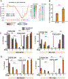
CDCP1 expression decreases in advanced prostate cancer. a CDCP1 gene expression in an integrated human prostate cancer cohort (n = 1321) of transcriptome profiles (PCTA) [33]. The x-axis denotes samples of benign, Gleason Sum < 7, Gleason Sum = 7, Gleason Sum > 7 prostate cancer. Numbers in parenthesis indicate samples in each group. Wilcoxon rank-sum tests p-values are shown above the box-plots. b CDCP1 protein expression levels in benign (B), primary tumor (PT), and metastases (M) from patients measure after staining of a TMA described in [36] by immunohistochemistry. The staining intensity of CDCP1 was separately scored in the membrane and cytoplasm using a categorical scoring scheme. Numbers in parenthesis indicate samples in each group, with each sample represented in duplicate or triplicate on the TMA. c Expression of CDCP1 mRNA in circulating tumor cells. mRNA expression levels of 10 genes were determined in CTCs from each of 9 patients with castrate-resistant prostate cancer (CRPC). A heatmap of qPCR cycle numbers of the Androgen Receptor (AR), AR variant 1 (V1), AR variant 7 (V7), keratin 8 (KRT8), Transmembrane Protease, Serine 2 (TMPRSS2), Kallikrein-3 (KLK3), 60S acidic ribosomal protein P0 (RPLP0). Patients 1–3 had detectable CDCP1 gene expression. d GSEA enrichment plot of EMT signature genes associated with loss of CDCP1 expression in CRPC from the PCTA (n = 260). The heat map displays the expression of leading edge genes from GSEA analysis in samples with high vs. low CDCP1 expression. *P <0.05 and **P < 0.01
Fig. 2
Fig. 2
TGFβ1 induces reversible loss of CDCP1 expression. a GSEA analysis of the hallmark TGFβ1 signature from 260 cases of metastatic prostate cancer [33]. Gene in the leading edge are indicated beside the graph. b FACS analysis of CDCP1 protein in E-cadherin-positive DU145 and PC3 prostate cancer cell lines. All cells are E-cadherin positive. The percentage of cells that are positive for CDCP1 is indicated on the y-axis. c, d Changes in CDCP1 and E-cadherin surface expression (c) or mRNA expression (d) in E+ DU145 and E+ PC3 cells after treatment with TGFβ1. TGFβ1 was removed from the culture medium on day 5 and cells grown for an additional 3 days. *P < 0.05 and **P < 0.01
Fig. 3
Fig. 3
CDCP1 loss changes cell behavior. a MTT assay of DU145 and PC3 CDCP1 knockdown (shCDCP1) or control hairpin (Control) cells suspended in a HEMA-coated 96-well plate for 24 h. b Soft agar colony formation assay of DU145 and PC3 shControl and CDCP1 knockdown (shCDCP1). Colonies were quantified using ImageJ. c Xenograft mouse model. 1 × 10⁶ DU145+/−CDCP1 were injected subcutaneously in 9-week-old male beige SCID mice. Tumors were harvested after 9 weeks and the volumes shown in the bar graph. d Adhesion assay of DU145 shControl (gray bars) and CDCP1 knockdown shCDCP1 (orange bars). Cells were adhered for 2 or 4 h to surfaces coated with Matrigel (Mtgl), Fibronectin (Fbn), or Collagen I (Col1). Adherent cell numbers per 20× field, averaged from eight measurements, are plotted on the y-axis. e Serum (10%)-induced migration assay of DU145 and PC3 through a 0.8 μM Transwell membrane. Migrated cells were stained using crystal violet and quantified with ImageJ. *P < 0.05 and **P < 0.01
Fig. 4
Fig. 4
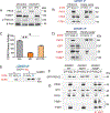
CDCP1 silencing inhibits inside-out activation of ITGB1/β1-integrin and activates c-SRC in suspended cells. a Immunofluorescent staining of CDCP1-positive (shControl) and negative (shCDCP1) DU145 cells on coverslips. The HUTS-4 antibody was used to determine the activation state of ITGB1/β1-integrin (green: active ITGB1*), total ITGB1/β1-integrin (red), and nuclei (blue). White bar = 10 μM. b FACS analysis with HUTS-4 antibody. The staining intensity with HUTS-4 is plotted on the x-axis. The y-axis reveals the number of cells. c Time course of ITGB1/β1-integrin inactivation Active ITGB1/β1-integrin (ITGB1*) and total ITGB1/β1-integrin (ITGB1) were measured after suspending DU145 CDCP1 knockdown cells for the times indicated. d Activation of ITGB1/β1-integrin in adherent and suspended (3 h) DU145 and PC3 cells. Cells were analyzed after CDCP1 (sh1) or scrambled control (Cnt) knockdown. e Active ITGB1/β1-integrin (ITGB1*) and total ITGB1/β1-integrin (ITGB1) in protein lysates from three control and CDCP1-silenced xenografts [–3]. Quantification normalized to total ITGB1/β1-integrin on y-axis. *P < 0.05 and **P < 0.01
Fig. 5
Fig. 5
De novo phosphorylation of CDCK5R1 after silencing of CDCP1. a Loss of CDK5R1-CDK5 complex formation in suspended CDCPl-silenced cells. Reciprocal immunoprecipitations of CDK5 or CDK5R1. WCL whole cell lysate, CAB IgG control, IP immunopre-cipitate. b ITGB1-CDK5 complex formation. Western blot of ITGB1/β1-integrin immunoprecipitation (ITGB1 IP) from DU145 cells with and without silencing of CDCP1 and probed for CDCP1, CDK5, and CDK5R1. c CDK5R1 immunoprecipitation of suspended DU145/ shCDCPl. CDK5R1 complexes from DU145/shControl (Cnt) or shCDCPl(shl) were probed with the 4G10 antibody. Western blot membranes were re-probed with antibodies reactive to PKCδ, SRC and CDK5R1. d Regulation of ITGB1/β1-integrin inside-out activity by c-SRC. DU145/shCDCP1 cells were treated with 10 μM Sar-acatinib or buffer and suspended for 3 h. Western blot probed with antibodies reactive to pTalin-S425, total Talin (Talin), HUTS-4 (ITGB1*), or total ITGB1/β1-integrin (ITGB1). e-f Re-adhesion of suspended DU145/shCDCP1 to fibronectin (e). Image of plate after adhesion of DU145/shCDCP1 cells with and without treatment with Vanadate treatment and quantification of adherent cell numbers (f). Western blot tyrosine phosphorylated proteins visualized with the 4G10 antibody. *P <0.05 and **P <0.01
Fig. 6
Fig. 6
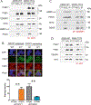
PKCδ binds phosphorylated CDK5R1/p35 via its C2 domain in CDCP1-silenced and suspended DU145 cells. a PKCδ (siPδ) silencing in suspended DU145/shCDCP1 leads to reactivation of ITGB1/β1-integrin. Equal amounts of whole lysates from DU145/shControl or DU145/shCDCP1 were analyzed by western blotting and probed with anti-PKCδ, HUTS-4 (ITGB1*), and anti-pTALIN. b Binding of GFPPKCδ-C2 domain to CDK5R1/p35. Western blots of CDK5R1 or IgG control (CAB) immunopreceipitation of DU145/shCDCP1 expressing either GFP-PKCδ-C2 domain (PKCδ-C2) or empty vector. Membranes were probed with antibodies reactive with PKCδ, CDK5, or HUTS-4. c Kinase assay of CDK5 in DU145 shControl (Cnt), shCDCP1 (sh1), and shCDCP1 + GFP-PKCδ-C2 overexpression (sh1 + C2). Kinase activity measured by incorporation of 32P into CDK5 substrate is plotted on the y-axis. d Overexpression of GFPtagged PKCδ kinase dead (PKCδ-KD) in DU145/shCDCP1. Western blots of CDK5R1 immunoprecipitates (IP), whole cell lysate (WCL), or IgG control (CAB) IPs from cells expressing GFP-PKCδ-KD or empty vector. Western blots were probed with antibodies detecting PKCδ, GFP, CDK5, or HUTS-4 (ITGB1*). e Inhibition of PKCδ with Go6983. The CDK5 and CDK5R1 complex formation is demonstrated with and without treatment with G06983. f–g pCDK5R1–PKCδ complex formation. f CDCP1-silenced cells were transfected with MYCtag-wt-CDK5R1, MYCtag-CDK5R1-Y231F, or MYCtag-CDK5R1-Y234F and co-transfected with GFP-PKCδ-C2 domain. Lysates were precipitated with the anti-MYCtag antibody and probed for GFP (g). Immunoprecipitation of cell expressing wild-type and mutant CDK5R1 as described in f. Membranes were probed with antibodies reactive with CDK5, MYC, PKCδ, HUTS-4 (ITGB1*) or total integrin (ITGB1). *P < 0.05 and **P < 0.01
Fig. 7
Fig. 7
Phosphorylation of CDK5-T77 by PKC5 leads to dissociation of CDK5-CDKR1 complexes. a CDK5-T77A complex formation. MYCtag-CDK5-T77A or MYCtag-wt-CDK5 were expressed in suspended DU145/shCDCP1. Western blots of MYCtag- immunoprecipitations (IP) and controls were probed with antibodies reactive with HUTS-4 (ITGB1*), total ITGB1/β1-integrin (ITGB1), CDK5R1, or MYCtag. b CDCP1-silenced DU145 cells transfected with wt-CDK5 (WT) and CDK5T77A (A77) or CDCP1-expressing DU145 cells transfected with wt-CDK5 (WT) or CDK5T77D (D77) were probed for ITGB1* (green), total integrin (red), or DAPI (blue). The staining was quantified using ImageJ. Staining intensities per cell are indicated on the y-axis. The number of cells analyzed is shown in parentheses. c CDK5–T77A complexes with CDK5R1. Western blot of CDK5R1 immunoprecipitation in DU145 cells described in a. Western blots were probed with antibodies reactive with pSRCY416, PKCδ, MYC, and CDK5. d CDK5–T77D complex formation. MYCtag-CDK5-T77D or MYCtag-wt-CDK5 were expressed in suspended DU145/shControl cells. Western blot as described in a. *P <0.05 and **P < 0.01
Fig. 8
Fig. 8
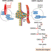
Model of loss of ITGB1/β1-integrin inside-out activation in detached prostate cancer cells with low CDCP1 expression

Similar articles

Cited by

References

    1. Wortmann A, He Y, Deryugina EI, Quigley JP, Hooper JD. The cell surface glycoprotein CDCP1 in cancer—insights, opportunities, and challenges. IUBMB Life. 2009;61:723–30. - PubMed
    1. Siva AC, Kirkland RE, Lin B, Maruyama T, McWhirter J, Yantiri-Wernimont F, et al. Selection of anti-cancer antibodies from combinatorial libraries by whole-cell panning and stringent subtraction with human blood cells. J Immunol Methods. 2008;330:109–19. - PubMed
    1. Siva AC, Wild MA, Kirkland RE, Nolan MJ, Lin B, Maruyama T, et al. Targeting CUB domain-containing protein 1 with a monoclonal antibody inhibits metastasis in a prostate cancer model. Cancer Res. 2008;68:3759–66. - PubMed
    1. Deryugina EI, Conn EM, Wortmann A, Partridge JJ, Kupriyanova TA, Ardi VC, et al. Functional role of cell surface CUB domain-containing protein 1 in tumor cell dissemination. Mol Cancer Res. 2009;7:1197–211. - PMC - PubMed
    1. Fukuchi K, Steiniger SC, Deryugina E, Liu Y, Lowery CA, Gloeckner C, et al. Inhibition of tumor metastasis: functional immune modulation of the CUB domain containing protein 1. Mol Pharm. 2010;7:245–53. - PMC - PubMed

Publication types

MeSH terms