. 2018 Feb 15;10(2):563-572.
eCollection 2018.

lncRNA MALAT1/miR-205-5p axis regulates MPP+-induced cell apoptosis in MN9D cells by directly targeting LRRK2

Affiliations

lncRNA MALAT1/miR-205-5p axis regulates MPP+-induced cell apoptosis in MN9D cells by directly targeting LRRK2

Qin Chen et al. Am J Transl Res. .

Abstract

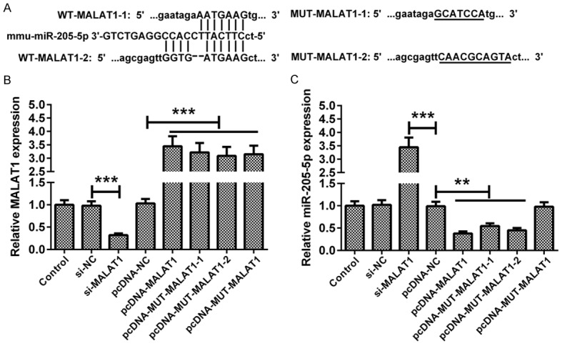
Metastasis-associated lung adenocarcinoma transcript 1 (MALAT1), as a long chain non-coding RNA (lncRNA), has been reported to be upregulated in Parkinson's disease (PD). However, the mechanisms underlying this process remain unknown. Hence, to investigate the role of MALAT1 in PD, N-methyl-4-phenylpyridinium (MPP+) was used to induce PD in vitro in the MN9D dopaminergic neuronal cell line and 1-methyl-4-phenyl-1,2,3,6-tetrahydropyridine (MPTP) was used to induce PD in vivo in C57BL/6 mice. Quantitative Real-Time PCR (qRT-PCR) and western blot assay showed that the expression levels of MALAT1 and leucine-rich repeat kinase (LRRK2) were increased, and that of miR-205-5p was decreased in the midbrains of mice in which PD was induced by MPTP. MALAT1 suppressed the expression of miR-205-5p in MN9D cells. The results of luciferase reporter assay indicated that LRRK2 was a direct target of miR-205-5p. Transfection with the miR-205-5p mimics decreased, whereas transfection with miR-205-5p inhibitor increased the expression levels of LRRK2 mRNA and protein. The cell counting kit-8 (CCK-8) and flow cytometry assays showed that overexpression of LRRK2 reduced the viability and promoted apoptosis in MN9D cells treated with MPP+. MALAT1 knockdown exerted a protective effect on the viability and apoptosis of MN9D cells treated with MPP+, which was abrogated by LRRK2 overexpression and miR-205-5p inhibition. Our study demonstrates that the MALAT1/miR-205-5p axis regulates MPP+-induced apoptosis in MN9D cells by targeting LRRK2, thereby improving our understanding of the molecular pathogenesis of PD.

Keywords: LRRK2; MALAT1; Parkinson’s disease; apoptosis; long non-coding RNA; miR-205-5p.


Conflict of interest statement

None.

Figures

Figure 1
Figure 1
Changes in MALAT1, miR-205-5p and LRRK2 expression in both in vivo and in vitro models of PD. Seven days after the last MPTP injection, mice were sacrificed and brain tissues were harvested. MN9D cells were treated with 100 μM MPP+ for 24 h. A-C. qRT-PCR was performed to determine the expression of MALAT1, miR-205-5p, and LRRK2 mRNA in mice in which PD was induced by MPTP, and in cells treated with MPP+. D. The result of western blot showed that the LRRK2 protein was overexpressed in the midbrains of MPTP-induced PD mice and in cells treated with MPP+. Data are expressed as the mean ± SD (n = 3). **P < 0.01, ***P < 0.001.
Figure 2
Figure 2
MALAT1 inhibits miR-205-5p expression. A. Bioinformatics analysis showed the combination of MALAT1 and miR-205-5p. WT-MALAT1-1 and WT-MALAT1-2 were the two target locations. B. pcDNA-MALAT1: WT-MALAT1-1 + WT-MALAT1-2. pcDNA-MUT-MALAT1-1: MUT-MALAT1-1 + WT-MALAT1-2. pcDNA-MUT-MALAT1-2: WT-MALAT1-1 + MUT-MALAT1-2. After transfection with different molecules, MN9D cells were cultured for 48 h. The regulatory effects of si-MALAT1, pcDNA-MALAT1, pcDNA-MUT-MALAT1-1, pcDNA-MUT-MALAT1-2 or pcDNA-MUT-MALAT1 were validated by qRT-PCR. C. The effects of MALAT1 knockdown and overexpression on the expression of miR-205-5p were assessed in MN9D cells. Data are expressed as the mean ± SD (n = 3). **P < 0.01, ***P < 0.001.
Figure 3
Figure 3
LRRK2 was a target gene of miR-205-5p. A. The putative miR-205-5p binding sites on LRRK2 WT 3’UTR and alignment of the seed sequence with MUT LRRK2 3’UTR. B. MN9D cells were co-transfected with a miR-205-5p mimic or inhibitor and WT-LRRK2 or MUT-LRRK2 luciferase reporter plasmids. 48 h after transfection, MN9D cells were harvested, and the luciferase activity was determined. The results showed that miR-205-5p suppressed the luciferase activity of WT LRRK2 3’UTR. C, D. At 24 h after transfection with a miR-205-5p mimic or inhibitor, MN9D cells were harvested and then subjected to qRT-PCR and a western blot analysis. Transient transfection with a miR-205-5p mimic in MN9D cells attenuated LRRK2 mRNA and protein expression, and transient transfection with miR-205-5p inhibitor increased LRRK2 mRNA and protein expression. Data are expressed as the mean ± SD (n = 3). **P < 0.01, ***P < 0.001.
Figure 4
Figure 4
LRRK2 promoted MPP+-induced apoptosis of MN9D cells. After transfection with si-LRRK2, pcDNA-LRRK2, or their corresponding controls, MN9D cells were treated with 100 μM MPP+ for 24 h. A. LRRK2 protein expression in MN9D cells was downregulated and upregulated by transfection with si-LRRK2 and pcDNA-LRRK2, respectively. B, D. The effects of LRRK2 overexpression and knockdown on the cell viability and apoptosis in MN9D cells, in the presence of MPP+. C. The representative images of Flow cytometry were shown. Data are expressed as the mean ± SD (n = 3). *P < 0.05, **P < 0.01, ***P < 0.001.
Figure 5
Figure 5
The effects of LRRK2 overexpression and miR-205-5p downregulation on the cell viability and apoptosis induced by si-MALAT1. MN9D cells were transfected with the indicated molecules and then treated with 100 μM MPP+ for 24 h. A, B. qRT-PCR analysis of MALAT1 and miR-205-5p expression in MN9D cells with different treatments. C. CCK-8 assay was performed to assess the viability of MN9D cells transfected with indicated molecules. D. Flow cytometry assay was conducted to evaluate the apoptosis of MN9D cells transfected with indicated molecules. Data are expressed as the mean ± SD (n = 3). *P < 0.05, **P < 0.01, ***P < 0.001.

References

    1. Ma CL, Su L, Xie JJ, Long JX, Wu P, Gu L. The prevalence and incidence of Parkinson’s disease in China: a systematic review and meta-analysis. J Neural Transm (Vienna) 2014;121:123–134. - PubMed
    1. Manza P, Zhang S, Li CS, Leung HC. Resting-state functional connectivity of the striatum in early-stage Parkinson’s disease: cognitive decline and motor symptomatology. Hum Brain Mapp. 2016;37:648–662. - PMC - PubMed
    1. Kalia LV, Lang AE. Parkinson’s disease. Lancet. 2015;386:896–912. - PubMed
    1. Lu C, Zhang J, Shi X, Miao S, Bi L, Zhang S, Yang Q, Zhou X, Zhang M, Xie Y, Miao Q, Wang S. Neuroprotective effects of tetramethylpyrazine against dopaminergic neuron injury in a rat model of Parkinson’s disease induced by MPTP. Int J Biol Sci. 2014;10:350–357. - PMC - PubMed
    1. Mercer TR, Dinger ME, Mattick JS. Long non-coding RNAs: insights into functions. Nat Rev Genet. 2009;10:155–159. - PubMed

LinkOut - more resources