

Bowel endometriosis treated with simultaneous ileocecal and rectal resection
- PMID: 29511528
- PMCID: PMC5829574
- DOI: 10.1093/jscr/rjy034
Bowel endometriosis treated with simultaneous ileocecal and rectal resection
Abstract
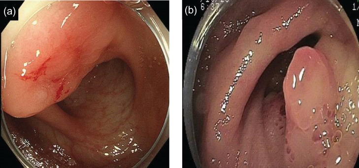
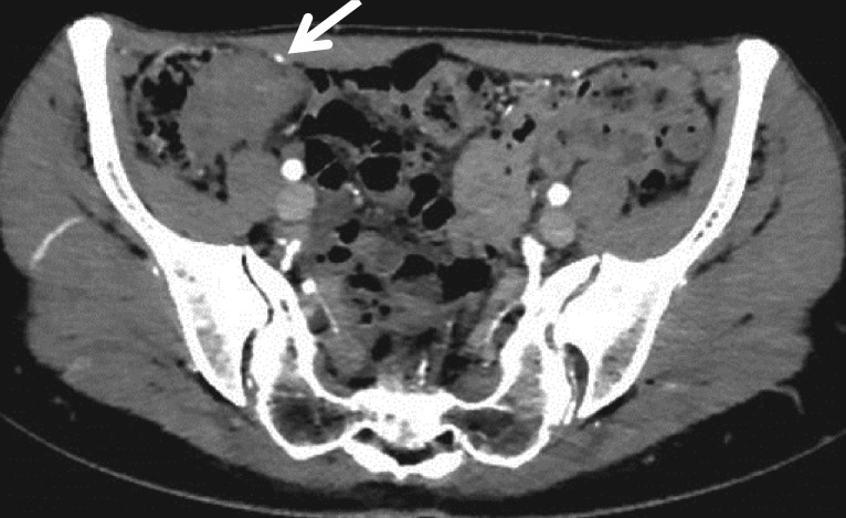
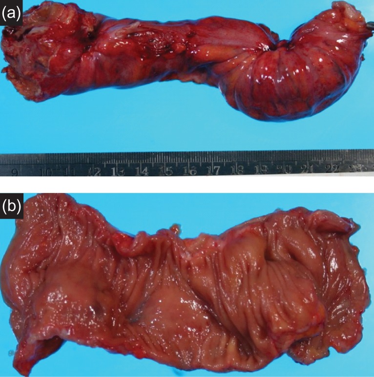
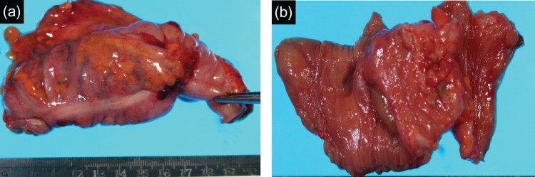
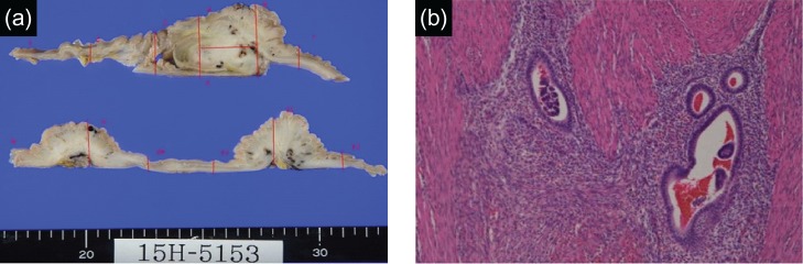
A 43-year-old female noticed hematochezia and lower-right abdominal pain during menstruation. Her family doctor detected a mass by computed tomography at the ileocecum. She was referred to our hospital and colonoscopy was performed. We observed extrinsic pressure resulting in mucosal change at the ileocecum. We also observed a submucosal tumor-like lesion at the rectosigmoid. We performed biopsy from both lesions, both were benign. Ileocecal resection and rectal low anterior resection were performed for diagnosis. Redness, induration and serosal dimpling were recognized at the ileocecum, rectosigmoid and upper rectum. All lesions had endometorial tissue in muscular layer, so pathological diagnosis was bowel endometriosis. Bowel endometriosis occurring in multiple parts and where two colectomies were performed simultaneously is very rare. To determine the optimal method of treatment for the bowel endometriosis, detailed preoperative examination must be performed, specifically complete surgical resection of the lesion for definite diagnosis.
Figures
Similar articles
-
Laparoscopic Double Discoid Resection With a Circular Stapler for Bowel Endometriosis.J Minim Invasive Gynecol. 2015 Sep-Oct;22(6):929-31. doi: 10.1016/j.jmig.2015.04.021. Epub 2015 Apr 29. J Minim Invasive Gynecol. 2015. PMID: 25937595
-
Laparoscopic discoid anterior rectal excision with the circular stapler for rectosigmoid endometriosis, performed by the gynecologic surgeon.J Minim Invasive Gynecol. 2015 Jan;22(1):8-9. doi: 10.1016/j.jmig.2014.08.003. Epub 2014 Aug 10. J Minim Invasive Gynecol. 2015. PMID: 25117838
-
Preoperative rectosigmoid endoscopic ultrasonography predicts the need for bowel resection in endometriosis.World J Gastroenterol. 2019 Feb 14;25(6):696-706. doi: 10.3748/wjg.v25.i6.696. World J Gastroenterol. 2019. PMID: 30783373 Free PMC article.
-
Rectal perforation caused by deep infiltrating endometriosis in non-pregnant woman: Case report and short review of the literature.Ann Ital Chir. 2019 Mar 5;8:S2239253X19029360. Ann Ital Chir. 2019. PMID: 30898991 Review.
-
[A case of endometrioid carcinoma arising from endometriosis of the rectum].Nihon Geka Gakkai Zasshi. 1992 Jun;93(6):651-3. Nihon Geka Gakkai Zasshi. 1992. PMID: 1630441 Review. Japanese.
References
-
- Dunselman GAJ, Vermeulen N, Becker C, Calhaz-Jorge C, D’Hooghe T, De Bie B, et al. . ESHRE guideline: management of women with endometriosis. Hum Reprod 2014;29:400–12. - PubMed
-
- Hudelist G, English J, Thomas AE, Tinelli A, Singer CF, Keckstein J. Diagnostic accuracy of transvaginal ultrasound for non invasive diagnosis of bowel endometriosis: systematic review and meta analysis. Ultrasound Obstet Gynecol 2011;37:257–63. - PubMed
-
- Takeuchi H, Kuwatsuru R, Kitade M, Sakurai A, Kikuchi I, Shimanuki H, et al. . A novel technique using magnetic resonance imaging jelly for evaluation of rectovaginal endometriosis. Fertil Steril 2005;83:442–7. - PubMed
Publication types
LinkOut - more resources
Full Text Sources
Other Literature Sources
