

Management of patients with high-risk pulmonary embolism: a narrative review
- PMID: 29511564
- PMCID: PMC5834898
- DOI: 10.1186/s40560-018-0286-8
Management of patients with high-risk pulmonary embolism: a narrative review
Abstract
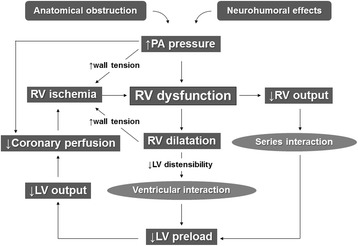
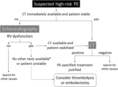
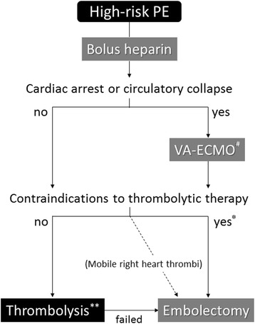
High-risk pulmonary embolism (PE) is a life-threatening disorder associated with high mortality and morbidity. Most deaths in patients with shock occur within the first few hours after presentation, and rapid diagnosis and treatment is therefore essential to save patients' lives. The main manifestations of major PE are acute right ventricular (RV) failure and hypoxia. RV pressure overload is predominantly related to the interaction between the mechanical pulmonary vascular obstruction and the underlying cardiopulmonary status. Computed tomography angiography allows not only adequate visualization of the pulmonary thromboemboli down to at least the segmental level but also RV enlargement as an indicator of RV dysfunction. Bedside echocardiography is an acceptable alternative under such circumstances. Although it does not usually provide a definitive diagnosis or exclude pulmonary embolism, echocardiography can confirm or exclude severe RV pressure overload and dysfunction. Extracorporeal membrane oxygenation support can be an effective procedure in patients with PE-induced circulatory collapse. Thrombolysis is generally accepted in unstable patients with high-risk PE; however, thrombolytic agents cannot be fully administered to patients with a high risk of bleeding. Conversely, catheter-directed treatment is an optimal treatment strategy for patients with high-risk PE who have contraindications for thrombolysis and is a minimally invasive alternative to surgical embolectomy. It can be performed with a minimum dose of thrombolytic agents or without, and it can be combined with various procedures including catheter fragmentation or embolectomy in accordance with the extent of the thrombus on a pulmonary angiogram. Hybrid catheter-directed treatment can reduce a rapid heart rate and high pulmonary artery pressure and can improve the gas exchange indices and outcomes. Surgical embolectomy is also performed in patients with contraindications for or an inadequate response to thrombolysis. Large hospitals having an intensive care unit should preemptively establish diagnostic and therapeutic protocols and rehearse multidisciplinary management for patients with high-risk PE. Coordination with a skilled team comprising intensivists, cardiologists, cardiac surgeons, radiologists, and other specialists is crucial to maximize success.
Keywords: Catheter-directed treatment; Multidisciplinary management; Pulmonary embolism; Surgical embolectomy; Thrombolytic therapy.
Conflict of interest statement
Not applicableNot applicableThe author declares that he has no competing interests.Springer Nature remains neutral with regard to jurisdictional claims in published maps and institutional affiliations.
Figures
References
-
- Goldhaber SZ. Pulmonary embolism. In: Mann D, Zipes D, Libby P, Bonow R, editors. Braunwald's heart disease: a textbook of cardiovascular medicine. tenth. Philadelphia: Saunders; 2015. pp. 1664–1681.
-
- Sakuma M, Nakamura M, Nakanishi N, Miyahara Y, Tanabe N, Yamada N, Kuriyama T, Kunieda T, Sugimoto T, Nakano T, Shirato K. Inferior vena cava filter is a new additional therapeutic option to reduce mortality from acute pulmonary embolism. Circ J. 2004;68:816–821. doi: 10.1253/circj.68.816. - DOI - PubMed
-
- Kasper W, Konstantinides S, Geibel A, Olschewski M, Heinrich F, Grosser KD, Rauber K, Iversen S, Redecker M, Kienast J. Management strategies and determinants of outcome in acute major pulmonary embolism: results of a multicenter registry. J Am Coll Cardiol. 1997;30:1165–1171. doi: 10.1016/S0735-1097(97)00319-7. - DOI - PubMed
Publication types
LinkOut - more resources
Full Text Sources
Other Literature Sources
