miR-373 regulates inflammatory cytokine-mediated chondrocyte proliferation in osteoarthritis by targeting the P2X7 receptor
- PMID: 29511609
- PMCID: PMC5832977
- DOI: 10.1002/2211-5463.12345
miR-373 regulates inflammatory cytokine-mediated chondrocyte proliferation in osteoarthritis by targeting the P2X7 receptor
Abstract
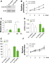
Inflammatory cytokines commonly initiate extreme changes in the synovium and cartilage microenvironment of osteoarthritis (OA) patients, which subsequently cause cellular dysfunction, especially in chondrocytes. It has been reported that induction of the purinergic P2X7 receptor (P2X7R) can regulate the expression of a variety of inflammatory factors, including interleukin (IL)-6 and -8, leading to OA pathogenesis. However, knowledge of the mechanism of upregulation of P2X7R in OA is still incomplete, and its role in chondrocyte proliferation is also not clear. It was reported previously that the expression of P2X7R was controlled by certain microRNAs, and so we tested the expression of several microRNAs and found that microRNA-373 (miR-373) was downregulated in the chondrocytes from OA patients. Regarding the mechanism of action, miR-373 inhibited chondrocyte proliferation by suppressing the expression of P2X7R, as well as inflammatory factors such as IL-6 and IL-8. Furthermore, the proliferative and pro-inflammatory effects of miR-373 on the chondrocytes could be suppressed by a P2X7R antagonist, further suggesting that miR-373 mediates chondrocyte proliferation and inflammation by targeting P2X7R. Generally, our results suggest a novel method for OA treatment by targeting the miR-373-P2X7R pathway.
Keywords: chondrocyte; miR‐373; osteoarthritis; purinergic P2X7 receptor.
Figures


Similar articles
-
Blocking of the P2X7 receptor inhibits the activation of the MMP-13 and NF-κB pathways in the cartilage tissue of rats with osteoarthritis.Int J Mol Med. 2016 Dec;38(6):1922-1932. doi: 10.3892/ijmm.2016.2770. Epub 2016 Oct 13. Int J Mol Med. 2016. PMID: 27748894
-
MicroRNA-142-3p Inhibits Chondrocyte Apoptosis and Inflammation in Osteoarthritis by Targeting HMGB1.Inflammation. 2016 Oct;39(5):1718-28. doi: 10.1007/s10753-016-0406-3. Inflammation. 2016. PMID: 27447821
-
Mechanical and IL-1β Responsive miR-365 Contributes to Osteoarthritis Development by Targeting Histone Deacetylase 4.Int J Mol Sci. 2016 Mar 23;17(4):436. doi: 10.3390/ijms17040436. Int J Mol Sci. 2016. PMID: 27023516 Free PMC article.
-
The P2X7 Receptor in Osteoarthritis.Front Cell Dev Biol. 2021 Feb 11;9:628330. doi: 10.3389/fcell.2021.628330. eCollection 2021. Front Cell Dev Biol. 2021. PMID: 33644066 Free PMC article. Review.
-
The role of cytokines in osteoarthritis pathophysiology.Biorheology. 2002;39(1-2):237-46. Biorheology. 2002. PMID: 12082286 Review.
Cited by
-
Experimental Study on the Correlation between miRNA-373 and HIF-1α, MMP-9, and VEGF in the Development of HIE.Biomed Res Int. 2021 May 3;2021:5553486. doi: 10.1155/2021/5553486. eCollection 2021. Biomed Res Int. 2021. Retraction in: Biomed Res Int. 2024 Jan 9;2024:9851358. doi: 10.1155/2024/9851358. PMID: 33997006 Free PMC article. Retracted.
-
CREB Ameliorates Osteoarthritis Progression Through Regulating Chondrocytes Autophagy via the miR-373/METTL3/TFEB Axis.Front Cell Dev Biol. 2022 Jun 9;9:778941. doi: 10.3389/fcell.2021.778941. eCollection 2021. Front Cell Dev Biol. 2022. PMID: 35756079 Free PMC article.
-
Non-Coding RNAs in Cartilage Development: An Updated Review.Int J Mol Sci. 2019 Sep 11;20(18):4475. doi: 10.3390/ijms20184475. Int J Mol Sci. 2019. PMID: 31514268 Free PMC article. Review.
-
The Role of Inflammation in the Pathogenesis of Osteoarthritis.Mediators Inflamm. 2020 Mar 3;2020:8293921. doi: 10.1155/2020/8293921. eCollection 2020. Mediators Inflamm. 2020. PMID: 32189997 Free PMC article. Review.
-
MicroRNA Alterations Induced in Human Skin by Diesel Fumes, Ozone, and UV Radiation.J Pers Med. 2022 Jan 28;12(2):176. doi: 10.3390/jpm12020176. J Pers Med. 2022. PMID: 35207665 Free PMC article.
References
-
- Johnson VL and Hunter DJ (2014) The epidemiology of osteoarthritis. Best Pract Res Clin Rheumatol 28, 5–15. - PubMed
-
- Felson DT, Lawrence RC, Dieppe PA, Hirsch R, Helmick CG, Jordan JM, Kington RS, Lane NE, Nevitt MC, Zhang Y et al (2000) Osteoarthritis: new insights. Part 1: the disease and its risk factors. Ann Intern Med 133, 635–646. - PubMed
-
- Mobasheri A, Matta C, Zakany R and Musumeci G (2015) Chondrosenescence: definition, hallmarks and potential role in the pathogenesis of osteoarthritis. Maturitas 80, 237–244. - PubMed
-
- Musumeci G, Szychlinska MA and Mobasheri A (2015) Age‐related degeneration of articular cartilage in the pathogenesis of osteoarthritis: molecular markers of senescent chondrocytes. Histol Histopathol 30, 1–12. - PubMed
-
- Musumeci G, Castrogiovanni P, Trovato FM, Imbesi R, Giunta S, Szychlinska MA, Loreto C, Castorina S and Mobasheri A (2015) Physical activity ameliorates cartilage degeneration in a rat model of aging: a study on lubricin expression. Scand J Med Sci Sports 25, e222–e230. - PubMed
LinkOut - more resources
Full Text Sources
Other Literature Sources

