Review
. 2018 Feb 2:10:1-19.
doi: 10.1016/j.pacs.2018.01.003. eCollection 2018 Jun.

Neonatal brain resting-state functional connectivity imaging modalities

Affiliations
Review

Neonatal brain resting-state functional connectivity imaging modalities

Ali-Reza Mohammadi-Nejad et al. Photoacoustics. .

Abstract

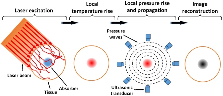
Infancy is the most critical period in human brain development. Studies demonstrate that subtle brain abnormalities during this state of life may greatly affect the developmental processes of the newborn infants. One of the rapidly developing methods for early characterization of abnormal brain development is functional connectivity of the brain at rest. While the majority of resting-state studies have been conducted using magnetic resonance imaging (MRI), there is clear evidence that resting-state functional connectivity (rs-FC) can also be evaluated using other imaging modalities. The aim of this review is to compare the advantages and limitations of different modalities used for the mapping of infants' brain functional connectivity at rest. In addition, we introduce photoacoustic tomography, a novel functional neuroimaging modality, as a complementary modality for functional mapping of infants' brain.

Keywords: Infants; Neonatal brain; Neuroimaging modalities; Photoacoustic tomography; Resting-state functional connectivity.


Figures

Fig. 1
Fig. 1
Overview of the pipeline for rs-FC data analysis. The processing steps are (1) data acquisition, (2) pre-processing, (3) region of interest (ROI) definition, (4) time-series extraction [49], (5) correlation analysis to generate weighted FC matrix, (6) thresholding to generate an adjacency matrix [50], and (7) network analysis.
Fig. 2
Fig. 2
Schematic of different resting state functional connectivity approaches including (1 st panel) In seed-based analysis, based on the time series of a seed voxel (or ROI), connectivity is calculated as the correlation of time series for all other voxels in the brain. The result of this analysis is a connectivity map showing Z-scores for each voxel (seed) indicating how well its time series correlates with the time series of other seeds. (2nd panel) In ICA, the functional data for different time points are reshaped to a single matrix (X). Then, based on the maximization of independence, this matrix is decomposed into two matrices: mixing matrix A, and source matrix S [54,55]. (3rd panel) In the graph theory the pipeline are as follow: (1) Extraction of the time-course of resting state data within each anatomical unit (ROI); (2) Calculation of a FC (i.e., network edge) correlation matrix between any pairs of nodes; (3) Thresholding the correlation matrix into a binary adjacency matrix; (4) Calculation of different graph theory metrics [56,57]. (4th panel) In clustering, after generating the initial connectivity map, based on a set of relevant characteristics, clustering algorithms attempt to group samples that are alike [58,59].
Fig. 3
Fig. 3
Formation of MRI based on the BOLD signal has several constituents: (1) a stimulus or modulation in background activity; (2) neuronal response; (3) synaptic or metabolic signaling; (4) complex relationship between neuronal activity and triggering a hemodynamic response (termed neurovascular coupling); (5) hemodynamic response itself [76]; and (6) the way in which this response is detected by an MRI scanner [77].
Fig. 4
Fig. 4
A basic EEG acquisition system is composed of (1) a set of electrodes to (2) extract the EEG time series from the scalp, (3) analog biomedical amplifiers with coupled analog low pass filters, analog-to-digital converters (A/D) [107], and (4) an interface with the data processing and display module. Brain waves measured by EEG mostly reflect electrical activity in the cortex, but include contributions from the whole brain.
Fig. 5
Fig. 5
MEG data acquisition and processing: (1) MEG time series are recorded by sensors [111] and after a (2) filtering procedure, (3) transformed to source-space time series by a beamformer algorithm [112]. (4) The resulting time series is then filtered in the alpha-band (8–12 Hz) and its phase and amplitude is extracted via Hilbert-envelope computation, resulting in 90 alpha-power time series [113]. (5) The model is constructed by taking the same AAL brain parcellation used for source-reconstruction of the MEG signal, and putting a model node in the centre of each brain area and construction an adjacency matrix for the FC.
Fig. 6
Fig. 6
After producing an appropriate PET tracer (1) and the radiotracer injection (2), (3) positrons are emitted within the subject’s body, combine with nearby electrons and annihilate [131]. The result is a pair of 511-KeV gamma photons released in opposite directions. PET scanners use pairs of radiation detectors to measure the nearly simultaneous, coincident interaction of the 511-KeV photons. The data recorded by the scanner is a collection of coincidence detections. In the reconstruction step (4), a mathematical procedure is implemented to convert the acquired data to tomographic images (5).
Fig. 7
Fig. 7
Schematic diagram of the fNIRS system. NIR-light are generated and guided to the human's head by optical fibers or cables. Another fiber bundle or cable directs diffusively reflected light from the head to detectors. A light detector captures the light resulting from the interaction with the chromophores (e.g. HbO, Hb), following a crescent-shaped path back to the surface of the skin.
Fig. 8
Fig. 8
Brain mapping using diffuse optical tomography. Optical Imaging: Brain activity is recorded by sending near infrared light into the brain and recording diffusely reflected light. The vascular response to the neural activity results in changes in the concentration of oxygenated and de-oxygenated hemoglobin (HbO and HbR) which results in changes in the optical absorption coefficient and so changes in the intensity of diffusely reflected light. DOT Image Reconstruction: Source and detector arrangement in a high-density DOT imaging array. Example first to fourth nearest neighbor source-detector pairs are shown. Forward light propagation model is constructed by registering the imaging array to the head model. 3D images of absorption coefficient changes are reconstructed. Image Analysis: HbO and HbR signals are generated by hemoglobin spectroscopy. Temporal filtering and spatial smoothing are applied to the images. Additional statistical analyses are performed to generate cortical maps of the brain activity [177].
Fig. 9
Fig. 9
Process of a photoacoustic tomography generation.

References

    1. Friston K.J., Frith C.D., Liddle P.F., Frackowiak R.S. Functional connectivity: the principal-component analysis of large (PET) data sets. J. Cereb. Blood Flow Metab. 1993;13:5–14. - PubMed
    1. Aslin R.N., Shukla M., Emberson L.L. Hemodynamic correlates of cognition in human infants. Annu. Rev. Psychol. 2015;66:349–379. - PMC - PubMed
    1. Smyser C.D., Snyder A.Z., Neil J.J. Functional connectivity MRI in infants: exploration of the functional organization of the developing brain. Neuroimage. 2011;56:1437–1452. - PMC - PubMed
    1. Vogel A.C., Power J.D., Petersen S.E., Schlaggar B.L. Development of the brain’s functional network architecture. Neuropsychol. Rev. 2010;20:362–375. - PMC - PubMed
    1. Smith-Collins A.P.R., Luyt K., Heep A., Kauppinen R.A. High frequency functional brain networks in neonates revealed by rapid acquisition resting state fMRI. Hum. Brain Mapp. 2015;36:2483–2494. - PMC - PubMed

LinkOut - more resources