

doi: 10.1016/j.dib.2018.01.091.
eCollection 2018 Apr.
Dataset on the effects of spermidine on linking number differences between histone H1-free and histone H1-bound circular polynucleosomes
Affiliations
- PMID: 29511714
- PMCID: PMC5832644
- DOI: 10.1016/j.dib.2018.01.091
Item in Clipboard
Dataset on the effects of spermidine on linking number differences between histone H1-free and histone H1-bound circular polynucleosomes
Data Brief.
.
Abstract
The data presented in this article are related to the research article entitled "Quantitative determination of linking number differences between circular polynucleosomes and histone H1-bound circular polynucleosomes" Zhang et al. (in press) [1]. DNA linking number differences between histone H1-free and histone H1-bound circular polynucleosomes at various spermidine concentrations was quantitatively determined by chloroquine-based gel electrophoretic analysis in this work, which provides information on the topological effects of histone H1 and spermidine on the linker DNA between nucleosomes.
Figures
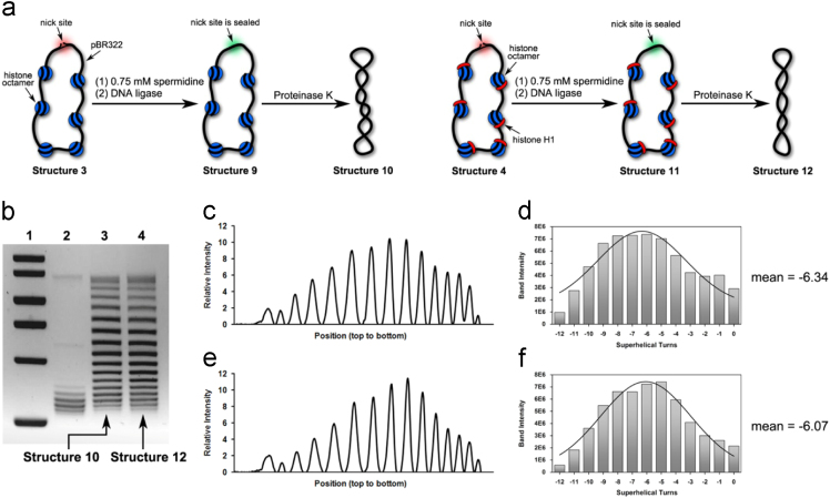
(a) Illustration of our preparations of Structure 10 and Structure 12 in the presence of 0.75 mM spermidine. (b) Chloroquine-based agarose gel electrophoretic analysis on Structure 10 and Structure 12. Lane 1: molecular weight markers; Lane 2: relaxed forms of pBR322; Lane 3: Structure 10 and Lane 4: Structure 12. (c) Densitometry tracing of gel electrophoretic results in Lane 3. (d) Plot of Gauss fit on the data shown in Fig. 1c, which gave rise to − 6.34 as its mean value of ΔLk (e) Densitometry tracing of gel electrophoretic results in Lane 4. (f) Plot of Gauss fit on the data shown in Fig. 1e, which gave rise to − 6.07 as its mean value of ΔLk.
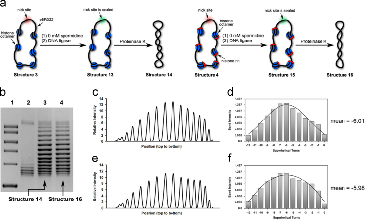
(a) Illustration of our preparations of Structure 14 and Structure 16 in the presence of 0 mM spermidine. (b) Chloroquine-based agarose gel electrophoretic analysis on Structure 14 and Structure 16. Lane 1: molecular weight markers; Lane 2: relaxed forms of pBR322; Lane 3: Structure 14 and Lane 4: Structure 16. (c) Densitometry tracing of gel electrophoretic results in Lane 3. (d) Plot of Gauss fit on the data shown in Fig. 2c, which gave rise to − 6.01 as its mean value of ΔLk (e) Densitometry tracing of gel electrophoretic results in Lane 4. (f) Plot of Gauss fit on the data shown in Fig. 2e, which gave rise to − 5.98 as its mean value of ΔLk.
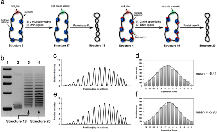
(a) Illustration of our preparations of Structure 18 and Structure 20 in the presence of 2 mM spermidine. (b) Chloroquine-based agarose gel electrophoretic analysis on Structure 18 and Structure 20. Lane 1: molecular weight markers; Lane 2: relaxed forms of pBR322; Lane 3: Structure 18 and Lane 4: Structure 20. (c) Densitometry tracing of gel electrophoretic results in Lane 3. (d) Plot of Gauss fit on the data shown in Fig. 3c, which gave rise to − 6.41 as its mean value of ΔLk (e) Densitometry tracing of gel electrophoretic results in Lane 4. (f) Plot of Gauss fit on the data shown in Fig. 3e, which gave rise to − 5.98 as its mean value of ΔLk.
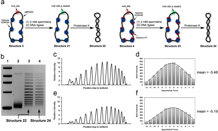
(a) Illustration of our preparations of Structure 22 and Structure 24 in the presence of 3 mM spermidine. (b) Chloroquine-based agarose gel electrophoretic analysis on Structure 22 and Structure 24. Lane 1: molecular weight markers; Lane 2: relaxed forms of pBR322; Lane 3: Structure 22 and Lane 4: Structure 24. (c) Densitometry tracing of gel electrophoretic results in Lane 3. (d) Plot of Gauss fit on the data shown in Fig. 4c, which gave rise to − 5.48 as its mean value of ΔLk (e) Densitometry tracing of gel electrophoretic results in Lane 4. (f) Plot of Gauss fit on the data shown in Fig. 4e, which gave rise to − 5.19 as its mean value of ΔLk.
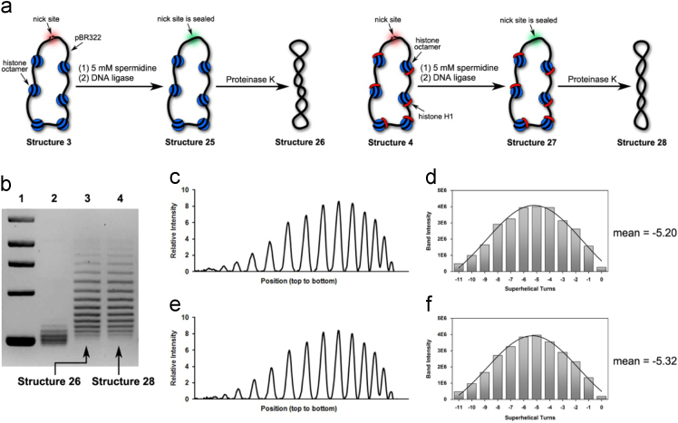
(a) Illustration of our preparations of Structure 26 and Structure 28 in the presence of 5 mM spermidine. (b) Chloroquine-based agarose gel electrophoretic analysis on Structure 26 and Structure 28. Lane 1: molecular weight markers; Lane 2: relaxed forms of pBR322; Lane 3: Structure 26 and Lane 4: Structure 28. (c) Densitometry tracing of gel electrophoretic results in Lane 3. (d) Plot of Gauss fit on the data shown in Fig. 5c, which gave rise to − 5.20 as its mean value of ΔLk (e) Densitometry tracing of gel electrophoretic results in Lane 4. (f) Plot of Gauss fit on the data shown in Fig. 5e, which gave rise to − 5.32 as its mean value of ΔLk.
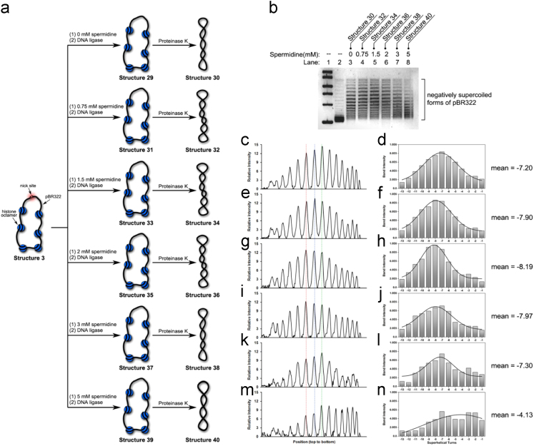
(a) Illustration of our preparations of Structure 30, Structure 32, Structure 34, Structure 36, Structure 38 and Structure 40 in the presence of increasing amount of spermidine. (b) This figure is same as Fig. 6(a) in Ref. . (c) Densitometry tracing of Lane 3. (d) Plot of Gauss fit on the data shown in (c), which gave rise to − 7.20 as its mean value of ΔLk. (e) Densitometry tracing of Lane 4. (f) Plot of Gauss fit on the data shown in (e), which gave rise to − 7.90 as its mean value of ΔLk. (g) Densitometry tracing of Lane 5. (h) Plot of Gauss fit on the data shown in (g), which gave rise to − 8.19 as its mean value of ΔLk. (i) Densitometry tracing of Lane 6. (j) Plot of Gauss fit on the data shown in (i), which gave rise to − 7.97 as its mean value of ΔLk. (k) Densitometry tracing of Lane 7. (l) Plot of Gauss fit on the data shown in (k), which gave rise to − 7.30 as its mean value of ΔLk. (m) Densitometry tracing of Lane 8. (n) Plot of Gauss fit on the data shown in (m), which gave rise to − 4.13 as its mean value of ΔLk.
Similar articles
-
Quantitative determination of linking number differences between circular polynucleosomes and histone H1-bound circular polynucleosomes.Bioorg Med Chem Lett. 2018 Feb 1;28(3):537-540. doi: 10.1016/j.bmcl.2017.10.072. Epub 2017 Oct 31. Bioorg Med Chem Lett. 2018. PMID: 29269213
-
Reconstitution of compact polynucleosomes and comparison of the functions of histones H1 and H5.J Biochem. 1984 Oct;96(4):1071-8. doi: 10.1093/oxfordjournals.jbchem.a134924. J Biochem. 1984. PMID: 6520112
-
Characterization of poly(ADP-ribose)--histone H1 complex formation in purified polynucleosomes and chromatin.Eur J Biochem. 1980 Dec;113(1):15-25. doi: 10.1111/j.1432-1033.1980.tb06133.x. Eur J Biochem. 1980. PMID: 7460942
-
Germline-specific H1 variants: the "sexy" linker histones.Chromosoma. 2016 Mar;125(1):1-13. doi: 10.1007/s00412-015-0517-x. Epub 2015 Apr 29. Chromosoma. 2016. PMID: 25921218 Review.
-
Linker histone subtypes and their allelic variants.Cell Biol Int. 2012 Nov 1;36(11):981-96. doi: 10.1042/CBI20120133. Cell Biol Int. 2012. PMID: 23075301 Review.
References
-
- Zhang H., Li T. Quantitative determination of linking number differences between circular polynucleosomes and histone H1-bound circular polynucleosomes. Bioorg. Med. Chem. Lett. 2018 (in press) 28, 2018, 537-540. - PubMed
-
- Zhang H., Li T. Presence of negative supercoiling in aggregates of histone H1-plasmidic polynucleosome complexes. Bioorg. Med. Chem. Lett. 2017;27:168–170. - PubMed
-
- Zhang H., Li T. Effects of spermidine and ATP on stabilities of chromatosomes and histone H1-depleted chromatosomes. Bioorg. Med. Chem. Lett. 2017;27:1149–1153. - PubMed
-
- Bates Andrew D., Maxwell A. The role of ATP in the reactions of type II DNA topoisomerases. Biochem. Soc. Trans. 2010;38:438–442. - PubMed
LinkOut - more resources
Full Text Sources
Other Literature Sources
