

Myocardial Extracellular Volume Quantification by Cardiovascular Magnetic Resonance and Computed Tomography
- PMID: 29511861
- PMCID: PMC5840231
- DOI: 10.1007/s11886-018-0961-3
Myocardial Extracellular Volume Quantification by Cardiovascular Magnetic Resonance and Computed Tomography
Abstract
Purpose of review: This review article discusses the evolution of extracellular volume (ECV) quantification using both cardiovascular magnetic resonance (CMR) and computed tomography (CT).
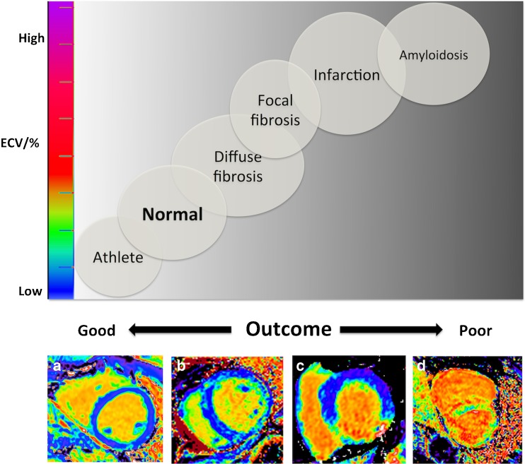
Recent findings: Visualizing diffuse myocardial fibrosis is challenging and until recently, was restricted to the domain of the pathologist. CMR and CT both use extravascular, extracellular contrast agents, permitting ECV measurement. The evidence base around ECV quantification by CMR is growing rapidly and just starting in CT. In conditions with high ECV (amyloid, oedema and fibrosis), this technique is already being used clinically and as a surrogate endpoint. Non-invasive diffuse fibrosis quantification is also generating new biological insights into key cardiac diseases. CMR and CT can estimate ECV and in turn diffuse myocardial fibrosis, obviating the need for invasive endomyocardial biopsy. CT is an attractive alternative to CMR particularly in those individuals with contraindications to the latter. Further studies are needed, particularly in CT.
Keywords: Cardiovascular magnetic resonance; Computed tomography; Extracellular volume; Tissue characterization.
Conflict of interest statement
Conflict of Interest
Paul R. Scully, James C. Moon, and Thomas A. Treibel declare that they have no conflict of interest.
Gorka Bastarrika reports other from Siemens Healthcare, General Electric, and Bayer.
Human and Animal Rights and Informed Consent
This article does not contain any studies with human or animal subjects performed by any of the authors.
Figures
References
-
- Kim RJ, Albert TSE, Wible JH, Elliott MD, Allen JC, Lee JC, Parker M, Napoli A, Judd RM, for the Gadoversetamide Myocardial Infarction Imaging Investigators Performance of delayed-enhancement magnetic resonance imaging with gadoversetamide contrast for the detection and assessment of myocardial infarction: an international, multicenter, double-blinded, randomized trial. Circulation. 2008;117:629–637. doi: 10.1161/CIRCULATIONAHA.107.723262. - DOI - PubMed
-
- Oliva MR, Mortele KJ. Iodinated contrast agents for cardiac CT. In: Di Carli MF, Lipton MJ, editors Card PET PETCT Imaging, New York, NY: Springer New York; 2007, p. 83–93. doi:10.1007/978-0-387-38295-1_6.
Publication types
MeSH terms
Substances
Grants and funding
LinkOut - more resources
Full Text Sources
Other Literature Sources
Medical
Research Materials
