Review
. 2018 Dec;12(4):517-521.
doi: 10.1007/s12105-018-0903-9. Epub 2018 Mar 6.

Spongiotic Gingival Hyperplasia Synchronously Involving Multiple Sites: Case Report and Review of the Literature

Affiliations
Review

Spongiotic Gingival Hyperplasia Synchronously Involving Multiple Sites: Case Report and Review of the Literature

Ioannis Siamantas et al. Head Neck Pathol. 2018 Dec.

Abstract

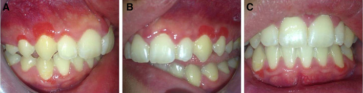
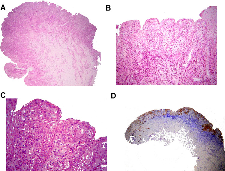
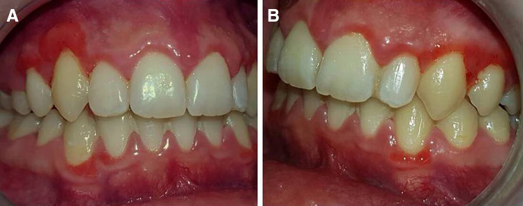
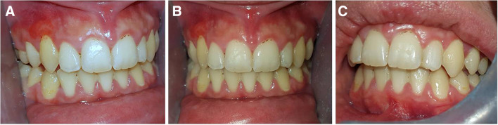
Localized juvenile spongiotic gingival hyperplasia (LJSGH) is a gingival lesion with unique clinicopathologic features that may involve synchronously multiple sites. We present a case with lesions clinically consistent with LJSGH in four jaw quadrants, confirmed by biopsy and review the English literature on multifocal LJSGH cases. A 19 year-old woman presented with circumscribed, erythematous overgrowths on the right and left maxillary and mandibular gingiva. With the provisional diagnosis of multifocal LJSGH, total excision of four maxillary lesions was performed. Clinical, microscopic and immunohistochemical examination with cytokeratin 19 confirmed the diagnosis of LJSGH in multiple sites. The excised lesions showed partial to complete recurrence after 4 months, while spontaneous regression of all but one lesion was observed after 15 months. Twenty cases with synchronous involvement of the gingiva of at least two teeth were previously reported. Their clinical features were comparable to that of solitary LJSGH. Only one case involved all four jaw quadrants. Spontaneous remission has not been documented before. The recognition of multiple lesions with clinicopathologic features diagnostic of LJSGH in the same adult patient argue against the designations "localized" and "juvenile". Recurrences are common, while remission might occur.

Keywords: Generalized; Gingivitis; Localized; Multifocal; Spongiotic gingival hyperplasia.


Conflict of interest statement

All Authors declares that they have no conflict of interest.

Figures

Fig. 1
Fig. 1
Clinical examination. Well-circumscribed, bright red, pedunculated overgrowths with papillary surface, on the labial marginal and attached gingiva of a maxillary right lateral incisor to first premolar, b maxillary left canine and first premolar, and c mandibular right lateral incisor and canine, and left canine
Fig. 2
Fig. 2
a Microscopic examination revealed mucosal fragments covered by non-keratinized stratified squamous epithelium, with papillary surface (hematoxylin and eosin stain, original magnification ×25). b Microscopically, the lesion was covered by non-keratinized stratified squamous epithelium, showing papillary surface, acanthosis, spongiosis, focally elongated and interconnecting rete pegs, as well as neutrophilic exocytosis (hematoxylin and eosin stain, original magnification ×100). c The lesional epithelium was characterized by acanthosis, spongiosis, focally elongated and interconnecting rete pegs and neutrophilic exocytosis. The underlying connective tissue was vascular with dense inflammatory infiltrations mostly by neutrophils, lymphocytes and plasma cells (hematoxylin and eosin stain, original magnification ×400). d Intense cytoplasmic positivity of CK19 in all cell layers of lesional epithelium, but only in the basal cell layer of the adjacent gingival epithelium (streptavidin-biotin-peroxidase, original magnification ×25)
Fig. 3
Fig. 3
Re-examination 4 months post biopsy. Recurrence of excised lesions, totally around the right upper canine and first premolar (a) and partly around the left (b). No change is noticed on lesions of the mandibular facial, marginal gingiva
Fig. 4
Fig. 4
Re-examination 15 months post biopsy. The lesion (a) around the maxillary right canine is unchanged; (b) around the mandibular right lateral incisor, right canine and left canine has significantly regressed; and (ac) around the maxillary right first premolar, left canine and left first premolar has disappeared

References

    1. Chang JY, Kessler HP, Wright JM. Localized juvenile spongiotic gingival hyperplasia. Oral Surg Oral Med Oral Pathol Oral Radiol Endod. 2008;106(3):411–418. doi: 10.1016/j.tripleo.2008.04.024. - DOI - PubMed
    1. Darling MR, Daley TD, Wilson A, Wysocki GP. Juvenile spongiotic gingivitis. J Periodontol. 2007;78(7):1235–1240. doi: 10.1902/jop.2007.060502. - DOI - PubMed
    1. Argyris PP, Nelson AC, Papanakou S, Merkourea S, Tosios KI, Koutlas IG. Localized juvenile spongiotic gingival hyperplasia featuring unusual p16INK4A labeling and negative human papillomavirus status by polymerase chain reaction. J Oral Pathol Med. 2015;44(1):37–44. doi: 10.1111/jop.12214. - DOI - PubMed
    1. de Freitas Silva BS, Silva Sant’Ana SS, Watanabe S, Vencio EF, Roriz VM, Yamamoto-Silva FP. Multifocal red bands of the marginal gingiva. Oral Surg Oral Med Oral Pathol Oral Radiol Endod. 2015;119(1):3–7. doi: 10.1016/j.oooo.2014.07.005. - DOI - PubMed
    1. MacNeill SR, Rokos JW, Umaki MR, Satheesh KM, Cobb CM. Conservative treatment of localized juvenile spongiotic gingival hyperplasia. Clin Adv Periodontics. 2011;1(3):199–204. doi: 10.1902/cap.2011.110003. - DOI

LinkOut - more resources