. 2018 Jun;41(6):3379-3393.
doi: 10.3892/ijmm.2018.3526. Epub 2018 Mar 1.

MicroRNA-155 inhibits the osteogenic differentiation of mesenchymal stem cells induced by BMP9 via downregulation of BMP signaling pathway

Affiliations

MicroRNA-155 inhibits the osteogenic differentiation of mesenchymal stem cells induced by BMP9 via downregulation of BMP signaling pathway

Hongxia Liu et al. Int J Mol Med. 2018 Jun.

Abstract

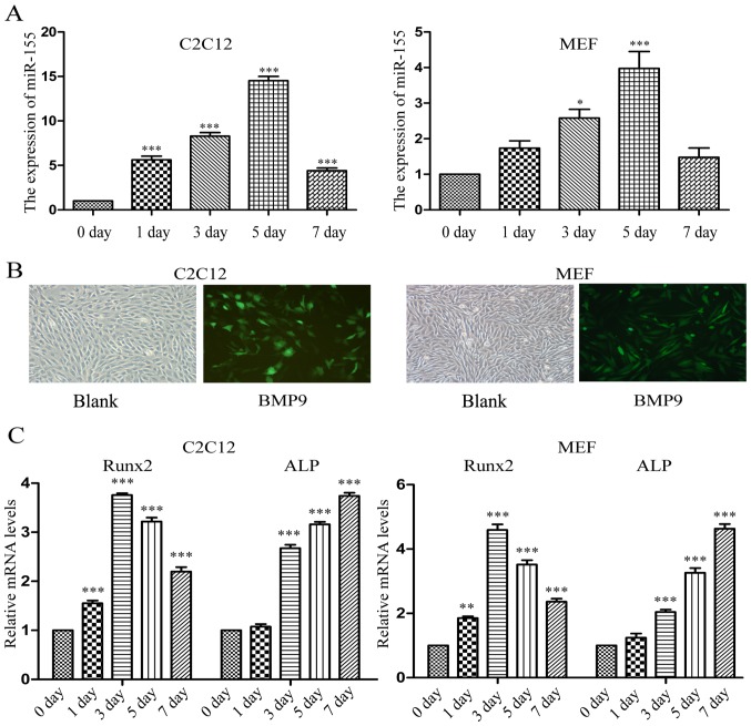
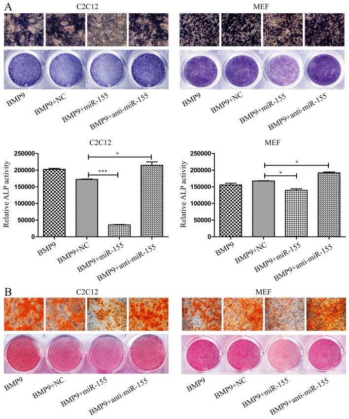
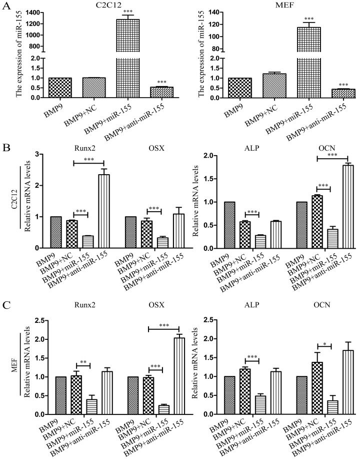
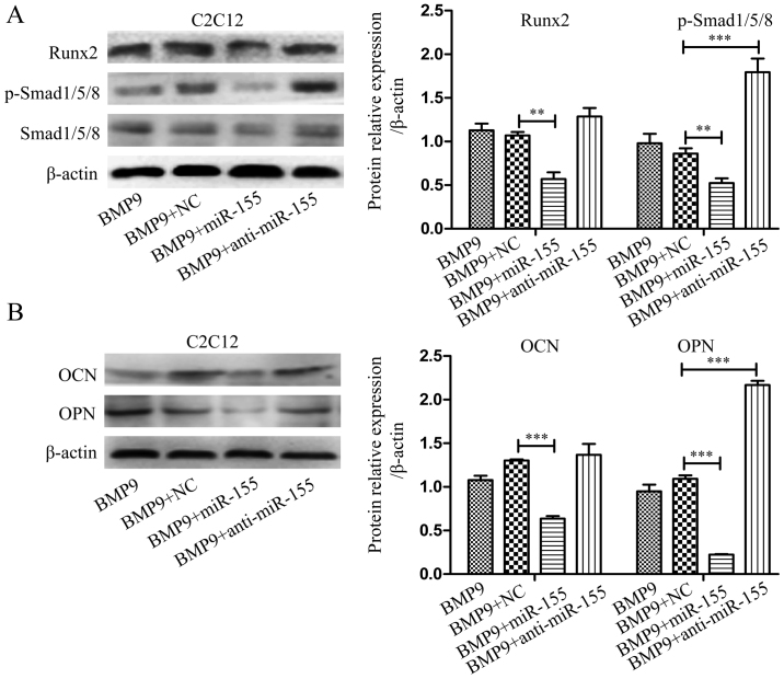
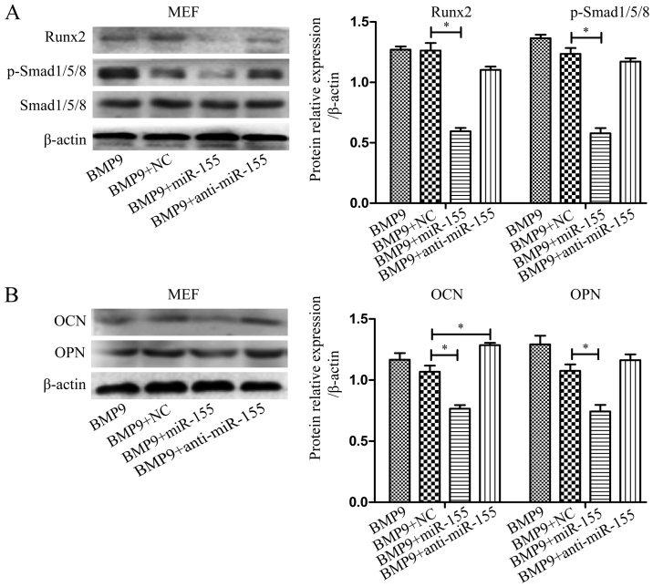
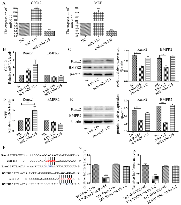
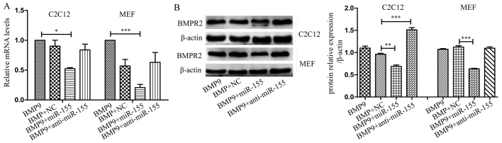
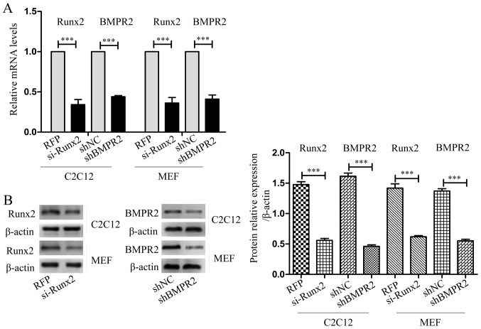
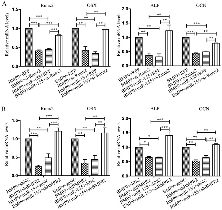
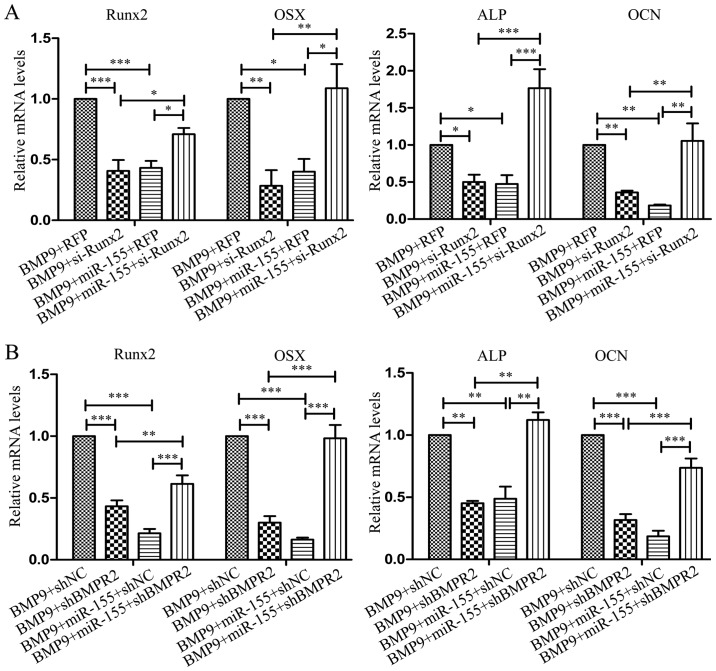
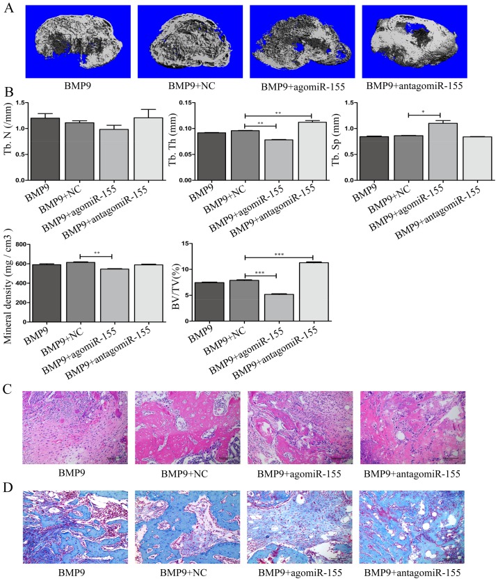
Previous studies have indicated that bone morphogenetic protein 9 (BMP9) can promote the osteogenic differentiation of mesenchymal stem cells (MSCs) and increase bone formation in bone diseases. However, the mechanisms involved remained poorly understood. It is necessary to investigate the specific regulatory mechanisms of osteogenic differentiation that were induced by BMP9. During the process of osteogenic differentiation induced by BMP9, the expression of microRNA-155 (miR-155) exhibited a tendency of increasing at first and then decreasing, which made us consider that miR-155 may have a modulatory role in this process, but the roles of this process have not been elucidated. This study aimed to uncover miR-155 capable of concomitant regulation of this process. mmu-miR-155 mimic (miR-155) was transfected into MSCs and osteogenesis was induction by using recombinant adenovirus expressing BMP9. Overexpressed miR-155 in MSCs led to a decrease in alkaline phosphatase (ALP) staining and Alizarin red S staining during osteogenic differentiation, and reduced the expression of osteogenesis-related genes, such as runt-related transcription factor 2 (Runx2), osterix (OSX), osteocalcin (OCN) and osteopontin (OPN). On protein levels, overexpressed miR-155 markedly decreased the expression of phosphorylated Smad1/5/8 (p-Smad1/5/8), Runx2, OCN and OPN. Luciferase reporter assay revealed Runx2 and bone morphogenetic protein receptor 9 (BMPR2) are two direct target genes of miR-155. Downregulation of the expression of Runx2 and BMPR2, respectively could offset the inhibitory effect of miR-155 in the osteogenesis of MSCs. In vivo, subcutaneous ectopic osteogenesis of MSCs in nude mice showed miR-155 inhibited osteogenic differentiation. In conclusion, our results demonstrated that miR-155 can inhibit the osteogenic differentiation induced by BMP9 in MSCs.


Figures

Figure 1
Figure 1
The expression level of miR-155 is increased at first and then decreased in the process of bone morphogenetic protein 9 (BMP9)-induced early osteogenesis. (A) The expression of miR-155 was detected by RT-qPCR. *p<0.05 and ***p<0.001 vs. the value at day 0. (B) BMP9 was transfected into C2C12 cells and mouse embryonic fibroblasts (MEF) cells efficiently. Magnification, ×100. White, white light used as the excitation light; green, blue light used as the excitation light. Multiple of infection (MOI): 5. (C) The expression of runt-related transcription factor 2 (Runx2) and alkaline phosphatase (ALP) in C2C12 cells and MEF cells in differentiation days induced by BMP9 were detected by RT-qPCR. **p<0.01 and ***p<0.001 vs. the value at day 0. Day: osteogenic differentiation day.
Figure 2
Figure 2
miR-155 inhibits the osteogenic differentiation of C2C12 cells and mouse embryonic fibroblasts (MEF) cells induced by bone morphogenetic protein 9 (BMP9). (A) Alkaline phosphatase (ALP) staining and ALP activity were performed after transfecting C2C12 cells and MEF cells with NC, miR-155 or anti-miR-155, induced osteogenesis by BMP9 for 7 days. (B) The effect of miR-155 on BMP9-induced matrix mineralization though Alizarin Red S staining on the 14th day of differentiation in C2C12 cells and MEF cells. Each staining was repeated three times for both types of the cells in this experiment, and one of the three independent results is shown. *p<0.05 and ***p<0.001.
Figure 3
Figure 3
miR-155 downregulates the mRNA expression levels of osteogenesis genes runt-related transcription factor 2 (Runx2), osterix (OSX), alkaline phosphatase (ALP) and osteocalcin (OCN). C2C12 cells and mouse embryonic fibroblasts (MEF) were transfected with NC, miR-155 or anti-miR-155, respectively, then induced osteogenesis by bone morphogenetic protein 9 (BMP9). (A) The expression of miR-155 by RT-qPCR in the third day of differentiation. (B) Runx2 and OSX were tested on the 3rd day of differentiation, ALP and OCN were tested on the 7th day of differentiation in C2C12 cells. (C) Runx2 and OSX were tested on the 3rd day of differentiation, ALP and OCN were tested on the 7th day of differentiation in MEF cells. *p<0.05, **p<0.01 and ***p<0.001.
Figure 4
Figure 4
miR-155 acts as an inhibitor of osteogenesis via suppressing BMP signaling during osteogenesis induced by bone morphogenetic protein 9 (BMP9) in C2C12 cells. (A) After treatment of C2C12 cells, the expression of p-Smad1/5/8 and runt-related transcription factor 2 (Runx2) were detected by western blotting. (B) The protein expression levels of osteocalcin (OCN) and OPN were measured by western blotting. β-actin was used as the internal controls, **p<0.01 and ***p<0.001.
Figure 5
Figure 5
miR-155 acts as an inhibitor of osteogenesis via suppressing BMP signaling during the osteogenesis induced by bone morphogenetic protein 9 (BMP9) in mouse embryonic fibroblasts (MEF) cells. (A) After treatment of MEF cells, the expression of p-Smad1/5/8 and runt-related transcription factor 2 (Runx2) was detected by western blotting. (B) The protein expression levels of osteocalcin (OCN) and osteopontin (OPN) were measured by western blotting. β-actin was used as the internal controls, *p<0.05.
Figure 6
Figure 6
miR-155 directly targets runt-related transcription factor 2 (Runx2) and BMPR2. C2C12 cells and mouse embryonic fibroblasts (MEF) were transfected with NC, miR-155 or anti-miR-155, respectively. (A) Detected the expression of miR-155 by RT-qPCR. (B) Tested the expression of Runx2 and BMPR2 in C2C12 cells by RT-qPCR. (C) The expression of Runx2 and BMPR2 in C2C12 cells by western blotting. (D) The expression of Runx2 and BMPR2 in MEF cells by RT-qPCR. (E) The expression of Runx2 and BMPR2 in MEF cells by western blotting. (F) Putative binding sites for miR-155 in the 3′-untranslated region (3′-UTR) of Runx2 and BMPR2 as predicted by TargetScan and PicTar. Mutated nucleotides in mutant Runx2 3′-UTR and BMPR2 3′-UTR seed sequences are represented in blue. (G) Luciferase reporter assays in HEK-293 cells. Cells were co-transfected with WT-Runx2, or MT-Runx2, or WT-BMPR2 or MT-BMPR2 reporter construct or miR-155 overnight. Forty-eight hours post transfection luciferase activities were tested. Results were normalized to the value of cells that transfected pMIR-REPORT only. β-actin was used as the internal controls for western blotting, **p<0.01 and ***p<0.001.
Figure 7
Figure 7
During the osteogenic differentiation induced by bone morphogenetic protein 9 (BMP9), miR-155 decreases the expression of BMPR2. (A) The mRNA expression of BMPR2 in C2C12 cells and mouse embryonic fibroblasts (MEF) cells by RT-qPCR. (B) The protein expression of BMPR2 in C2C12 cells and MEF cells by western blot. β-actin was used as the internal controls, *p<0.05, **p<0.01 and ***p<0.001.
Figure 8
Figure 8
Interference of runt-related transcription factor 2 (Runx2) and BMPR2 in C2C12 cells and mouse embryonic fibroblasts (MEF), respectively. (A) The interference efficiency of si-Runx2 and shBMPR2 in C2C12 cells and mouse embryonic fibroblasts (MEF) after transfection by RT-qPCR. (B) The interference efficiency of si-Runx2 and shBMPR2 in C2C12 cells and MEF cells after transfection by western blotting. β-actin was used as the internal controls, ***p<0.001.
Figure 9
Figure 9
Knockdown of runt-related transcription factor 2 (Runx2) and BMPR2 separately reduced the effect of miR-155 in C2C12 cells. (A and B) C2C12 cells were co-transfected with RFP, or si-Runx2, or shNC, or shBMPR2, or miR-155, then induced osteogenic differentiation by bone morphogenetic protein 9 (BMP9) for 3 days to test the expression of Runx2, osterix (OSX), and induced for 7 days to test the expression of alkaline phosphatase (ALP), osteocalcin (OCN) used RT-qPCR. *p<0.05, **p<0.01 and ***p<0.001.
Figure 10
Figure 10
Knockdown runt-related transcription factor 2 (Runx2) and BMPR2 separately reduced the effect of miR-155 in mouse embryonic fibroblasts (MEF) cells. (A and B) MEF cells were co-transfected with RFP or si-Runx2 or shNC or shBMPR2 and miR-155, then induced osteogenic differentiation by BMP9 for 3 days to test the expression of Runx2, osterix (OSX), was induced for 7 days to test the expression of alkaline phosphatase (ALP) and osteocalcin (OCN) by RT-qPCR. *p<0.05, **p<0.01 and ***p<0.001.
Figure 11
Figure 11
miR-155 repressed ectopic bone formation induced by bone morphogenetic protein 9 (BMP9) in vivo. Ectopic bones were harvested 6 weeks after treated-mouse embryonic fibroblasts (MEF) cells were injected subcutaneously in 4-week-old female nude mice. (A) µCT with representative image of the ectopic bones. Scale bar, 1 mm. (B) µCT quantification of the ectopic bones from 10-week-old female nude mice, which were injected with transfected-MEF cells at 4 weeks. Parameters include: trabecular number, thickness, separation, volume and BMD. n=3, *p<0.05, **p<0.01 and ***p<0.001. (C) Histological images of 6-week ectopic bones from each group upon H&E staining. Scale bars, 100 µm. (D) Histological images of 6-week ectopic bones from each group upon Masson's trichrome staining. Scale bars, 100 µm.

References

    1. Luu HH, Song WX, Luo X, Manning D, Luo J, Deng ZL, Sharff KA, Montag AG, Haydon RC, He TC. Distinct roles of bone morphogenetic proteins in osteogenic differentiation of mesenchymal stem cells. J Orthop Res. 2007;25:665–677. doi: 10.1002/jor.20359. - DOI - PubMed
    1. Beederman M, Lamplot JD, Nan G, Wang J, Liu X, Yin L, Li R, Shui W, Zhang H, Kim SH, et al. BMP signaling in mesenchymal stem cell differentiation and bone formation. J Biomed Sci Eng. 2013;6:32–52. doi: 10.4236/jbise.2013.68A1004. - DOI - PMC - PubMed
    1. Bidart M, Ricard N, Levet S, Samson M, Mallet C, David L, Subileau M, Tillet E, Feige JJ, Bailly S. BMP9 is produced by hepatocytes and circulates mainly in an active mature form complexed to its prodomain. Cell Mol Life Sci. 2012;69:313–324. doi: 10.1007/s00018-011-0751-1. - DOI - PMC - PubMed
    1. Brown MA, Zhao Q, Baker KA, Naik C, Chen C, Pukac L, Singh M, Tsareva T, Parice Y, Mahoney A, et al. Crystal structure of BMP-9 and functional interactions with pro-region and receptors. J Biol Chem. 2005;280:25111–25118. doi: 10.1074/jbc.M503328200. - DOI - PubMed
    1. Wang RN, Green J, Wang Z, Deng Y, Qiao M, Peabody M, Zhang Q, Ye J, Yan Z, Denduluri S, et al. Bone Morphogenetic Protein (BMP) signaling in development and human diseases. Genes Dis. 2014;1:87–105. doi: 10.1016/j.gendis.2014.07.005. - DOI - PMC - PubMed

MeSH terms