Review
. 2018 Apr 1;98(2):881-917.
doi: 10.1152/physrev.00017.2017.

The Biology of Regeneration Failure and Success After Spinal Cord Injury

Affiliations
Review

The Biology of Regeneration Failure and Success After Spinal Cord Injury

Amanda Phuong Tran et al. Physiol Rev. .

Abstract

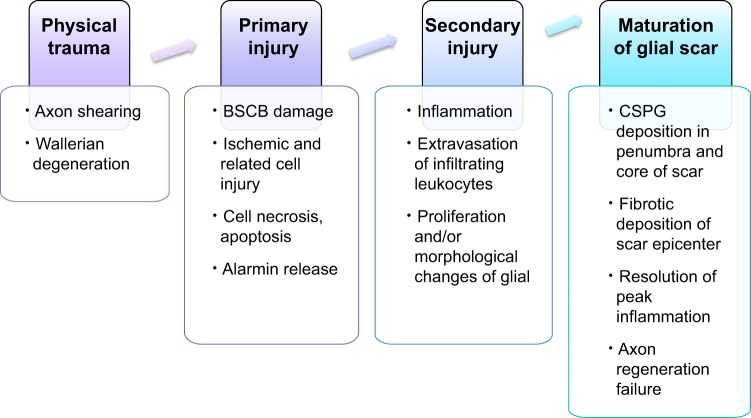
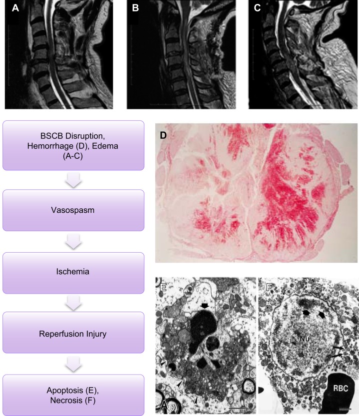
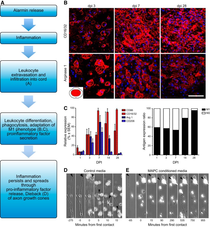
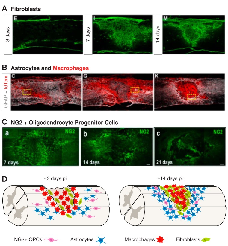
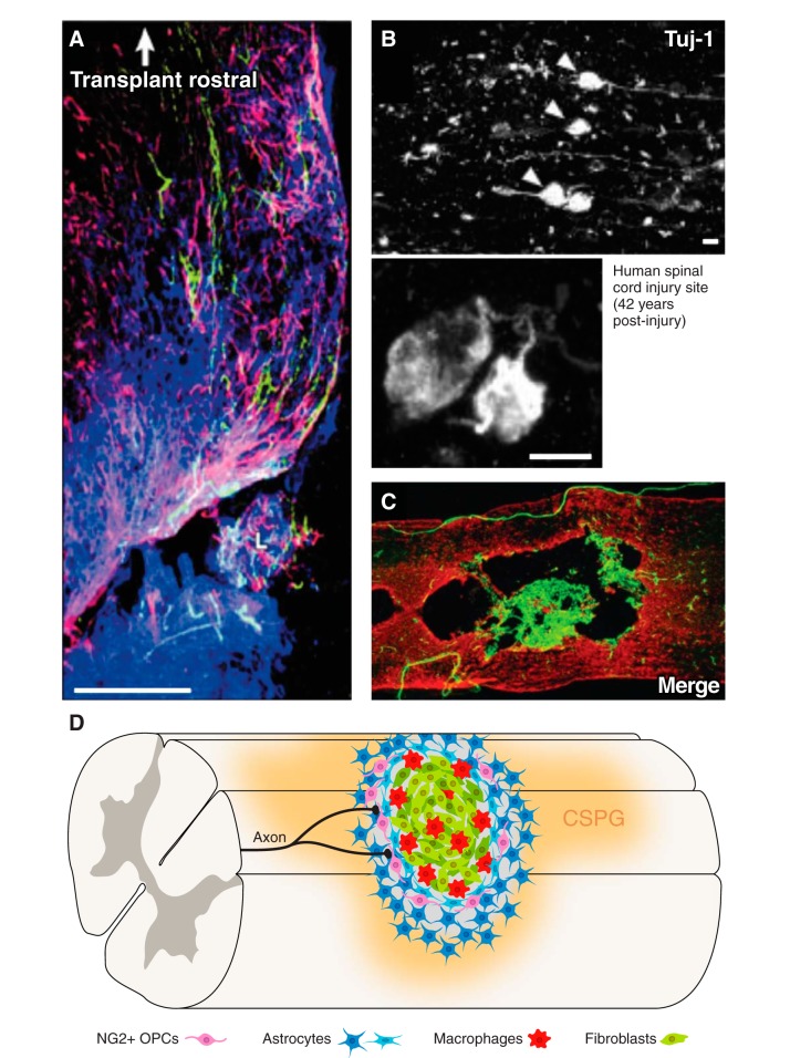
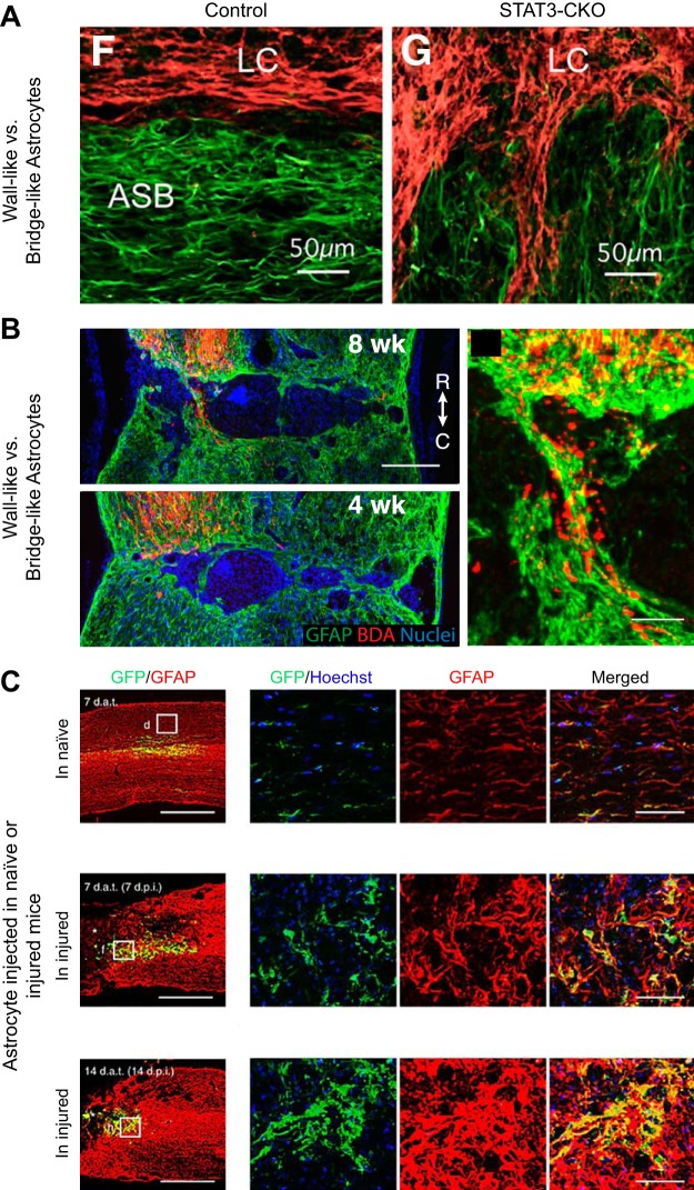
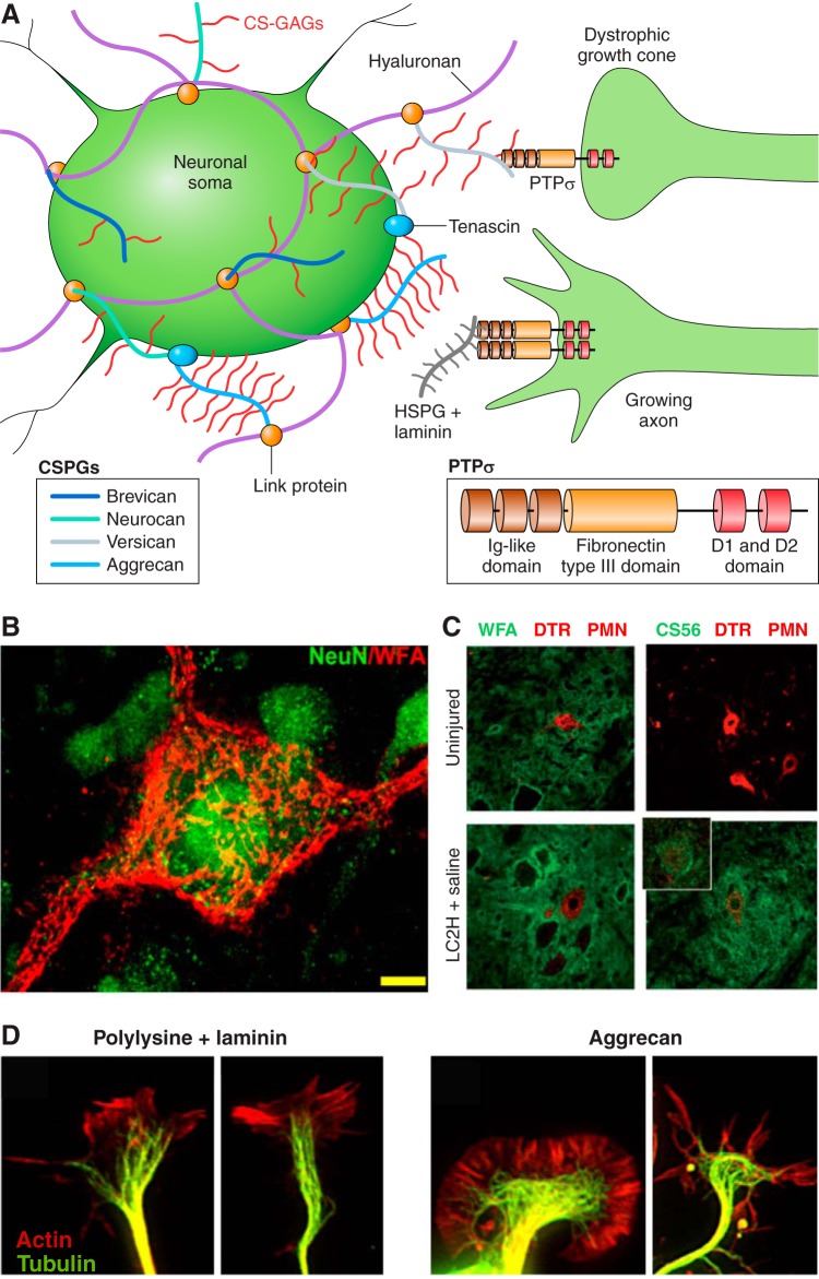
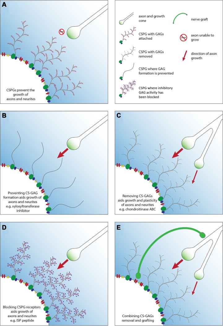
Since no approved therapies to restore mobility and sensation following spinal cord injury (SCI) currently exist, a better understanding of the cellular and molecular mechanisms following SCI that compromise regeneration or neuroplasticity is needed to develop new strategies to promote axonal regrowth and restore function. Physical trauma to the spinal cord results in vascular disruption that, in turn, causes blood-spinal cord barrier rupture leading to hemorrhage and ischemia, followed by rampant local cell death. As subsequent edema and inflammation occur, neuronal and glial necrosis and apoptosis spread well beyond the initial site of impact, ultimately resolving into a cavity surrounded by glial/fibrotic scarring. The glial scar, which stabilizes the spread of secondary injury, also acts as a chronic, physical, and chemo-entrapping barrier that prevents axonal regeneration. Understanding the formative events in glial scarring helps guide strategies towards the development of potential therapies to enhance axon regeneration and functional recovery at both acute and chronic stages following SCI. This review will also discuss the perineuronal net and how chondroitin sulfate proteoglycans (CSPGs) deposited in both the glial scar and net impede axonal outgrowth at the level of the growth cone. We will end the review with a summary of current CSPG-targeting strategies that help to foster axonal regeneration, neuroplasticity/sprouting, and functional recovery following SCI.


Figures

FIGURE 1.
FIGURE 1.
Overview of spinal cord injury pathophysiology. Spinal cord injury can be divided among four progressive stages: physical trauma, primary injury, secondary injury, which ultimately creates a chronically axon-inhibitory structure called the glial scar.
FIGURE 2.
FIGURE 2.
Primary injury. Primary injury caused by direct trauma induces blood spinal cord barrier (BSCB) disruption among other disruptions including ischemia and reperfusion injury. AC: single (A), multi-level edema (B), and hemorrhage and surrounding edema (C) visualized in human spinal cord injury seen through sagittal T2 MRI with C1–C6 injuries. (From Bozzo et al. J Neurotrauma 28: 1401–1411, 2011. Copyright Mary Ann Liebert, Inc.) D: traumatic injury induces hemorrhaging following BSCB disruption as seen in a human case of C4–5 at 3 days following SCI. (From Tator and Koyanagi. J Neurosurgery 86: 483–492, 1997. TheJNS.org) E and F: ischemia and reperfusion injury result in neuronal and glial apoptosis marked by dense cellular condensation (E) and necrosis marked by cytoplasmic blebbing (F) as seen through EM imaging of the spinal cord. [From Liu et al. (237), with permission from Society for Neuroscience conveyed through Copyright Clearance Center, Inc.]
FIGURE 3.
FIGURE 3.
Secondary injury. Secondary injury is marked by inflammation initiated by physical injury and release of alarmins. A–C: leukocyte infiltration through a compromised BSCB and subsequent differentiation can be seen in the lesion of the mouse spinal cord after injury. While both M1 (CD16/32+) and M2 (arginase 1) macrophages are present, the M1 type dominates and persists. [From Kigerl et al. (201), with permission from Society for Neuroscience conveyed through Copyright Clearance Center, Inc.] D and E: M1-like macrophages may induce further damage to axons through dieback, or contact-based retraction of the axon to the soma as seen with peripheral rat axons in vitro. However, immune modulatory treatments such as MAPC stem cells may push macrophages towards an M2-like phenotype which does not cause dieback in vitro even upon axon contact. [From DePaul et al. (89), with permission from Nature Publishing Group.]
FIGURE 4.
FIGURE 4.
Increased cell proliferation following spinal cord injury. Inflammation following spinal cord injury drives proliferation of many cell types. A–C: quantification of fate-mapped cells in hemisected mouse spinal cords including ependymal (FoxJ1-CreER), astrocyte (Cx30-CreER), and oligodendrocytes progenitor cells (Olig2-CreER) shows proliferation 2 wk and 4 mo following injury. In addition to proliferating, some cell types differentiate including ependymal cells, which differentiate into astrocytes and mature oligodendrocytes. [From Barnabé-Heider et al. (21), with permission from Elsevier.]
FIGURE 6.
FIGURE 6.
The glial scar is composed of multiple cell types. Cells become activated, proliferate, and together form the glial scar as seen in the following sagittal mouse sections. A: fibroblasts visualized using Col1alpha1 promoter at 3, 7, and 14 days following mouse SCI originate from blood vessels and proliferate to form fibrotic component of glial scar. B: astrocytes (GFAP, white) and hematogenous macrophages (tdTomato driven by lysM promoter, red) at 5, 7, and 14 days following mouse contusive SCI. [A and B from Zhu et al. (444), with permission from Elsevier.] C: NG2 staining of oligodendrocyte progenitor cells in mouse dorsal column crush at 7, 14, and 21 days. [From Filous et al. (113), with permission from Society for Neuroscience conveyed through Copyright Clearance Center, Inc.] D: cartoon depicted progressive glial scar formation. Activated and proliferating fibroblasts and macrophages occupy the lesion core of the glial scar by around 14 days post SCI. Activated astrocytes and NG2+ oligodendrocytes occupy the lesion penumbra.
FIGURE 7.
FIGURE 7.
The axon-inhibitory mature glial scar. The mature glial scar becomes a chronically axon-inhibitory structure. A: transplanted dorsal root ganglion neurons (green) are stalled by the glial scar as visualized by GFAP (red) with a gradient of CSPG (CS-56, blue) in rat. [From Davies et al. (85), with permission from Society for Neuroscience conveyed through Copyright Clearance Center, Inc.] B: dystrophic growth cones (arrows and inset, TuJ1, white) are stalled by the glial scar as long as 42 yr in a human case of spinal cord injury. [From Ruschel et al. (330), with permission from AAAS.] C: sagittal section of rat spinal cord 56 days after contusive injury with GFAP (red) and PDGFR-β (green) depicting fibroblasts within the lesion core. [From Zhu et al. (445). Copyright Mary Ann Liebert, Inc.] D: cartoon of mature glial scar depicts reactive astrocytes including palisading astrocytes and NG2+ oligodendrocytes at the lesion penumbra. The lesion core is occupied by macrophages and fibroblasts. Axons become dystrophic as they approach the gradient of CSPGs.
FIGURE 5.
FIGURE 5.
Astrocyte plasticity following spinal cord injury. Astrocytes are a heterogeneous population of glia that are highly plastic and whose phenotype also depends on environmental factors. A: astrocytes (GFAP, green) activated by the post-injury inflamed environment (14 days after mouse crush SCI) become “wall-like” to wall-off fibroblasts (fibronectin, red) of the lesion core (LC) at the astrocyte scar border (ASB). Astrocytes prevented from becoming activated through STAT3-KO, however, become more “bridge-like” instead at the scar border. [From Wanner et al. (408), with permission from Society for Neuroscience conveyed through Copyright Clearance Center, Inc.] B: astrocytes (GFAP, green) in AAV-shPTEN mice create bridges for BDA-labeled cortical spinal tract axons (red) to cross the lesion by 8 wk following crush injury. [From Zukor et al. (447), with permission from Society for Neuroscience conveyed through Copyright Clearance Center, Inc.] C: astrocytes grown in vitro (GFP, green) and transplanted into naive, 7, or 14 days post spinal cord contused mice adopt inflamed “wall-like” or axon-conducive “bridge-like” phenotypes based on their environments as visualized through surrounding astrocyte (GFAP, red) staining. [From Hara et al. (157), with permission from Macmillan Publishers Ltd.]
FIGURE 8.
FIGURE 8.
Chondroitin sulfate proteoglycans and the growth cone. Chondroitin sulfate proteoglycans (CSPGs) of the glial scar inhibit axon outgrowth through the protein tyrosine phosphatase sigma receptor (PTPσ). A: CSPGs consist of a protein core with varying glycosaminoglycans. CSPGs attach to hyaluronan through linker proteins to form the perineuronal net (PNN) surrounding the soma of select neurons. The glycosaminoglycan segment of CSPGs binds to transmembrane receptor PTPσ to contribute to receptor monomerization causing growth cone dystrophy. Heparan sulfate proteoglycans binding to PTPσ promote their oligomerization to allow for axon growth. B: the PNN is visualized through WFA staining of glycosaminoglycans. [From Massey et al. (251), with permission from Society for Neuroscience conveyed through Copyright Clearance Center, Inc.] C: CSPGs visualized through CS56 antibody staining (green) are upregulated following C2 hemisecton and surround dextran amine Texas Red (DTR)-labeled phrenic motor neurons (PMN). [From Alilain et al. (5), with permission from Macmillan Publishers Ltd.] D: axonal growth cones on polylysine and laminin are visualized through actin (red) and tubulin (green) staining. Dystrophic growth cones are visualized through tubulin and actin staining of adult mouse dorsal root ganglion neurons on aggrecan. (From Hur et al. Proc Natl Acad Sci USA 108: 5057–5062, 2011, with permission from PNAS.)
FIGURE 9.
FIGURE 9.
SCI repair strategies involving CSPG modification. A: CSPGs within the ECM and PNNs are upregulated following spinal cord injury. This molecule creates a physical and chemical barrier to the growth of axons. B: preventing the formation of CSPGs through processes such as xylosyltransferase inhibition, the use of xyloside, or N-acetylgalactosaminyltransferase-1 deletion removes the inhibitory component from the ECM enabling axonal growth. C: the catabolism of CS-GAGs through use of the bacterial enzyme ChABC removes the major inhibitory component of the glial scar and ECM facilitating axonal growth. The enzyme additionally facilitates plasticity through axonal and neuronal sprouting and synaptogenesis while encouraging neuronal protection. D: the inhibition of the CSPG receptor through the use of drugs such as ISP and ILP masks the inhibitory signals from the proteoglycans facilitating axonal and neuronal growth. E: combination treatment strategies may further facilitate repair following SCI, here demonstrating the use of ChABC with a peripheral nerve graft to facilitate axonal growth, sprouting, and plasticity as well as the formation of functional axonal connections.

References

    1. Abnet K, Fawcett JW, Dunnett SB. Interactions between meningeal cells and astrocytes in vivo and in vitro. Brain Res Dev Brain Res 59: 187–196, 1991. doi:10.1016/0165-3806(91)90099-5. - DOI - PubMed
    1. Afshari FT, Kwok JC, Fawcett JW. Astrocyte-produced ephrins inhibit Schwann cell migration via VAV2 signaling. J Neurosci 30: 4246–4255, 2010. doi:10.1523/JNEUROSCI.3351-09.2010. - DOI - PMC - PubMed
    1. Agrawal SK, Fehlings MG. Mechanisms of secondary injury to spinal cord axons in vitro: role of Na+, Na+-K+-ATPase, the Na+-H+ exchanger, and the Na+-Ca2+ exchanger. J Neurosci 16: 545–552, 1996. - PMC - PubMed
    1. Agrawal SK, Nashmi R, Fehlings MG. Role of L- and N-type calcium channels in the pathophysiology of traumatic spinal cord white matter injury. Neuroscience 99: 179–188, 2000. doi:10.1016/S0306-4522(00)00165-2. - DOI - PubMed
    1. Alilain WJ, Horn KP, Hu H, Dick TE, Silver J. Functional regeneration of respiratory pathways after spinal cord injury. Nature 475: 196–200, 2011. doi:10.1038/nature10199. - DOI - PMC - PubMed

Publication types

Substances