. 2018 Jun 1;119(6):2176-2185.
doi: 10.1152/jn.00378.2017. Epub 2018 Mar 7.

Systemic inflammation inhibits serotonin receptor 2-induced phrenic motor facilitation upstream from BDNF/TrkB signaling

Affiliations

Systemic inflammation inhibits serotonin receptor 2-induced phrenic motor facilitation upstream from BDNF/TrkB signaling

Ibis M Agosto-Marlin et al. J Neurophysiol. .

Abstract

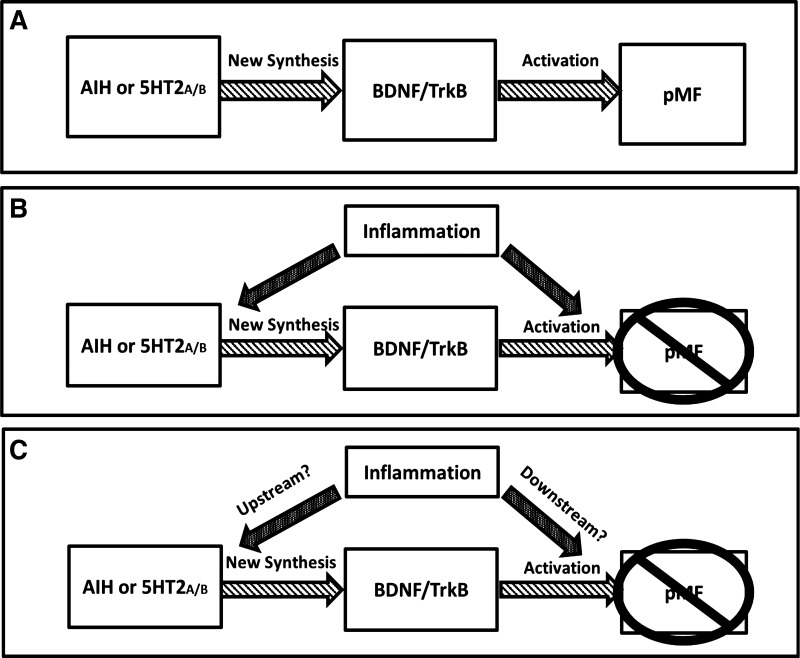
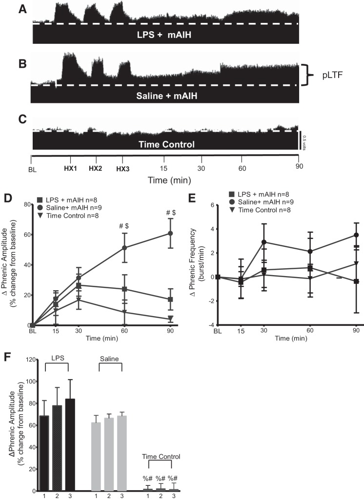
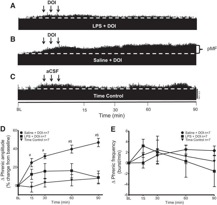
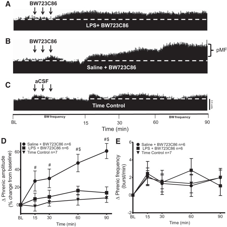
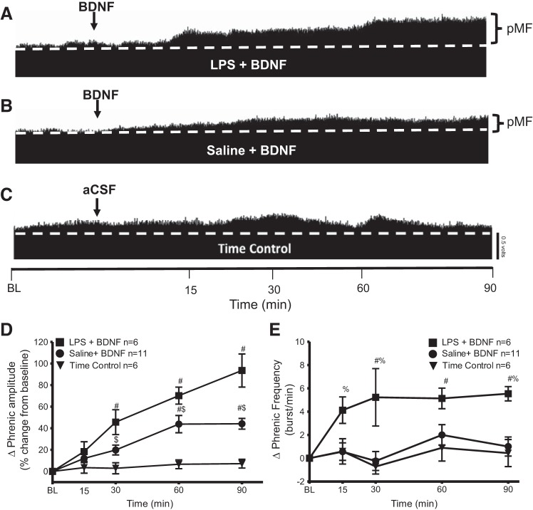
Although systemic inflammation induced by even a low dose of lipopolysaccharide (LPS, 100 μg/kg) impairs respiratory motor plasticity, little is known concerning cellular mechanisms giving rise to this inhibition. Phrenic motor facilitation (pMF) is a form of respiratory motor plasticity elicited by pharmacological agents applied to the cervical spinal cord, or by acute intermittent hypoxia (AIH; 3, 5-min hypoxic episodes); when elicited by AIH, pMF is known as phrenic long-term facilitation (pLTF). AIH consisting of moderate hypoxic episodes (mAIH, arterial Po2 = 35-55 mmHg) elicits pLTF via the Q pathway to pMF, a mechanism that requires spinal serotonin (5HT2) receptor activation and new brain-derived neurotrophic factor (BDNF) protein synthesis. Although mild systemic inflammation attenuates mAIH-induced pLTF via spinal p38 MAP kinase activation, little is known concerning how p38 MAP kinase activity inhibits the Q pathway. Here, we confirmed that 24 h after a low LPS dose (100 μg/kg ip), mAIH-induced pLTF is greatly attenuated. Similarly, pMF elicited by intrathecal cervical injections of 5HT2A (DOI; 100 μM; 3 × 6 μl) or 5HT2B receptor agonists (BW723C86; 100 μM; 3 × 6 μl) is blocked 24 h post-LPS. When pMF was elicited by intrathecal BDNF (100 ng, 12 μl), pMF was actually enhanced 24 h post-LPS. Thus 5HT2A/2B receptor-induced pMF is impaired downstream from 5HT2 receptor activation, but upstream from BDNF/TrkB signaling. Mechanisms whereby LPS augments BDNF-induced pMF are not yet known. NEW & NOTEWORTHY These experiments give novel insights concerning mechanisms whereby systemic inflammation undermines serotonin-dependent, spinal respiratory motor plasticity, yet enhances brain-derived neurotrophic factor (BDNF)/TrkB signaling in phrenic motor neurons. These insights may guide development of new strategies to elicit functional recovery of breathing capacity in patients with respiratory impairment by reducing (or bypassing) the impact of systemic inflammation characteristic of clinical disorders that compromise breathing.

Keywords: 5HT2A; 5HT2B; hypoxia; intermittent; long-term facilitation; motor neuron; phrenic; respiratory plasticity; serotonin.


Figures

Fig. 1.
Fig. 1.
Possible mechanisms whereby inflammation abolishes the Q pathway to phrenic motor facilitation (pMF). A: simple depiction of the serotonin-dependent Q pathway to pMF. Serotonin2A/B receptors activated by acute intermittent hypoxia (AIH) or intrathecal agonist injections cause new brain-derived neurotrophic factor (BDNF) synthesis and BDNF/TrkB signaling leading to pMF. B: pMF elicited by serotonin-dependent receptor activation is abolished or attenuated by systemic inflammation through unknown mechanisms; the goal of this study is to reveal steps in this cellular cascade that are undermined by mild systemic inflammation. C: possible mechanisms whereby inflammation impairs the Q pathway to pMF. Inflammation may affect pMF by inhibiting mechanisms upstream and/or downstream: either 1) 5HT2A/B receptor activation or function (upstream mechanisms); and/or 2) BDNF/TrkB signaling (downstream mechanisms).
Fig. 2.
Fig. 2.
Systemic inflammation induced by LPS (100 μg/kg; 24 h postinjection) attenuates phrenic long-term facilitation (pLTF) elicited by moderate acute intermittent hypoxia (mAIH). A–C: representative traces of compressed integrated phrenic neurograms during and following mAIH. A: LPS-injected group was exposed to mAIH, and no longer exhibits pLTF. B: in contrast, the saline-injected group exposed to mAIH exhibits robust pLTF. C: time controls (LPS or saline groups without mAIH exposure) do not show any consistent, time-dependent changes in phrenic burst amplitude. HX1, HX2, and HX3 are hypoxia 1, hypoxia 2, and hypoxia 3, respectively; BL, baseline. D: group data for phrenic burst amplitude (percent change from baseline). LPS + mAIH (n = 8) and saline + mAIH (n = 9) were compared with time controls (n = 8). LPS + mAIH was significantly reduced vs. saline + mAIH at 60 and 90 min. LPS + mAIH rats were not significantly different from time controls at any time, but were significantly different from saline + mAIH at 60 and 90 min. Significance is P ≤ 0.05: #significance vs. time control; $significance vs. LPS. E: group data for phrenic burst frequency (bursts/min). LPS + mAIH (n = 8) and saline + mAIH (n = 9) were compared with time controls (n = 8). There were no significant differences between any group or time point (P > 0.05). F: there is no progressive augmentation of the short-term hypoxic phrenic response during mAIH + saline or mAIH + LPS. The numbers represent hypoxic episodes (1–3). Significance is P ≤ 0.05: %significance vs. saline; #significance vs. LPS.
Fig. 3.
Fig. 3.
Systemic inflammation (24 h) induced by LPS (100 μg/kg) inhibits phrenic motor facilitation (pMF) elicited by 5HT2A agonist DOI. A–C: representative traces of compressed integrated phrenic neurograms after intrathecal administration of the 5HT2A receptor agonist DOI. A: LPS-injected group given intrathecal DOI did not show significant pMF. B: in contrast, the saline group given intrathecal DOI exhibits robust pMF. C: time controls given intrathecal vehicle (20% DMSO/80% aCSF) do not show a time-dependent change in amplitude vs. baseline (BL) values. D: group data for phrenic burst amplitude (percent change from baseline). LPS + DOI (n = 7) and saline + DOI (n = 7) were compared with time control (vehicle 20% DMSO/80% aCSF; triangle, n = 7). pMF following LPS + DOI was significant from saline + DOI at 60 and 90 min. There was no significant difference at any time between LPS + DOI and time control (P > 0.05). Significance is P ≤ 0.05: #significant difference vs. time control; $significance difference vs. LPS. Arrows represent intrathecal injections (3, 6-µl injections, 5-min intervals). E: group data for phrenic burst frequency (bursts/min). LPS + DOI (n = 7) and saline + DOI (n = 7), were compared with time control (vehicle 20% DMSO/80% aCSF) (n = 7) protocols. There was no significant difference between any group at any time (P > 0.05).
Fig. 4.
Fig. 4.
LPS (100 μg/kg)-induced inflammation (24 h) inhibits phrenic motor facilitation (pMF) elicited by the 5HT2B agonist BW723C86. A–C: representative traces of compressed integrated phrenic neurograms after intrathecal BW723C86 injections. A: LPS + BW723C86 did not elicit pMF. B: saline + BW723C86 exhibited robust pMF. C: time controls given intrathecal vehicle (20% DMSO/80% aCSF) do not show time-dependent changes in phrenic burst amplitude. D: group data for phrenic burst amplitude (percent change from baseline). LPS + BW723C86 (n = 6) and saline + BW723C86 (n = 6) were compared with 20% DMSO/80% aCSF (n = 7). LPS + BW723C86 was significant to saline + BW723C86 at 90 min post-intrathecal injection. There were no significant differences at any time between LPS + BW723C86 and time controls (P > 0.05). Significance is P ≤ 0.05: #significant difference vs. time control; $significant difference vs. LPS. Arrows represent intrathecal drug injections (3, 6-µl injections, 5-min intervals). E: group data for phrenic burst frequency (bursts/min). LPS + BW723C86 (n = 6) and saline + BW723C86 (n = 6) were compared with 20% DMSO/80% aCSF (n = 7). There were no significant differences between groups at any time (P > 0.05).
Fig. 5.
Fig. 5.
Systemic inflammation (24 h) by LPS (100 μg/kg) enhances brain-derived neurotrophic factor (BDNF)-induced phrenic motor facilitation (pMF). A–C: representative traces of compressed integrated phrenic neurograms after intrathecal BDNF. A: LPS + BDNF shows increased phrenic burst amplitude vs. baseline (BL) values, demonstrating robust pMF. B: saline + BDNF also exhibits pMF. C: time control group given intrathecal vehicle (aCSF + 0.1% BSA) does not show time-dependent changes in phrenic burst amplitude. Arrow indicates intrathecal BDNF injection (12 µl, 100 ng). D: group data for phrenic burst amplitude (percent change from baseline). LPS + BDNF (n = 6) and saline + BDNF (n = 11) were compared with time control (n = 6) protocols. After intrathecal BNDF, there is a significant increase from baseline values for at least 90 min. LPS + BDNF was significantly greater than saline + BDNF at 30, 60, and 90 min, demonstrating that LPS enhanced BDNF-induced pMF. Significance is P ≤ 0.05: #significant difference vs. time control; $significant difference vs. LPS. E: group data for phrenic burst frequency (bursts/min). LPS + BDNF (n = 6) and saline + BDNF (n = 11) were compared with time control (n = 6) protocols. LPS + BDNF group increased more than saline + BDNF and time controls at 15, 30, 60, and 90 min postinjection. Significance is P ≤ 0.05: #significant difference vs. time control; %significant difference vs. saline treatment.

References

    1. Abel T, Havekes R, Saletin JM, Walker MP. Sleep, plasticity and memory from molecules to whole-brain networks. Curr Biol 23: R774–R788, 2013. doi: 10.1016/j.cub.2013.07.025. - DOI - PMC - PubMed
    1. Allen NJ, Barres BA. Signaling between glia and neurons: focus on synaptic plasticity. Curr Opin Neurobiol 15: 542–548, 2005. doi: 10.1016/j.conb.2005.08.006. - DOI - PubMed
    1. Andero R, Choi DC, Ressler KJ. BDNF-TrkB receptor regulation of distributed adult neural plasticity, memory formation, and psychiatric disorders. Prog Mol Biol Transl Sci 122: 169–192, 2014. doi: 10.1016/B978-0-12-420170-5.00006-4. - DOI - PubMed
    1. Arsenault M, Ladouceur A, Lehmann A, Rainville P, Piché M. Pain modulation induced by respiration: phase and frequency effects. Neuroscience 252: 501–511, 2013. doi: 10.1016/j.neuroscience.2013.07.048. - DOI - PubMed
    1. Bach KB, Mitchell GS. Hypoxia-induced long-term facilitation of respiratory activity is serotonin dependent. Respir Physiol 104: 251–260, 1996. doi: 10.1016/0034-5687(96)00017-5. - DOI - PubMed

Publication types

MeSH terms

Substances

LinkOut - more resources