. 2018 Mar 15;555(7696):377-381.
doi: 10.1038/nature25975. Epub 2018 Mar 7.

Human hippocampal neurogenesis drops sharply in children to undetectable levels in adults

Affiliations

Human hippocampal neurogenesis drops sharply in children to undetectable levels in adults

Shawn F Sorrells et al. Nature. .

Abstract

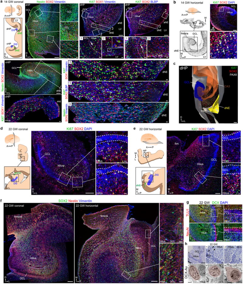
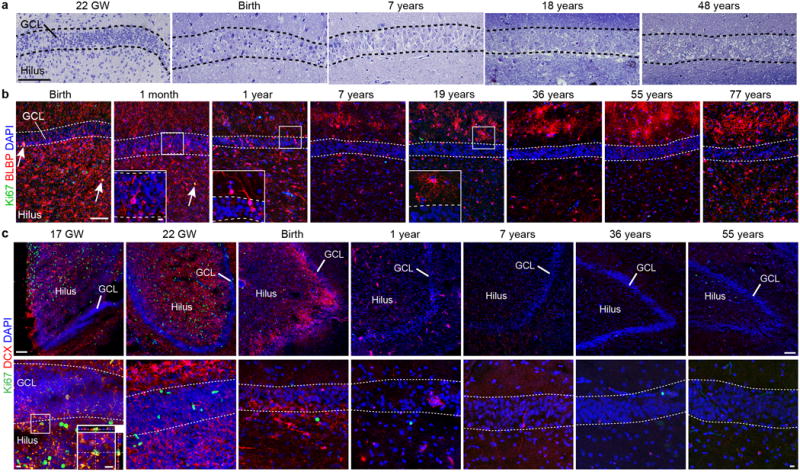
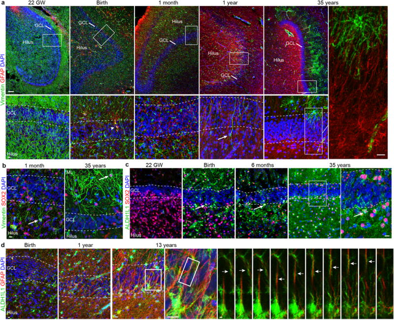
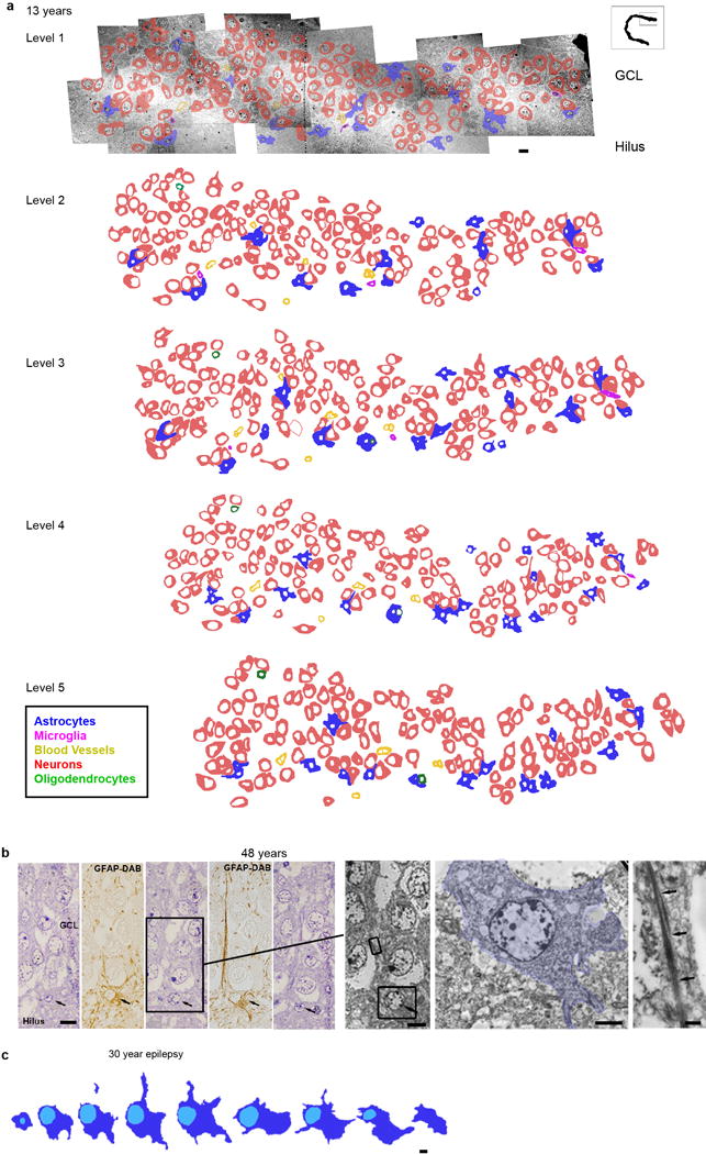
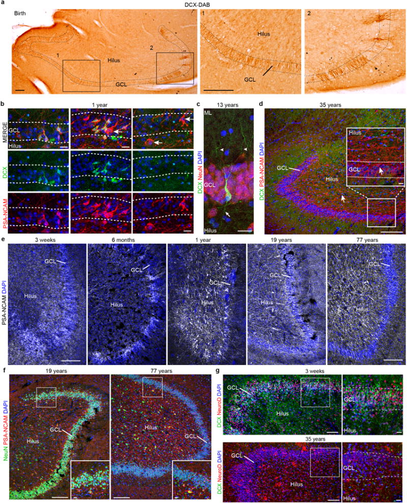
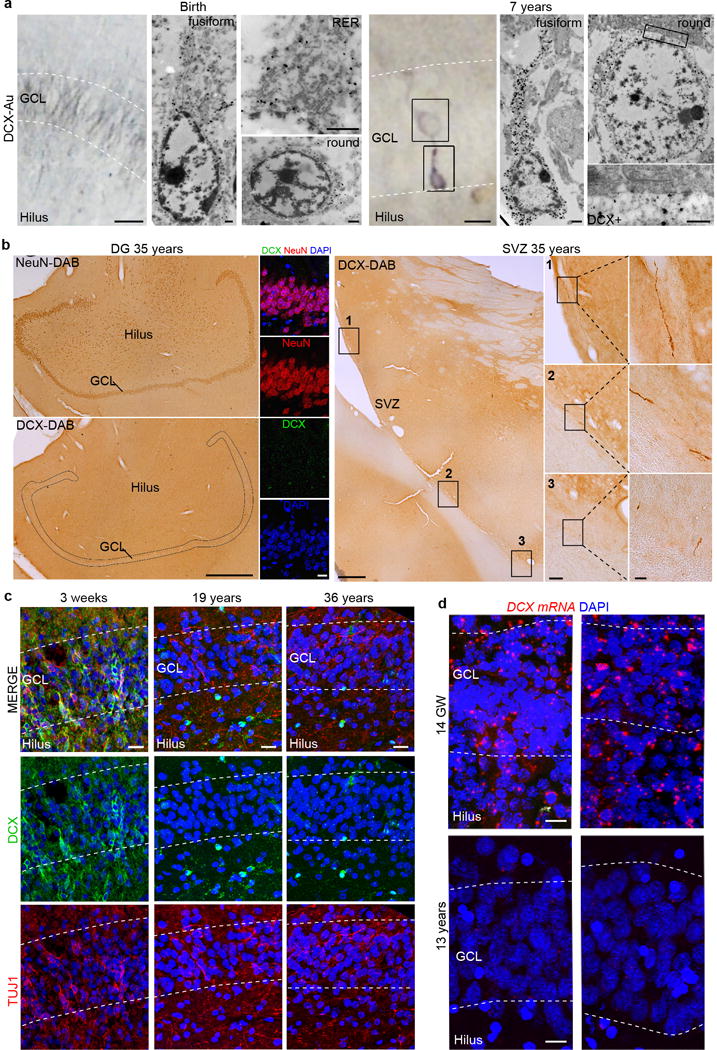
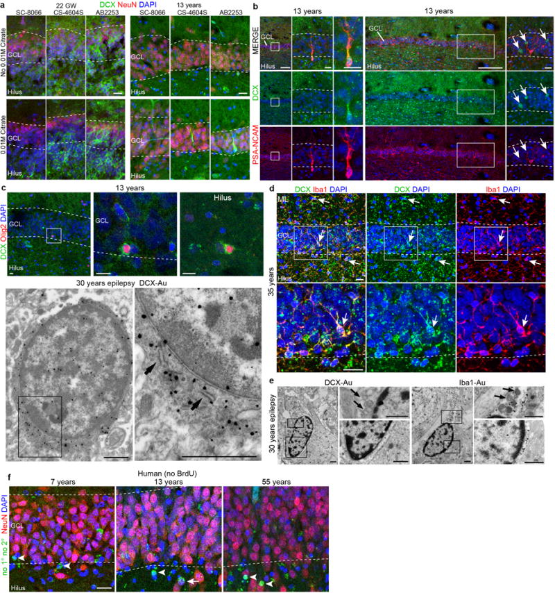
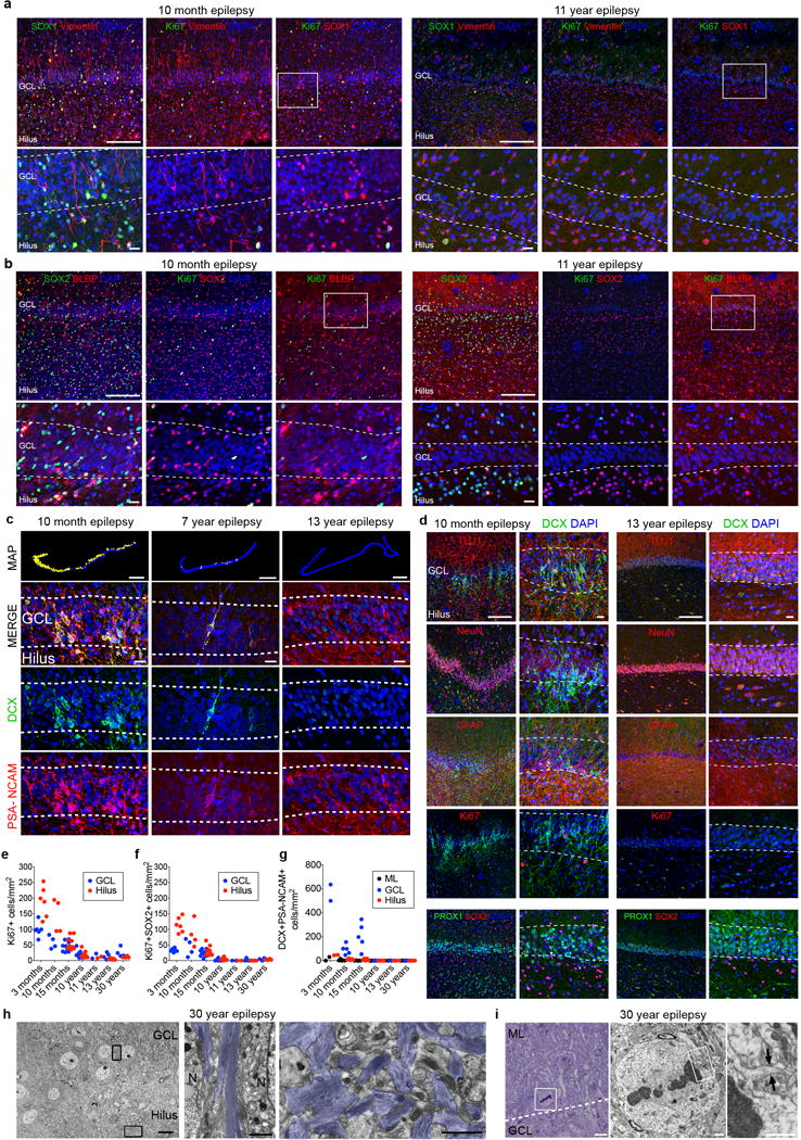
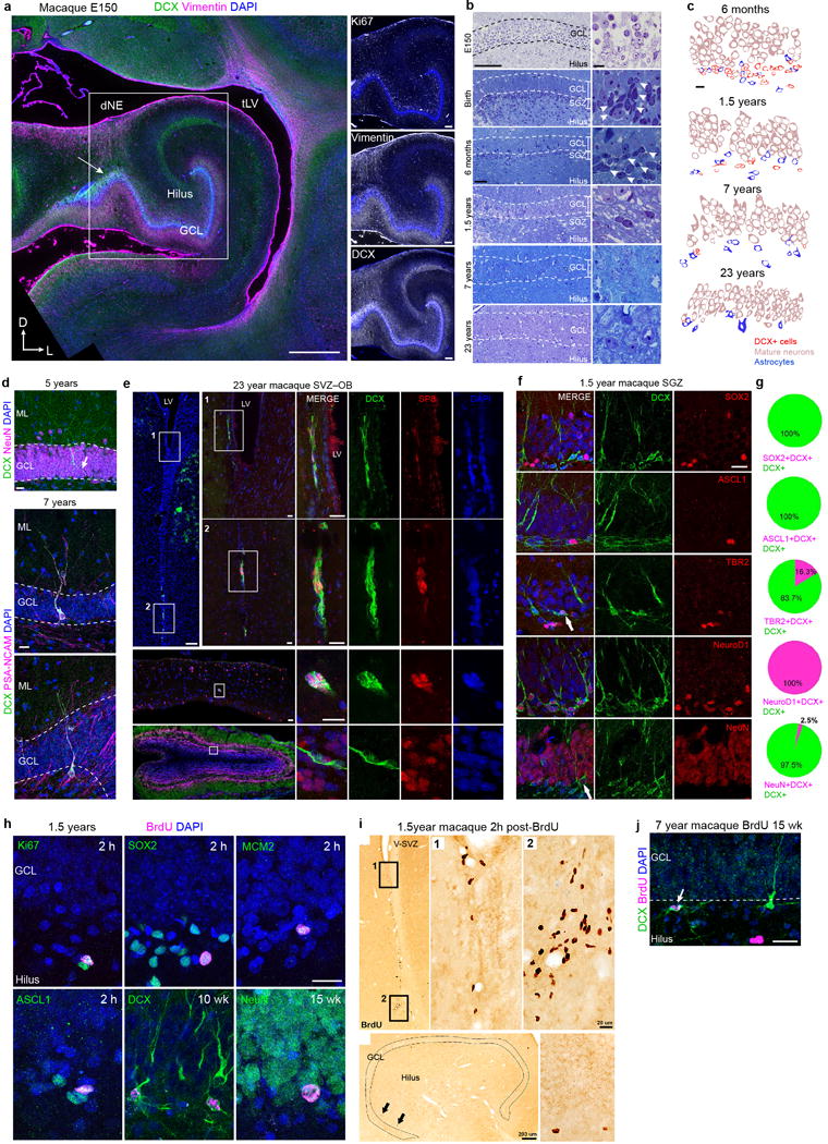
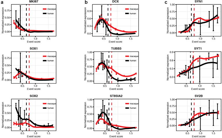
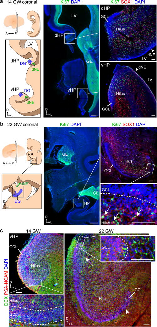
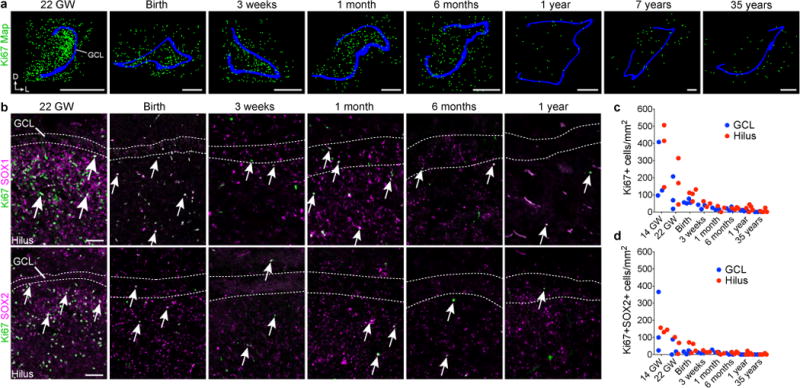
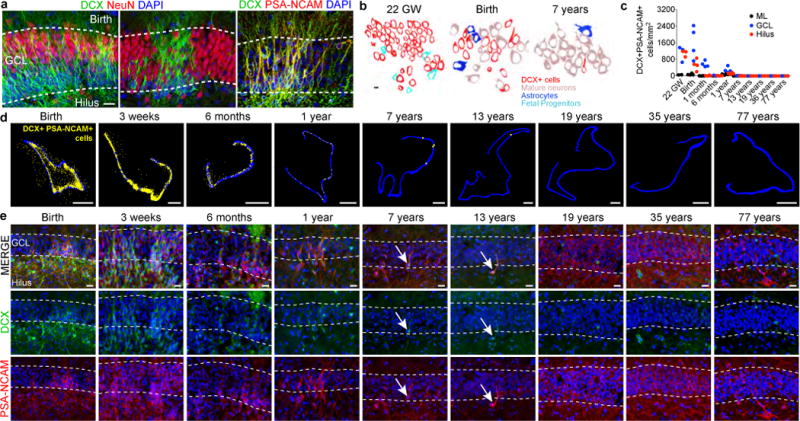
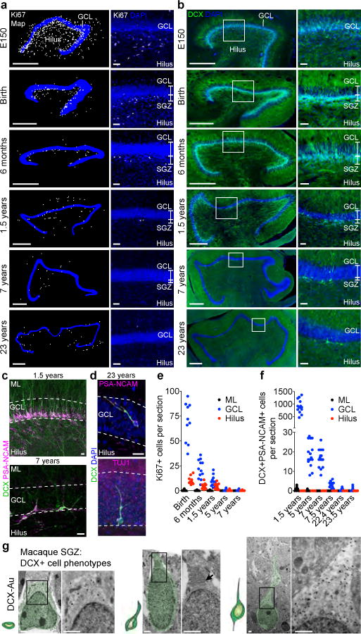
New neurons continue to be generated in the subgranular zone of the dentate gyrus of the adult mammalian hippocampus. This process has been linked to learning and memory, stress and exercise, and is thought to be altered in neurological disease. In humans, some studies have suggested that hundreds of new neurons are added to the adult dentate gyrus every day, whereas other studies find many fewer putative new neurons. Despite these discrepancies, it is generally believed that the adult human hippocampus continues to generate new neurons. Here we show that a defined population of progenitor cells does not coalesce in the subgranular zone during human fetal or postnatal development. We also find that the number of proliferating progenitors and young neurons in the dentate gyrus declines sharply during the first year of life and only a few isolated young neurons are observed by 7 and 13 years of age. In adult patients with epilepsy and healthy adults (18-77 years; n = 17 post-mortem samples from controls; n = 12 surgical resection samples from patients with epilepsy), young neurons were not detected in the dentate gyrus. In the monkey (Macaca mulatta) hippocampus, proliferation of neurons in the subgranular zone was found in early postnatal life, but this diminished during juvenile development as neurogenesis decreased. We conclude that recruitment of young neurons to the primate hippocampus decreases rapidly during the first years of life, and that neurogenesis in the dentate gyrus does not continue, or is extremely rare, in adult humans. The early decline in hippocampal neurogenesis raises questions about how the function of the dentate gyrus differs between humans and other species in which adult hippocampal neurogenesis is preserved.


Conflict of interest statement

Competing financial interests. The authors declare no competing financial interests.

Figures

Extended Data Figure 1
Extended Data Figure 1. Additional marker and ultrastructural analysis of early fetal development of the human dentate gyrus
a–c, 14 GW human brain a, schematic of dHP and vHP in a coronal section. Precursor cells labeled with Nestin, SOX2 and Vimentin are organized in ribbons between the dNE and GCL. Ki67+ cells expressing SOX1 and Vimentin or SOX2 and BLBP are present in the GCL and hilus (inset 1), along the wall of the LV (inset 2), and between the GCL and the dNE (inset 3). The dNE is located at the edge of the ammonic neuroepithelium (aNE) closest to the fimbria. A similar organization is present in the vHP where Nestin+SOX2+Vimentin+ cells connect the dNE to the developing GCL. Ki67+SOX1+Vimentin+ cells are present in a strip along the ventricular wall and fill the region between the dNE and the GCL. b, (left) 14 GW hemisphere, Nissl-stained horizontal sections. (right) Ki67+ cells expressing SOX2 (arrows). c, 3D reconstruction of the dHP showing field of Ki67+ and SOX2+ cells between the dNE and GCL. d–h, 22 GW human brain, coronal (d) and horizontal (e) sections; the hilus and GCL contain Ki67+SOX2+ cells, (insets 1,2) as well as (f) Nestin+SOX2+Vimentin+ cells. These populations are asymmetrically distributed; sparse in the medial (proximal) GCL and hilus (inset 1) but abundant in the lateral (distal) GCL and hilus (inset 2). g, DCX+TUJ1+ cells and NeuN+ cells in the DG at 22 GW. NeuN+ GCL neurons in the distal GCL (arrow). h, Toluidine blue-stained semithin section (top) and TEM micrographs at 22 GW showing the ultrastructural characteristics of DCX immunogold labeled cells (pseudocolored, bottom). Insets of the semithin section show the proximal (1) and distal (2) ends of the GCL. Most DCX+ cells in the hilus and the proximal GCL have scant cytoplasm, few organelles, and a small, irregular nucleus (i-ii); some in the hilus have an elongated, fusiform morphology (i). Some DCX+ cells in the GCL have mature neuronal characteristics such as a round nucleus, more cytoplasm, ribosomes, rough endoplasmic reticulum and mitochondria (iii); this cell type was more common in the distal GCL. Scale bars: 200 μm (a–h lower magnifications), 20μm (a–h higher magnifications), 2 μm (h, TEM).
Extended Data Figure 2
Extended Data Figure 2. A coalesced proliferative SGZ does not form in the human DG; additional marker expression
a, Toluidine blue counterstained semithin sections of the human GCL from fetal to adult ages. Note that a discrete cellular layer does not form next to the GCL and the small dark cells characteristic of SGZ precursors are not present (compare to Extended Fig. 9b in the macaque). b, BLBP+ cells are distributed broadly in the DG from birth to one year, many of these cells have an elongated morphology (see insets) and some co-express Ki67 (arrows) at birth and one month. By 7 years and in adults, most BLBP is present in the ML in stellate protoplasmic astrocytes. c, DCX+Ki67+ cells in the GCL are rare at 17 GW (orthogonal views, inset) but were abundant in the GE at the same age (not shown). DCX+Ki67+ cells were absent in the GCL from 22 GW to 55 years. Scale bars: 100 μm (a–c), 10μm (a–c, insets).
Extended Data Figure 3
Extended Data Figure 3. Additional marker expression for astroglial cells and progenitor cells in the human DG at different ages
a, Vimentin+ and GFAP+ cells in the hippocampus from 22 gestational weeks to 35 years. Vimentin is widely expressed during fetal and early postnatal development and is mostly restricted to protoplasmic astrocytes in the molecular layer in adults. GFAP is not expressed at 22 gestational weeks, but at birth a few vimentin+GFAP+ cells are present in the hilus and GCL (arrowhead). Interestingly, some vimentin+GFAP− cells with a radial morphology (arrow) are observed in samples at 1 year of age, but not at the other ages. In adults, GFAP and vimentin are not co-expressed (right, high-magnification of thin GFAP+vimentin− fibres within the GCL). b, Vimentin+Sox2+ simpler elongated cells in the hilus at 1 month (arrow) and protoplasmic astrocytes in the molecular layer at 35 years of age (arrow). c, SOX2+ cells are abundant in the GCL and hilus at 22 gestational weeks, and co-express ALDH1L1 in the brain at birth and in older individuals (arrows). d, At birth, there are few ALDH1L1+GFAP+ cells in the DG, but by 13 years of age many stellate astrocytes express both of these markers. Right, z stack of radial GFAP+ processes that are surrounded by ALDH1L1 staining. Scale bars, 100 μm (a (top row)), 10 μm (a (bottom row and insets), b–d) and 2 μm (d (z-stack)).
Extended Data Figure 4
Extended Data Figure 4. EM analysis of cell types in the DG of a 13 year old and adult human brain; absence of SGZ precursor cells or immature neurons
a, Reconstruction of 5 ultrathin sections (separated by 1.5 μm) from the 13 year old GCL with outlines of cell membranes. Colors corresponding to the different cell types defined by their ultrastructural characteristics are indicated in the key. No clusters or isolated cells with young neuronal ultrastructure were found. Cells associated in small groups were identified as astrocytes, oligodendrocytes or microglia. b–c, reconstructions of astroglial cells next to the GCL searching for possible examples of RA in the adult human DG. b, example of an astrocyte with radial morphology in the adult GCL. Five serial semithin sections of this astrocyte (black arrows) next to the GCL of a 48 year old DG ; alternating semithin sections show that this cell is GFAP+. This cell extends a thin radial fiber through the GCL, but has multiple processes (stellate morphology) in the hilus. Boxed area shows the ultrastructure from the indicated semi-thin section of this astrocyte (pseudo-coloured in blue) and the bundles of intermediate filaments present in the expansion (arrows). c, Another example of a serially reconstructed astrocyte in the DG of a 30 year old epileptic case (separated by 1.4 μm) showing a short radial expansion and processes into the hilus. Scale bars: 10 μm (a, b semithin and TEM), 5 μm (c), 2 μm (b, soma), 500 nm (b, intermediate filaments).
Extended Data Figure 5
Extended Data Figure 5. Young neurons are present in the infant but not the adult human DG
a, DAB staining in the hippocampus at birth reveals many young neurons in the GCL. b, DCX+PSA-NCAM+ cells are distributed in clusters across the GCL at 1 year of age. Most PSA-NCAM+ cells are DCX+, but some are DCX−PSA-NCAM+ (arrows). c, In the samples from a 13-year-old individual, DCX+ cells have a more mature neuronal morphology. The cell shown is NeuN+ and has dendrites in the molecular layer (arrowheads) and an axon projecting into the hilus (arrow). d, At 35 years of age, the DG does not contain DCX+PSA-NCAM+ cells, but does contain many DCX−PSA-NCAM+ cells that do not have the morphology of young neurons. e, PSA-NCAM+ staining in the human DG from 3 weeks to 77 years; in adults, these cells have a more mature neuronal morphology and are localized in the hilus. f, PSA-NCAM+ cells in the DG are NeuN+ in samples of 19- and 77-year-old individuals. g, At 3 weeks of age, the GCL and hilus were filled with clusters of DCX+NeuroD+ cells, and many of the DCX− GCL neurons were NeuroD+. At 35 years, no DCX+NeuroD+ cells were observed; antibody labelling for NeuroD was non-specific. Scale bars, 200 μm (a, d–g), 20 μm (b, c, and d, f, g (inset)).
Extended Data Figure 6
Extended Data Figure 6. DCX+ young neurons in the developing human brain, but not in the adult
a, TEM micrographs of DCX immunogold staining at birth and 7 years of age. At birth, the GCL contains small DCX+ cells with little cytoplasm, rough endoplasmic reticulum (RER) cisternae and a fusiform or round nucleus. At 7 years of age, DCX+ cells closer to the hilus have characteristics of immature neurons, including few organelles and a long expansion towards the GCL. DCX+ cells located within the GCL have mature neuron characteristics, including a large, round nucleus, rough endoplasmic reticulum, mitochondria and microtubules consistent with a more mature neuronal morphology (see Extended Data Fig. 5). At higher magnification, the more mature-appearing DCX-labelled cells are adjacent to DCX− GCL neurons. b, No DCX+ cells in the hilus and GCL (stained by NeuN antibodies; left insets) were found in the brain of a 35-year-old individual that showed exceptional preservation. In this sample, rare DCX+ cells with the features of young migratory neurons were present in the ventricular–subventricular zone (right insets). SVZ, subventricular zone. c, DCX+TUJ1+ cells were present in the GCL and hilus at 3 weeks of age, but were not detected in the adult DG. d, RNA-scope detection of DCX mRNA revealed many cells in the DG at 14 gestational weeks, but weakly labelled cells distributed throughout in the DG and other regions of the hippocampus at 13 years of age. Scale bars: 1 mm (b (left)), 100 μm (b (middle right inset)), 20 μm (b (right insets), c), 10 μm (d), 5 μm (a (left)) and 500 nm (a (right, TEM)).
Extended Data Figure 7
Extended Data Figure 7. DCX+/PSA-NCAM- glial cells in the adult human hippocampus
a, Comparison of citrate antigen retrieval using three DCX antibodies from this study (SC-8066, CS-4604S and AB2253) in the GCL obtained from individuals at 22 gestational weeks and 13 years of age. The 13 year old DCX+ cell (Extended Data Fig. 5c) is shown in the lower right panel and adjacent sections were stained with the other antibodies. b, Example of a DCX+PSA-NCAM+ neuron in the GCL and DCX+PSA-NCAM− staining in the sample from the 13-year-old individual (arrows). c, Examples of DCX+OLIG2+ cells in the GCL and hilus of the 13-year-old individual. Immunogold-labelled DCX+ cells viewed by TEM had single short endoplasmic reticulum cisternae (arrows), a very irregular contoured membrane and a round nucleus with condensed chromatin characteristic of oligodendrocytes. d, In some samples (see Extended Data Fig. 5g, bottom right inset), we found DCX+ immunoreactivity in many small multipolar cells. This staining was not limited to the hilus, or GCL, but was present in cells across the tissue and co-localized with the microglial marker IBA1 (arrows). e, TEM micrographs of DCX and IBA1 immunogold-labelled cells in an adult DG of a 30-year-old individual with epilepsy. DCX+ and IBA1+ cells have similar characteristics: elongated nucleus with clumps of chromatin beneath the nuclear envelope and throughout the nucleoplasm, irregular contour and the presence of lysosomes and lipofucsin (arrows). Note that these features are typical of microglial cells. f, Human hippocampus stained with NeuN followed by processing for BrdU detection (with no primary or secondary antibodies) shows round fluorescent signal (arrowheads indicate signal that is NeuN−) occasionally overlapping with NeuN staining (arrow). Scale bars, 200 μm (b (left column and wide column)), 20 μm (a, b (left-middle columns, right column), d, f), 10 μm (c (top row)) and 1 μm (c (bottom row), e).
Extended Data Figure 8
Extended Data Figure 8. Neurogenesis declines in epilepsy cases from infancy into childhood
a, Ki-67+SOX1+vimentin+ cells are located in the hilus and GCL at 10 months but are not present at 11 years of age. b, Ki-67+SOX2+BLBP+ cells are located in the hilus and GCL at 10 months but are not present at 11 years of age. c, Maps of DCX+PSA-NCAM+ cells (yellow dots) and representative immunostaining at 10 months, 7 years and 13 years (bottom rows). d, In the 10 month-old epileptic DG of a patient with epilepsy, DCX+ cells co-expressing PSA-NCAM or TUJ1 are distributed throughout the NeuN+PROX1+ GCL, but do not co-express Ki-67 or GFAP. In the 13-year-old patient with epilepsy, DCX+ cells co-expressing PSA-NCAM or TUJ1 were not present. Few Ki-67+ cells were visible throughout the DG. e–g, Quantification of Ki-67+ (e), Ki-67+SOX2+ (f) and DCX+PSA-NCAM+ (g) cells in the DG of surgically resected hippocampuses. h, TEM micrographs of the brain of a 30-year-old patient with epilepsy showing astroglial expansions with high number of intermediate filaments (blue) ensheathing GCL neuronal bodies. A dense network of astrocytic expansions in the hilus, containing dense bundles of intermediate filaments (blue), fills the region proximal to the GCL with no evidence of SGZ progenitor cells. i, Mitotic cells are very rare and not restricted to the hilus or GCL. A toluidine-blue-stained 1.5-μm section from the DG of a 30-year-old brain shows a dividing cell in the molecular layer, adjacent to the GCL. The TEM micrograph shows the dividing cell in metaphase with a light cytoplasm, few organelles and an irregular contour with a small expansion (arrows), which are characteristic of astrocytes (shown at higher magnification). N, neuron. For quantifications, staining replicates (≥3) are shown by dots (each age, n = 1). Scale bars, 1 mm (c (maps)), 200 μm (a, b (top), d (left)), 20 μm (a–c (bottom), d (right)) and 10 μm (h, i (left)), 1 μm (h, i (middle and right)).
Extended Data Figure 9
Extended Data Figure 9. Development of the macaque DG and evidence for the presence of a proliferative SGZ and postnatal neurogenesis with a sharp decline in adulthood
a, The E150 macaque hippocampus has many Ki67+ cells in the SGZ as well as Vimentin+ fibers and DCX+ cells between the dNE and the GCL (arrow). b, Toluidine blue-stained semi-thin sections of the macaque DG reveal small and darkly condensed nuclei (arrows) in the SGZ from fetal ages (E150) to 1.5 years; few are visible at 7 and these cells are very rare in the 23 year macaque DG (compare with human data in Extended Data Fig. 2a). c, Profiles of the cellular populations in the macaque DG at 6 months, 1.5 years, 7 years, and 23 years of age. As in the human DG, DCX+ cells decrease dramatically with age and have scarce cytoplasm and a smaller nucleus compared to mature granule neurons. d, (top) Example of a DCX+NeuN+ cell with mature dendrites in the macaque GCL at 5 years of age. (middle, bottom) Two examples of DCX+PSA-NCAM+ cells with dendritic arborization present in the macaque GCL at 7.5 years of age. An axonal extension (arrow) into the hilus is visible. e, The 23 year old macaque V-SVZ and olfactory bulb (OB) contain some DCX+SP8+ cells with morphology of young neurons, but similar cells are rare in the 23 year old GCL in the hippocampus (ig. 4b, d) f, DCX+ cells in the 1.5 year old macaque SGZ express transcription factors in common with those in the mouse SGZ. g, Percentages of DCX+ cells expressing markers in (f). h, Immunostains of BrdU+ cells and cell proliferation (Ki67, MCM2), progenitor cell markers (Sox2, Ascl1) or DCX, in the 1.5 year-old macaque sacrificed at 2 hours after BrdU injection. BrdU+DCX+ and BrdU+NeuN+ cells could be identified at 10 or 15 weeks after BrdU exposure, respectively. i, DAB-staining for BrdU in the 1.5 year old macaque V-SVZ and SGZ, 2 hours after BrdU. j, Example of a rare DCX+BrdU+ cell in the 7.5 year old macaque. Scale bars: 1 mm (a left) 200 μm (a right, e top left, bottom left, i, left), 100 μm (b, left) 20 μm (d, e middle left and right, f,h, i right, j), 10 μm (b, right, c).
Extended Data Figure 10
Extended Data Figure 10. Decline in markers associated with neurogenesis in the macaque and human hippocampus (gene expression profiling)
a, markers of dividing or precursor cells b, markers of young neurons c, markers of mature neurons. Human RNAseq (brainspan.org) and macaque expression profiling (data set from Bakken et al. 2016) developmental data from hippocampus for the indicated genes. Human data are averaged over biological replicates by developmental period (as defined by Kang et al. 2011). Normalized data are plotted on the same developmental event scale. Loess-fit curves are displayed with data points (mean +/− sem). Dashed lines indicate birth.
Figure 1
Figure 1. Early fetal development of the human dentate gyrus
a, Left, schematic of one hemisphere at 14 gestational weeks (GW), coronal section showing the lateral ventricle (LV) and ganglionic eminence (GE). Middle and right, immunofluorescence images. Ki-67+SOX1+ cells were found in the dorsal (dHP) and ventral (vHP) hippocampus. A, anterior; D, dorsal, L, lateral; P, posterior. b, Left, schematic of one hemisphere at 22 gestational weeks, coronal section. Middle and right, SOX1+Ki-67+ cells in the hilus and GCL (arrows). c, Distribution of DCX+PSA-NCAM+ cells at 14 gestational weeks and 22 gestational weeks. The arrow indicates the end of the GCL most proximal to the dNE. The distal end of the GCL contained fewer young neurons (arrowhead). Scale bars, 1 mm (a, b (middle)) and 100 μm (a, b (right), c). Staining was replicated at least three times (n = 1 at 14 gestational weeks; n = 3 at 22 gestational weeks).
Figure 2
Figure 2. Human DG proliferation declines sharply during infancy and a layer of proliferating progenitors does not form in the SGZ
a, Maps of Ki-67+ (green) cells in the DG from samples of individuals that were between 22 gestational weeks and 35 years of age; GCL in blue. b, Ki-67+SOX1+ and Ki-67+SOX2+ cells (arrows) are distributed across the hilus and GCL and the number of double-positive cells decreases between 22 gestational weeks and 1 year of age. c, d, Quantification of Ki-67+ (c) and Ki-67+SOX2+ (d) cells in the hilus and GCL. For quantifications, dots indicate staining replicates (≥3) (each age n = 1). Scale bars, 1 mm (a) and 100 μm (b).
Figure 3
Figure 3. Young neurons decline in the human DG from infancy into childhood
a, DCX+ cells at birth are distributed in a continuous field (left) or tight clusters (middle) and express PSA-NCAM (right). b, Outlines of cell types in the GCL at 22 gestational weeks, birth and 7 years of age. c, Quantification of DCX+PSA-NCAM+ cells in the DG. d, Maps of DCX+PSA-NCAM+ cells (yellow dots; GCL, blue outline). e, DCX+PSA-NCAM+ cells in the DG (birth to 77 years) are rare by 7 and 13 years of age (arrows). For quantifications, dots indicate staining replicates (≥3) (each age, n = 1). Scale bars, 1 mm (d), 20 μm (a, e) and 5 μm (b).
Figure 4
Figure 4. An SGZ forms during macaque development but new neurons are rare in adults
a, b, Maps and immunostaining of Ki-67+ cells (a) and DCX+ cells (b) in the macaque SGZ (from E150 to 23 years of age). c, DCX+PSA-NCAM+ cells in the SGZ (1.5 and 7 years). d, DCX+PSA-NCAM+ or DCX+TUJ1+ cells (23 years). e, f, Quantification of Ki-67+ cells (e) and DCX+PSA-NCAM+ cells (f) in the macaque GCL, hilus and molecular layer (ML). n = 1 animal per age; dots indicate staining replicates (≥3). g, Immunogold (DCX–Au) transmission electron microscopy of neurons (light green overlay) at different stages of maturation. Left, small DCX+ cell; middle, DCX+ cell with a short process, mitochondria and prominent endoplasmic reticulum (arrow); right, large DCX+ cell with round soma, few organelles and an expansion into the GCL. Scale bars, 500 μm (a, b (left)), 50 μm (a, b (right)), 20 μm (c, d) and 1 μm (g).

Comment in

References

    1. Altman J, Das GD. Autoradiographic and histological evidence of postnatal hippocampal neurogenesis in rats. J Comp Neurol. 1965;124:319–335. - PubMed
    1. Kornack DR, Rakic P. Continuation of neurogenesis in the hippocampus of the adult macaque monkey. Proc Natl Acad Sci U S A. 1999;96:5768–5773. - PMC - PubMed
    1. Seri B, García-Verdugo JM, McEwen BS, Alvarez-Buylla A. Astrocytes give rise to new neurons in the adult mammalian hippocampus. J Neurosci. 2001;21:7153–7160. - PMC - PubMed
    1. van Praag H, et al. Functional neurogenesis in the adult hippocampus. Nature. 2002;415:1030–1034. - PMC - PubMed
    1. Patzke N, et al. In contrast to many other mammals, cetaceans have relatively small hippocampi that appear to lack adult neurogenesis. Brain Struct Funct. 2015;220:361–383. - PMC - PubMed

Publication types

MeSH terms