

Functional near-infrared spectroscopy during optic flow with and without fixation
- PMID: 29513720
- PMCID: PMC5841770
- DOI: 10.1371/journal.pone.0193710
Functional near-infrared spectroscopy during optic flow with and without fixation
Abstract
Background and purpose: Individuals with visual vertigo describe symptoms of dizziness, disorientation, and/or impaired balance in environments with conflicting visual and vestibular information or complex visual stimuli. Physical therapists often prescribe habituation exercises using optic flow to treat these symptoms, but there are no evidence-based guidelines for delivering optic flow and it is unclear how the brain processes such stimuli. The purposes of this study were to use functional near-infrared spectroscopy (fNIRS) to explore cerebral activation during optic flow, and determine if visual fixation had a modulating effect on brain activity.
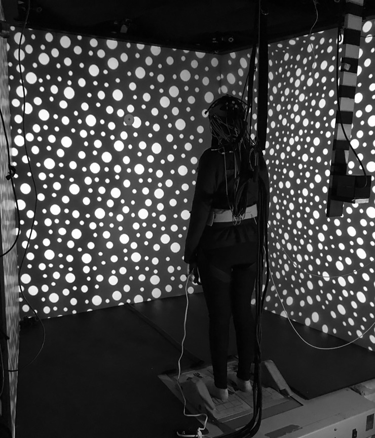
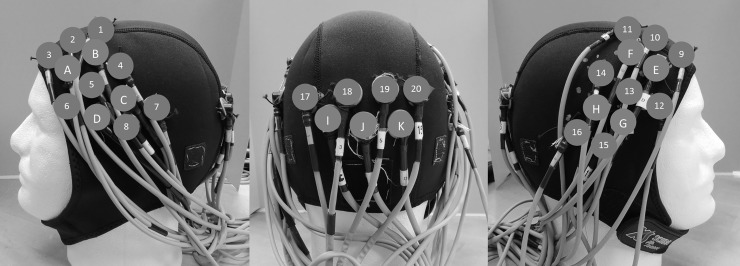
Methods: Fifteen healthy participants (7 males and 8 females; mean age 41 years old) stood in a virtual reality environment and viewed optic flow moving unidirectionally in the yaw plane with and without fixation. Changes in cerebral activation were recorded from the bilateral fronto-temporo-parietal and occipital lobes using fNIRS.
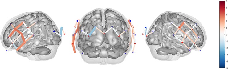
Results: Cerebral activation was greater with visual motion than while viewing a stationary scene. Greater cerebral activation in the bilateral fronto-temporo-parietal lobes was observed when optic flow was viewed with fixation.
Discussion and conclusions: Optic flow activates the bilateral fronto-temporo-parietal regions of the cerebral cortex. This activation is greater while viewing optic flow and a fixation target, providing preliminary evidence supporting the use of a fixation target during habituation exercises.
Conflict of interest statement
Figures
Similar articles
-
Changes in cerebral activation in individuals with and without visual vertigo during optic flow: A functional near-infrared spectroscopy study.Neuroimage Clin. 2018 Sep 5;20:655-663. doi: 10.1016/j.nicl.2018.08.034. eCollection 2018. Neuroimage Clin. 2018. PMID: 30211002 Free PMC article.
-
Visually induced motion sickness with radial displays: effects of gaze angle and fixation.Aviat Space Environ Med. 2007 Jul;78(7):659-65. Aviat Space Environ Med. 2007. PMID: 17679562
-
Cortical correlates of vestibulo-ocular reflex modulation: a PET study.Brain. 2003 Jul;126(Pt 7):1562-78. doi: 10.1093/brain/awg165. Epub 2003 May 21. Brain. 2003. PMID: 12805122
-
A semi-immersive virtual reality incremental swing balance task activates prefrontal cortex: a functional near-infrared spectroscopy study.Neuroimage. 2014 Jan 15;85 Pt 1:451-60. doi: 10.1016/j.neuroimage.2013.05.031. Epub 2013 May 17. Neuroimage. 2014. PMID: 23684867
-
[Balint's syndrome--visual disorientation].Ugeskr Laeger. 1992 May 18;154(21):1492-4. Ugeskr Laeger. 1992. PMID: 1598720 Review. Danish.
Cited by
-
Observation and motor imagery balance tasks evaluation: An fNIRS feasibility study.PLoS One. 2022 Mar 23;17(3):e0265898. doi: 10.1371/journal.pone.0265898. eCollection 2022. PLoS One. 2022. PMID: 35320324 Free PMC article.
-
iVR-fNIRS: studying brain functions in a fully immersive virtual environment.Neurophotonics. 2024 Apr;11(2):020601. doi: 10.1117/1.NPh.11.2.020601. Epub 2024 Apr 4. Neurophotonics. 2024. PMID: 38577629 Free PMC article. Review.
-
Self-Motion Perception Influences Postural Sway More than Environmental Motion Perception.bioRxiv [Preprint]. 2025 May 7:2025.05.02.651511. doi: 10.1101/2025.05.02.651511. bioRxiv. 2025. PMID: 40655001 Free PMC article. Preprint.
-
A Brain Network Analysis Model for Motion Sickness in Electric Vehicles Based on EEG and fNIRS Signal Fusion.Sensors (Basel). 2024 Oct 14;24(20):6613. doi: 10.3390/s24206613. Sensors (Basel). 2024. PMID: 39460093 Free PMC article.
-
Multiple levels of contextual influence on action-based timing behavior and cortical activation.Sci Rep. 2023 May 2;13(1):7154. doi: 10.1038/s41598-023-33780-1. Sci Rep. 2023. PMID: 37130838 Free PMC article.
References
-
- Staab JP. Chronic Subjective Dizziness. CONTINUUM: Lifelong Learning in Neurology. 2012;18:1118–41. doi: 10.1212/01.CON.0000421622.56525.58 - DOI - PubMed
-
- Rábago CA, Wilken JM. Application of a mild traumatic brain injury rehabilitation program in a virtual realty environment: a case study. Journal of Neurologic Physical Therapy. 2011;35:185–93. doi: 10.1097/NPT.0b013e318235d7e6 - DOI - PubMed
-
- Hall C, Herdman S, Whitney S, Cass S, Clendaniel R, Fife T, et al. Vestibular rehabilitation for peripheral vestibular hypofunction: An evidence-based clinical practice guideline: From the AMerican Physical Therapy Association Neurology Section. Journal of Neurologic Physical Therapy. 2016;40:124–55. doi: 10.1097/NPT.0000000000000120 - DOI - PMC - PubMed
-
- Szturm T, Ireland D, Lessing-Turner M. Comparison of different exercise programs in the rehabilitation of patients with chronic peripheral vestibular dysfunction. Journal of Vestibular Research. 1994;4:461–79. - PubMed
Publication types
MeSH terms
Grants and funding
LinkOut - more resources
Full Text Sources
Other Literature Sources
Medical
