. 2018 Jun;287(3):833-843.
doi: 10.1148/radiol.2018172300. Epub 2018 Mar 7.

MR Imaging of Rectal Cancer: Radiomics Analysis to Assess Treatment Response after Neoadjuvant Therapy

Affiliations

MR Imaging of Rectal Cancer: Radiomics Analysis to Assess Treatment Response after Neoadjuvant Therapy

Natally Horvat et al. Radiology. 2018 Jun.

Abstract

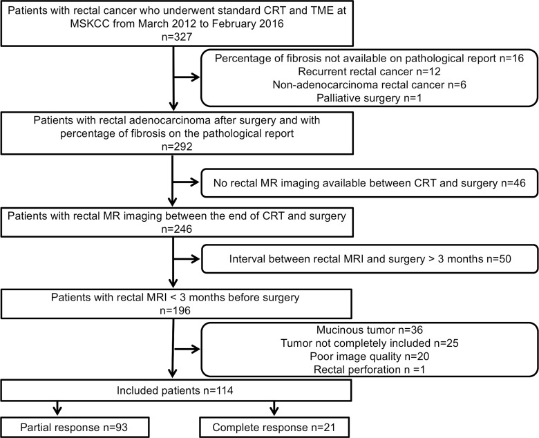
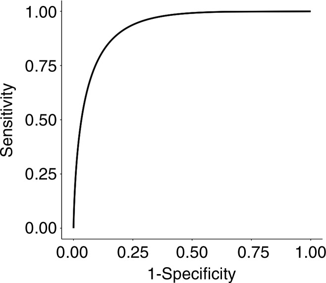
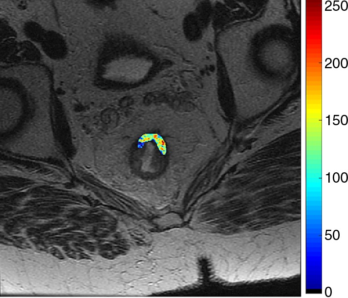
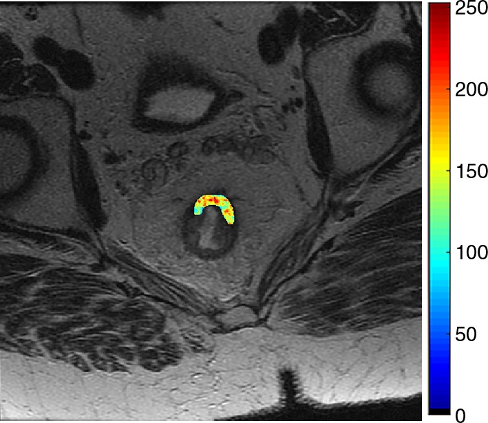
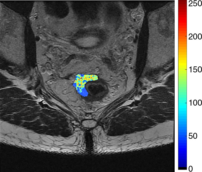
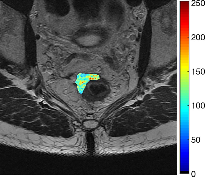
Purpose To investigate the value of T2-weighted-based radiomics compared with qualitative assessment at T2-weighted imaging and diffusion-weighted (DW) imaging for diagnosis of clinical complete response in patients with rectal cancer after neoadjuvant chemotherapy-radiation therapy (CRT). Materials and Methods This retrospective study included 114 patients with rectal cancer who underwent magnetic resonance (MR) imaging after CRT between March 2012 and February 2016. Median age among women (47 of 114, 41%) was 55.9 years (interquartile range, 45.4-66.7 years) and median age among men (67 of 114, 59%) was 55 years (interquartile range, 48-67 years). Surgical histopathologic analysis was the reference standard for pathologic complete response (pCR). For qualitative assessment, two radiologists reached a consensus. For radiomics, one radiologist segmented the volume of interest on high-spatial-resolution T2-weighted images. A random forest classifier was trained to separate the patients by their outcomes after balancing the number of patients in each response category by using the synthetic minority oversampling technique. Statistical analysis was performed by using the Wilcoxon rank-sum test, McNemar test, and Benjamini-Hochberg method. Results Twenty-one of 114 patients (18%) achieved pCR. The radiomic classifier demonstrated an area under the curve of 0.93 (95% confidence interval [CI]: 0.87, 0.96), sensitivity of 100% (95% CI: 0.84, 1), specificity of 91% (95% CI: 0.84, 0.96), positive predictive value of 72% (95% CI: 0.53, 0.87), and negative predictive value of 100% (95% CI: 0.96, 1). The diagnostic performance of radiomics was significantly higher than was qualitative assessment at T2-weighted imaging or DW imaging alone (P < .02). The specificity and positive predictive values were significantly higher in radiomics than were at combined T2-weighted and DW imaging (P < .0001). Conclusion T2-weighted-based radiomics showed better classification performance compared with qualitative assessment at T2-weighted and DW imaging for diagnosing pCR in patients with locally advanced rectal cancer after CRT. © RSNA, 2018 Online supplemental material is available for this article.


Figures

Figure 1:
Figure 1:
Flowchart summarizes patient accrual. CRT = chemotherapy–radiation therapy, MSKCC = Memorial Sloan Kettering Cancer Center, TME = total mesorectal excision.
Figure 2:
Figure 2:
Feature importance plot shows mean decrease in Gini impurity. Features that most reduce Gini impurity are those that result in the least misclassification.
Figure 3:
Figure 3:
Graph shows receiver operating characteristic curve for random forest classifier in differentiating pathologic complete response versus pathologic partial response by using repeated fivefold cross-validation.
Figure 4a:
Figure 4a:
Images show radiomics analysis in a 60-year-old man with rectal adenocarcinoma after completion of chemotherapy–radiation therapy with surgically proven complete response. (a) High-spatial-resolution axial oblique T2-weighted image demonstrates treated area with low signal intensity (arrow). (b–e) Illustration of intensity-based texture features overlaid on high-spatial-resolution axial T2-weighted images as follows: (b) contrast, (c) Gabor 45 contrast, and (d) homogeneity.
Figure 4b:
Figure 4b:
Images show radiomics analysis in a 60-year-old man with rectal adenocarcinoma after completion of chemotherapy–radiation therapy with surgically proven complete response. (a) High-spatial-resolution axial oblique T2-weighted image demonstrates treated area with low signal intensity (arrow). (b–e) Illustration of intensity-based texture features overlaid on high-spatial-resolution axial T2-weighted images as follows: (b) contrast, (c) Gabor 45 contrast, and (d) homogeneity.
Figure 4c:
Figure 4c:
Images show radiomics analysis in a 60-year-old man with rectal adenocarcinoma after completion of chemotherapy–radiation therapy with surgically proven complete response. (a) High-spatial-resolution axial oblique T2-weighted image demonstrates treated area with low signal intensity (arrow). (b–e) Illustration of intensity-based texture features overlaid on high-spatial-resolution axial T2-weighted images as follows: (b) contrast, (c) Gabor 45 contrast, and (d) homogeneity.
Figure 4d:
Figure 4d:
Images show radiomics analysis in a 60-year-old man with rectal adenocarcinoma after completion of chemotherapy–radiation therapy with surgically proven complete response. (a) High-spatial-resolution axial oblique T2-weighted image demonstrates treated area with low signal intensity (arrow). (b–e) Illustration of intensity-based texture features overlaid on high-spatial-resolution axial T2-weighted images as follows: (b) contrast, (c) Gabor 45 contrast, and (d) homogeneity.
Figure 5a:
Figure 5a:
Images show radiomics analysis in a 49-year-old man with rectal adenocarcinoma after completion of chemotherapy–radiation therapy with surgically proven residual tumor and 90% of fibrosis. (a) High-spatial-resolution axial oblique T2-weighted image demonstrates treated area with low signal intensity (arrow). (b–e) Illustration of intensity-based texture features overlaid on high-spatial-resolution axial T2-weighted images as follows: (b) contrast, (c) Gabor 45 contrast, and (d) homogeneity.
Figure 5b:
Figure 5b:
Images show radiomics analysis in a 49-year-old man with rectal adenocarcinoma after completion of chemotherapy–radiation therapy with surgically proven residual tumor and 90% of fibrosis. (a) High-spatial-resolution axial oblique T2-weighted image demonstrates treated area with low signal intensity (arrow). (b–e) Illustration of intensity-based texture features overlaid on high-spatial-resolution axial T2-weighted images as follows: (b) contrast, (c) Gabor 45 contrast, and (d) homogeneity.
Figure 5c:
Figure 5c:
Images show radiomics analysis in a 49-year-old man with rectal adenocarcinoma after completion of chemotherapy–radiation therapy with surgically proven residual tumor and 90% of fibrosis. (a) High-spatial-resolution axial oblique T2-weighted image demonstrates treated area with low signal intensity (arrow). (b–e) Illustration of intensity-based texture features overlaid on high-spatial-resolution axial T2-weighted images as follows: (b) contrast, (c) Gabor 45 contrast, and (d) homogeneity.
Figure 5d:
Figure 5d:
Images show radiomics analysis in a 49-year-old man with rectal adenocarcinoma after completion of chemotherapy–radiation therapy with surgically proven residual tumor and 90% of fibrosis. (a) High-spatial-resolution axial oblique T2-weighted image demonstrates treated area with low signal intensity (arrow). (b–e) Illustration of intensity-based texture features overlaid on high-spatial-resolution axial T2-weighted images as follows: (b) contrast, (c) Gabor 45 contrast, and (d) homogeneity.

References

    1. Heald RJ, Ryall RD. Recurrence and survival after total mesorectal excision for rectal cancer. Lancet 1986;1(8496):1479–1482. - PubMed
    1. Krook JE, Moertel CG, Gunderson LL, et al. . Effective surgical adjuvant therapy for high-risk rectal carcinoma. N Engl J Med 1991;324(11):709–715. - PubMed
    1. Gastrointestinal Tumor Study Group . Prolongation of the disease-free interval in surgically treated rectal carcinoma. N Engl J Med 1985;312(23):1465–1472. - PubMed
    1. Adjuvant therapy for patients with colon and rectum cancer. Consens Statement 1990;8(4):1–25. - PubMed
    1. Sauer R, Becker H, Hohenberger W, et al. . Preoperative versus postoperative chemoradiotherapy for rectal cancer. N Engl J Med 2004;351(17):1731–1740. - PubMed

Publication types