Review
. 2018 Aug:188:97-117.
doi: 10.1016/j.pharmthera.2018.03.002. Epub 2018 Mar 4.

The role of TAM family receptors and ligands in the nervous system: From development to pathobiology

Affiliations
Review

The role of TAM family receptors and ligands in the nervous system: From development to pathobiology

Bridget Shafit-Zagardo et al. Pharmacol Ther. 2018 Aug.

Abstract

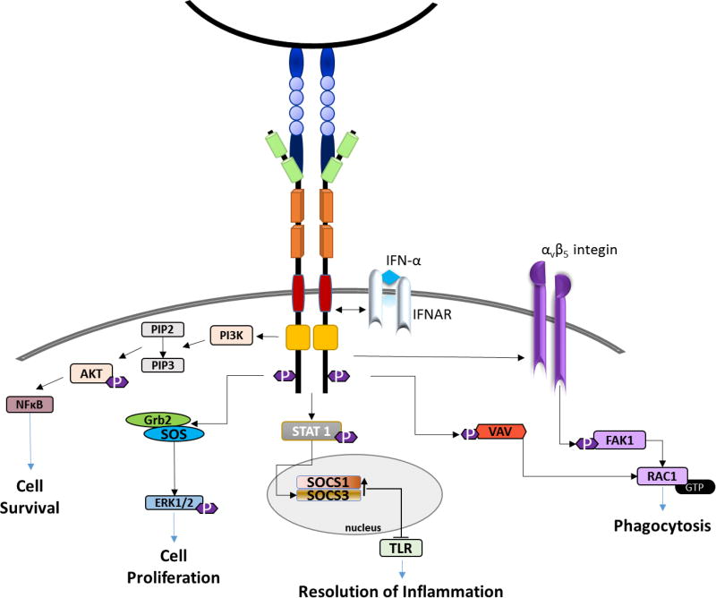
Tyro3, Axl, and Mertk, referred to as the TAM family of receptor tyrosine kinases, are instrumental in maintaining cell survival and homeostasis in mammals. TAM receptors interact with multiple signaling molecules to regulate cell migration, survival, phagocytosis and clearance of metabolic products and cell debris called efferocytosis. The TAMs also function as rheostats to reduce the expression of proinflammatory molecules and prevent autoimmunity. All three TAM receptors are activated in a concentration-dependent manner by the vitamin K-dependent growth arrest-specific protein 6 (Gas6). Gas6 and the TAMs are abundantly expressed in the nervous system. Gas6, secreted by neurons and endothelial cells, is the sole ligand for Axl. ProteinS1 (ProS1), another vitamin K-dependent protein functions mainly as an anti-coagulant, and independent of this function can activate Tyro3 and Mertk, but not Axl. This review will focus on the role of the TAM receptors and their ligands in the nervous system. We highlight studies that explore the function of TAM signaling in myelination, the visual cortex, neural cancers, and multiple sclerosis (MS) using Gas6-/- and TAM mutant mice models.

Keywords: Axl and Mertk receptor tyrosine kinase family; Gas6; Homeostasis in the nervous system; Inflammation; Myelination; Phagocytosis; ProS1; Tyro3.


Conflict of interest statement

Conflict of Interest

The authors have no actual or potential conflict of interest including any financial, personal or other relationships with individuals or organizations within three years of initiating the work that could inappropriately influence, or be perceived to influence, the study design or data interpretation.

Figures

Figure 1
Figure 1. Structural characterization of the TAM receptors and their ligands
TAM receptors have immunoglobulin (Ig)-like domains (green) and fibronectin repeats (orange) in the extracellular domain. A single pass transmembrane domain (red), an intracellular cytoplasmic domain containing a consensus kinase domain (yellow) and sites of tyrosine autophosphorylation (purple). The consensus sites for recruitment of signaling molecules including p85 subunit of PI3 kinase (pYXXM), Grb2 pYXN, and reported phosphatases are also shown. The structure of Gas6 and ProS1 is designated in blue. The ligands contain an amino terminal Gla domain that is γ-carboxylated in the presence of Vitamin K, four EGF-like repeats, and a carboxy-terminal sex hormone-binding globulin-like (SHBG-like) module that can bind and activate the TAM receptors.
Figure 2
Figure 2. TAM receptor signaling cascade
TAM receptors regulate cell survival/proliferation, inflammation and phagocytosis. The Axl receptor interacts with multiple SH2-containing signaling molecules, including Grb2 and PI3K, as well as STAT1 and αv β5-integin. Axl interaction with the interferon (IFN)-α/β receptor results in the phosphorylation and activation of STAT1, and increased SOCS1 and SOCS3 transcription. This reduces Toll-like receptor (TLR) activation and decreases the expression of proinflammatory molecules. Axl and Mertk are both important for phagocytosis of cell and myelin debris.
Figure 3
Figure 3. Forskolin and rhGas6 enhance MBP+ oligodendrocyte process ensheathment of axons
MBP+ OLs (green) ensheathing multiple neurofilament positive (NF+) axons (red) in co-cultures containing forskolin (5µM) (A–D), and forskolin and rhGas6 (2.6 nM) (E-H). Images showing MBP+ OLs wrapping NF+ axons were captured by confocal microscopy and Volocity software. Mann-Whitney p=0.015
Figure 4
Figure 4. Relative to WT mice, Gas6−/−Axl−/− DKO mice have swollen axons, fewer myelinated axons, and glial aggregates in the corpus callosum at 6-weeks cuprizone treatment+3-weeks recovery
Electron micrographs of WT mice (A) show abundant remyelinated axons relative to the DKO mice (B,C). The arrow (B) points to a swollen axon containing autophagosomes, autophagolysosomes, and multivesicular bodies. Abnormal myelin figure (arrowhead) and aggregated membranous structure (asterisk) consistent with a zebra body is shown in (C).
Figure 5
Figure 5. Gas6-AAV9 reduces clinical scores during acute but not chronic EAE
A. Immunofluorescent staining and microscopy show GFP staining in the cervical spinal cord of male mice injected IV with GFP-AAV2/9. Bar is 50 µm. B. Mice were sensitized with MOG35–55 peptide and Ptx on day 0, and Ptx on day 2. Six days after the second Ptx injection, equivalent AAV9 viral particles were injected IV into the tail vein in a 0.2ml volume. Mice were scored on a 0–5 scale. There was no significant difference in the onset of disease of the groups of mice. Therefore, data from two experiments was normalized to the onset of clinical signs of disease once a clinical score of 1 was obtained. *significance p<0.05, Mann-Whitney. C. qRT-PCR demonstrates increased Gas6 expression in liver, but not in brain or spinal cord at day 32 post-MOG injection. D. SMI32 staining of ventral spinal cord of GFP-AAV9 and Gas6-AAV9 mice shows no significant difference in SMI32+ axonal swellings; p>0.05. Visualization is by DAB. Bar is 200 µm. E. Quantification of the mean number of SMI32+ axonal swellings in ventral lumbar spinal cords of multiple GFP-AAV9 (n=4) and Gas6-AAV9 mice (n=5) mice; p>0.05. Gas6 and GFP were cloned into AAV2/9, grown and purified by the Penn Vector Core University of Pennsylvania. The vector was obtained by Dr. Brian Kaspar are driven by the chicken beta actin CMV hybrid promoter. AAV2/9 contains AAV2’s inverted terminal repeats with the AAV9 capsid.
Figure 6
Figure 6. Administration of rhGas6 to Axl−/− mice is not beneficial
Axl−/−mice (n=8/group)) were sensitized with MOG35–55 peptide. Six days after the second pertussis toxin injection, minipumps were connected to the cannula, inserted under the scapula,

References

    1. Abbott NJ, Patabendige AA, Dolman DE, Yusof SR, Begley DJ. Structure and function of the blood-brain barrier. Neurobiol Dis. 2010;37:13–25. - PubMed
    1. Abbott NJ, Ronnback L, Hansson E. Astrocyte-endothelial interactions at the blood-brain barrier. Nat Rev Neurosci. 2006;7:41–53. - PubMed
    1. Abu-Safieh L, Alrashed M, Anazi S, Alkuraya H, Khan AO, Al-Owain M, Al-Zahrani J, Al-Abdi L, Hashem M, Al-Tarimi S, Sebai MA, Shamia A, Ray-Zack MD, Nassan M, Al-Hassnan ZN, Rahbeeni Z, Waheeb S, Alkharashi A, Abboud E, Al-Hazzaa SA, Alkuraya FS. Autozygome-guided exome sequencing in retinal dystrophy patients reveals pathogenetic mutations and novel candidate disease genes. Genome Res. 2013;23:236–247. - PMC - PubMed
    1. Adembri C, Massagrande A, Tani A, Miranda M, Margheri M, De Gaudio R, Pellegrini-Giampietro DE. Carbamylated erythropoietin is neuroprotective in an experimental model of traumatic brain injury. Crit Care Med. 2008;36:975–978. - PubMed
    1. Agnello D, Bigini P, Villa P, Mennini T, Cerami A, Brines ML, Ghezzi P. Erythropoietin exerts an anti-inflammatory effect on the CNS in a model of experimental autoimmune encephalomyelitis. Brain Res. 2002;952:128–134. - PubMed

Publication types