. 2018 Mar 6;27(3):549-558.e4.
doi: 10.1016/j.cmet.2018.01.015.

Paracrine Interactions within the Pancreatic Islet Determine the Glycemic Set Point

Affiliations

Paracrine Interactions within the Pancreatic Islet Determine the Glycemic Set Point

Rayner Rodriguez-Diaz et al. Cell Metab. .

Abstract

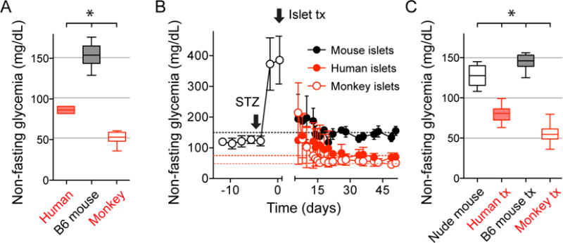
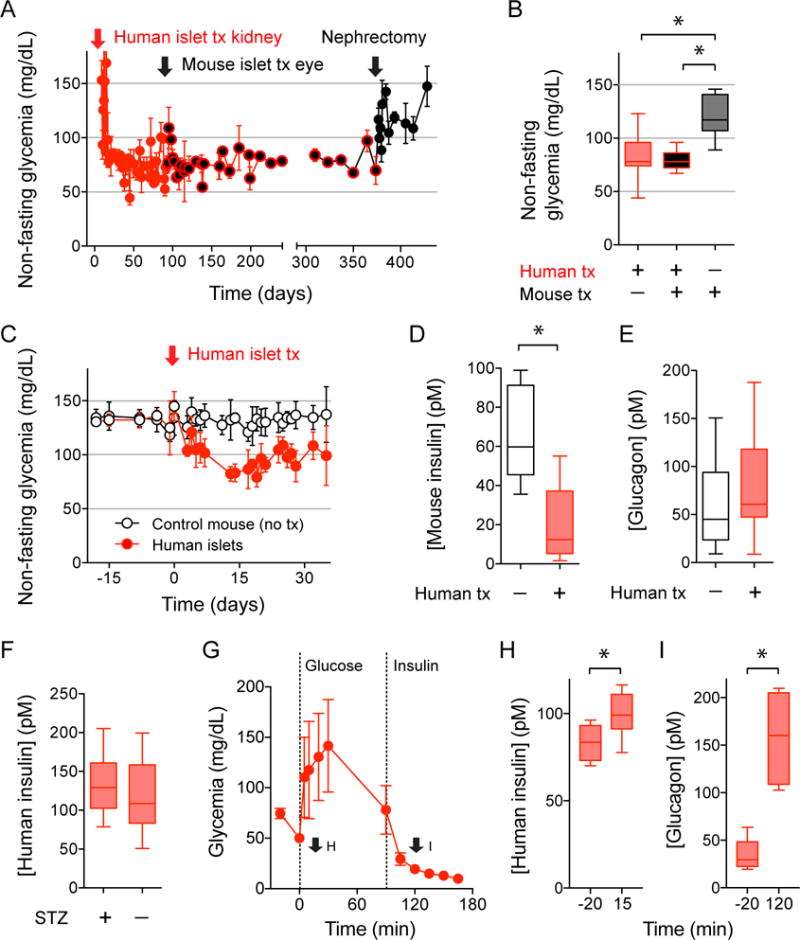
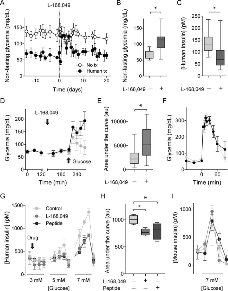
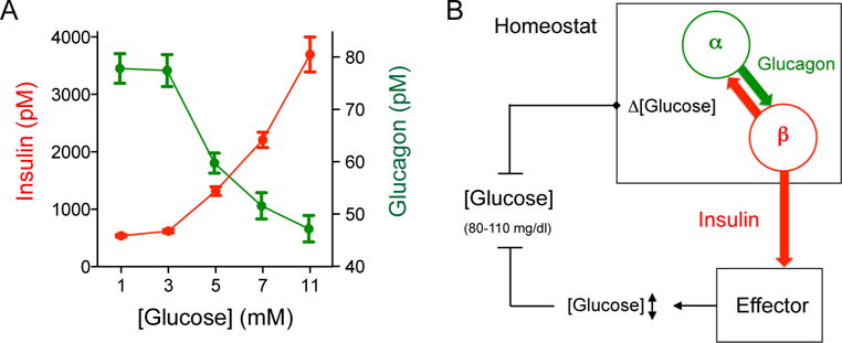
Every animal species has a signature blood glucose level or glycemic set point. These set points are different, and the normal glycemic levels (normoglycemia) of one species would be life threatening for other species. Mouse normoglycemia can be considered diabetic for humans. The biological determinants of the glycemic set point remain unclear. Here we show that the pancreatic islet imposes its glycemic set point on the organism, making it the bona fide glucostat in the body. Moreover, and in contrast to rodent islets, glucagon input from the alpha cell to the insulin-secreting beta cell is necessary to fine-tune the distinctive human set point. These findings affect transplantation and regenerative approaches to treat diabetes because restoring normoglycemia may require more than replacing only the beta cells. Furthermore, therapeutic strategies using glucagon receptor antagonists as hypoglycemic agents need to be reassessed, as they may reset the overall glucostat in the organism.

Keywords: alpha cell; beta cell; glucagon; glucose homeostasis; glycemia; glycemic set point; human pancreatic islet; humanized mouse; insulin secretion; paracrine signaling.


Conflict of interest statement

COMPETING FINANCIAL INTERESTS

P.-O.B. is cofounder and CEO of Biocrine, an unlisted biotech company that is using the anterior chamber of the eye technique as a research tool. B.L., I.L., and M.H.A. are consultants for the same company.

A.P. is currently employed at the National Institutes of Health (NIH). The opinions expressed in this article are the author’s own and do not necessarily reflect the views of the National Institutes of Health, the Department of Health and Human Services, or the United States government.

Figures

Figure 1
Figure 1. Pancreatic islet grafts transfer the glycemic set point of the islet donor species to recipient mice
(A) Non-fasting glycemic levels of humans (n = 5), C57Bl6J mice (n = 20), and cynomolgus monkeys (n = 11) were significantly different. (B) Nude mice rendered diabetic with streptozotocin (STZ) transplanted with islets from humans (n = 47 recipient mice), monkeys (n = 22), and C57Bl6J mice (n = 8) became normoglycemic with glycemic levels of the islet donor species. Curves are shown as average ± SD. (C) Quantification of results shown in B. All glycemic values were significantly different from each other. Data are shown as box and whisker plots and compared with one-way ANOVA followed by Tukey’s multiple comparison tests. Asterisks denote significance (P < 0.05).
Figure 2
Figure 2. Human islet grafts impose their glycemic set point
(A and B) Non-fasting glycemia of diabetic nude mice transplanted with human islets under the kidney capsule (red arrow) and then with islets from C57Bl6J mice into the eye (black arrow; n = 11 recipient mice). Human islets grafts were later removed by nephrectomy, which changed glycemic values to mouse levels (quantified in B). Data are shown as average ± SD (A) or box and whisker plots (B) and compared with one-way ANOVA followed by Tukey’s multiple comparison tests. Asterisks denote significance (P < 0.05). (C-E) Non-fasting glycemia shows that non-diabetic nude mice transplanted with human islets (black symbols, n = 5) acquired the human glycemic set point (C). Endogenous release of mouse insulin was inhibited in the presence of human islet grafts (D), but plasma glucagon levels were not affected (E). (F) Human insulin plasma levels in transplanted mice without endogenous islets (STZ-treated, STZ+) and with endogenous islets (STZ−; 15 measurements in 5 mice). Asterisks denote significance (P < 0.05, Student’s t-tests). (G-I) Intraperitoneal glucose tolerance test (4g/kg glucose) followed by an insulin tolerance test (0.75 U/kg insulin) performed in diabetic nude mice transplanted with human islets (G; n = 6 mice) show adequate insulin and glucagon responses to the glucose challenge (H) and the induced hypoglycemia (I), respectively. Hormone plasma levels were measured at the time points indicated in G (arrows). Asterisks denote significance (P < 0.05, Student’s t-tests).
Figure 3
Figure 3. Glycemic levels depend on donor species but are independent of transplanted islet mass
(A) Z-stacks of confocal images of the eyes of nude mice transplanted with 500, 300, 150, or 75 islet equivalents (islet backscatter shown in green and blood vessels in red. Asterisks indicates pupils; images acquired at day 70 after transplantation). (B and C) Non-fasting glycemic values show that transplanting different numbers of islets from C75Bl6J mice into diabetic nude mice reversed diabetes and produced similar levels of glycemia (quantified in B, n = 3 mice per group). Note, however, that recipient mice with a smaller mass of transplanted islets needed longer to return to normoglycemia. (D) Islet graft volumes of mice shown in A-C estimated by measuring islet backscatter (green) at days 35 (solid bars) and 70 (patterned bars) after transplantation. Mice receiving 500 islets had significantly more islet mass than those receiving 75 islets (P < 0.05, ANOVA followed by multiple comparison test). Over time, there was a small increase in islet volume in mice transplanted with fewer islets. (E) Mouse graft insulin contents were different for the four groups of mice at day 70 (gray scale columns; 500 significantly different from 75; P < 0.05, ANOVA followed by multiple comparison test), but plasma insulin concentrations (red symbols) were similar. (F) Photograph of nude mouse kidneys transplanted with 1000 or 2000 human islets. Arrows point at islet grafts, which were used for quantifications of mass in I and J. (G and H) Transplantation of 1000 and 2000, but not 500, human islets reversed diabetes in recipient nude mice (n = 3 mice per group). Mice with successful islet engraftment showed human normoglycemic values that were independent of the number of transplanted islets (quantified in H). (I) Human graft insulin contents were different for the two groups of mice at day 30 (P < 0.05, Student’s t-tests), but plasma insulin concentrations (red symbols) were similar. (J) In mice transplanted with different human (red symbols) and mouse (grey symbols) islet masses, graft insulin content did not correlate with target glycemia (slopes of regression lines not significantly different from 0), indicating that, once above the marginal mass required to achieve glucose homeostasis, islet mass does not impact the glycemic set point.
Figure 4
Figure 4. Modulating nervous input to islet grafts affects glycemia in nude mice transplanted with mouse islets, but not in mice transplanted with human islets
(A and B) Maximal projections of Z-stacks of confocal images of intraocular human islet grafts 90 days after transplantation showing immunostaining for the parasympathetic and sympathetic axon markers vAChT and TH, respectively. Notice that parasympathetic axons of the iris do not turn into the graft and that vAChT is present in endocrine cells. By contrast, some sympathetic axons reach into the islet parenchyma along blood vessels (labeled for αSMA). These staining patterns resemble those of islets in the native human pancreas (Rodriguez-Diaz et al, 2011a, 2011b). A’ and B’ are higher magnification images of regions denoted by boxes in A and B. (C) In contrast to human islet grafts, C57Bl6J mouse islet grafts showed a high density of parasympathetic axons in the islet parenchyma (see also Rodriguez-Diaz et al, 2012). Scale bars, 50 μm (A-C) and 10 μm (A’ and B’). (D) Non-fasting glycemic values show that modulating nervous input to human islet grafts via the pupillary light reflex with ambient illumination did not change glycemic levels. By contrast, increased nervous input reduced glycemic levels in mice with intraocular mouse islet grafts, but not in mice with mouse islets transplanted under the kidney capsule (P < 0.05, Student’s t-tests). Values obtained > 2 months after transplanting 1000 human islets into both eyes (n ≥ 12 mice per group) or 300 C7Bl6J mouse islet into the right eye or the kidney of diabetic nude mice (n = 4-5 mice per group).
Figure 5
Figure 5. Glycemic values set by insulin secretion from human islet grafts require glucagon input from neighboring alpha cells
(A-C) Injection of the human-specific glucagon receptor antagonist L169,049 (50 mg/kg, ip, day 0) increased glycemia in nude mice with human islets transplanted into the eye (glycemic levels quantified and compared to levels before treatment in B; n = 6 mice). During treatment with L169,049, human insulin plasma levels were significantly reduced in recipient mice (C). (D and E) Local application of L169,049 to the eye (4 mM) made transplanted mice glucose intolerant in intaperitoneal glucose tolerance tests (2 g/kg glucose, quantification as area under the curve of glucose excursion shown in E, n = 8 mice). (F) Local application of the muscarinic antagonist tropicamide (0.5%, 17 mM) did not affect glucose tolerance (4 g/kg glucose) in mice transplanted with human islets (n = 3 mice). Data are shown as average ± SEM and compared with Student’s t-test. Asterisks denote significance (P < 0.05). (G-I) Perifusion assays to measure insulin secretion showing that the glucagon receptor antagonists L-168,49 (50 nM) and des-His1-[Glu9]-Glucagon (1-29) amide (1μM) diminished glucose-stimulated (3, 5, and 7 mM) insulin secretion from human islets (G and H), but not from mouse islets (I). Antagonists were added at 3 mM glucose concentration (arrow) and maintained throughout the experiment. H shows a quantification (area under curve during stimulation with 5 and 7 mM glucose concentration) of experiments shown in G. Asterisks denote statistical significance (n = 4 islet human donors; P < 0.05; ANOVA followed by multiple comparisons).
Figure 6
Figure 6. Alpha cells and beta cells of the human islet cooperate to maintain the glycemic set point
(A) Glucose concentration-response relationship for insulin and glucagon secretion from human islets. Values were obtained in dynamic perifusions and represent the secretory levels during step increases in glucose concentration (n = 4 human pancreas preparations). Notice that glucagon and insulin secretion overlap substantially around 5 mM glucose concentration (equivalent to 90 mg/dl). Glucagon secretion at 5 mM was significantly different from that at 11 mM glucose concentration (one-way ANOVA followed by Tukey’s multiple comparison tests). (B) Cartoon depicting our view of the glucose homeostat. Fluctuations in glucose concentration (Δ [glucose]) around the glycemic set point are sensed by alpha and beta cells that continuously influence each other to fine-tune insulin secretion. Insulin serves as the control signal that regulates glucose uptake in effector organs (e.g. liver, muscles, and adipose tissue) to maintain normoglycemia. Without paracrine glucagon input to beta cells, the glucose homeostat fails to achieve target glycemic levels.

References

    1. Abdulreda MH, Rodriguez-Diaz R, Caicedo A, Berggren PO. Liraglutide Compromises Pancreatic β Cell Function in a Humanized Mouse Model. Cell Metab. 2016;23:541–546. - PMC - PubMed
    1. Almaça J, Molina J, Arrojo e Drigo R, Abdulreda MH, Jeon WB, Berggren PO, Caicedo A, Nam HG. Young capillary vessels rejuvenate aged pancreatic islets. Proc Natl Acad Sci USA. 2014;111:17612–17617. - PMC - PubMed
    1. Bertuzzi F, Berra C, Socci C, Davalli AM, Calori G, Freschi M, Piemonti L, De Nittis P, Pozza G, Pontiroli AE. Glucagon improves insulin secretion from pig islets in vitro. J Endocrinol. 1995;147:87–93. - PubMed
    1. Bosco D, Armanet M, Morel P, Niclauss N, Sgroi A, Muller Y, Giovannoni L, Parnaud G, Berney T. Unique arrangement of alpha- and beta-cells in human islets of Langerhans. Diabetes. 2010;59:1202–1210. - PMC - PubMed
    1. Bottino R, Inverardi L, Valente U, Ricordi C. Serum-free medium and pyruvate improve survival and glucose responsiveness of islet beta cells in culture. Transplant Proc. 1997;29:1978. - PubMed

Publication types

LinkOut - more resources