. 2018 Mar 6;22(10):2521-2529.
doi: 10.1016/j.celrep.2018.02.047.

ERRγ Promotes Angiogenesis, Mitochondrial Biogenesis, and Oxidative Remodeling in PGC1α/β-Deficient Muscle

Affiliations

ERRγ Promotes Angiogenesis, Mitochondrial Biogenesis, and Oxidative Remodeling in PGC1α/β-Deficient Muscle

Weiwei Fan et al. Cell Rep. .

Abstract

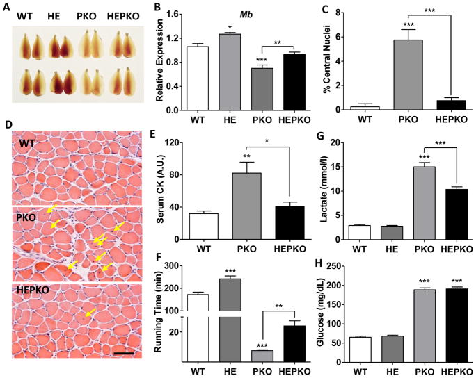
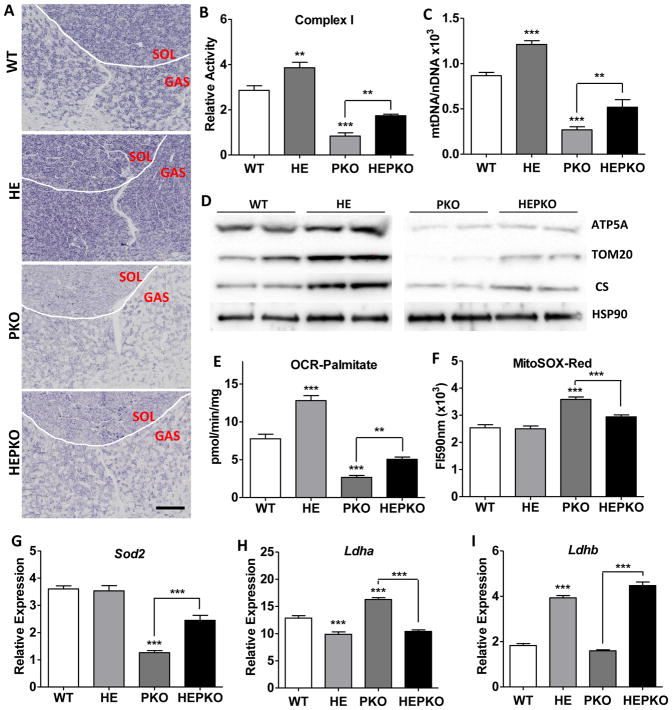
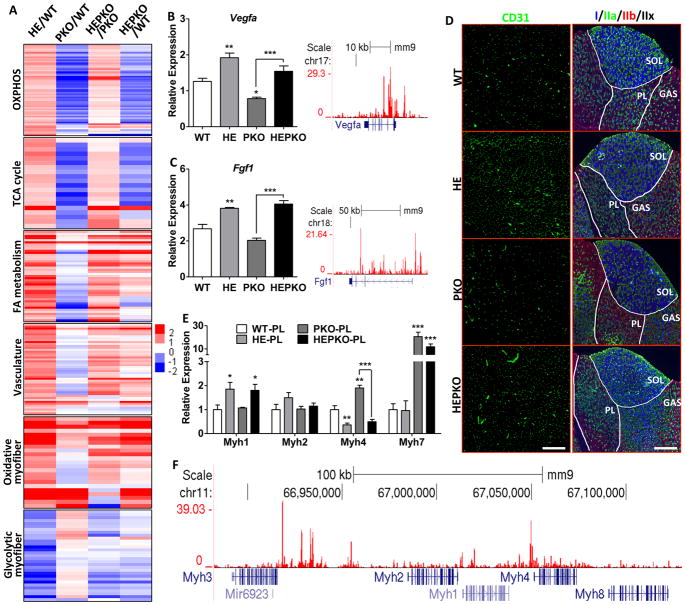
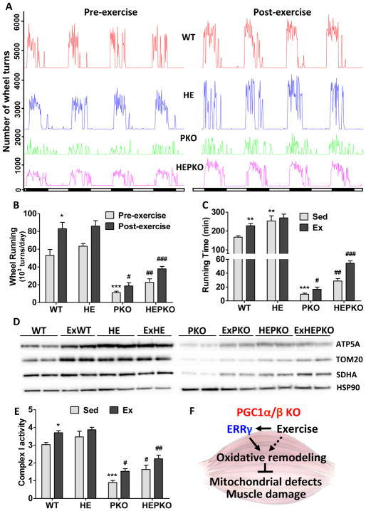
PGC1α is a pleiotropic co-factor that affects angiogenesis, mitochondrial biogenesis, and oxidative muscle remodeling via its association with multiple transcription factors, including the master oxidative nuclear receptor ERRγ. To decipher their epistatic relationship, we explored ERRγ gain of function in muscle-specific PGC1α/β double-knockout (PKO) mice. ERRγ-driven transcriptional reprogramming largely rescues muscle damage and improves muscle function in PKO mice, inducing mitochondrial biogenesis, antioxidant defense, angiogenesis, and a glycolytic-to-oxidative fiber-type transformation independent of PGC1α/β. Furthermore, in combination with voluntary exercise, ERRγ gain of function largely restores mitochondrial energetic deficits in PKO muscle, resulting in a 5-fold increase in running performance. Thus, while PGC1s can interact with multiple transcription factors, these findings implicate ERRs as the major molecular target through which PGC1α/β regulates both innate and adaptive energy metabolism.

Keywords: ERR; PGC1; estrogen related receptor; exercise; fatty acid oxidation; glycolysis; mitochondria; muscle; muscle damage; vasculature.


Figures

Figure 1
Figure 1. ERRγ improves running defect and muscle damage in PGC1-null mice
(A) Images of Tibialis anterior (TA) muscle from wild-type (WT), ERRγ transgenic (HE), muscle-specific PGC1α/β knockout (PKO), and HEPKO mice; (B) Relative expression of Myoglobin (Mb) in plantaris muscle; (C–D) Percentage centralized-nuclei in muscle and representative H&E staining (E) serum creatine kinase (CK) levels in sedentary mice; (F) running time during low-speed endurance test; (G) blood lactate levels after 6 minutes of endurance running (upon failure of the first mouse); and (H) blood glucose level at failure in endurance test. n=5. Data represent mean ± SEM, *p < 0.05, **p < 0.01, ***p < 0.001. Scale bar, 50μm. See also Figure S1.
Figure 2
Figure 2. ERRγ improves mitochondrial energetic defects in PGC1-null muscle
(A) Immunohistochemical staining for mitochondrial complex I activity in cryosections from soleus (SOL) and gastrocnemius (GAS); (B) mitochondrial complex I activity measured in isolated mitochondrial from quadriceps muscle; (C) relative mtDNA copy numbers in plantaris; (D) western blots showing the levels of mitochondrial proteins ATP5A, TOM20, and CS, as well as a cytoplasmic control HSP90 in plantaris; (E) oxygen consumption rates measured in freshly isolated quadriceps mitochondria using palmitoylcarnitine as substrate; (F) ROS measured as MitoSOX fluorescence intensity in freshly isolated quadriceps mitochondria; and (G–I) Relative expression levels of Sod2 (G), Ldha (H), and Ldhb (I) in plantaris. n=5. Data represent mean ± SEM, *p < 0.05, **p < 0.01, ***p < 0.001. Scale bar, 500μm. See also Figure S2.
Figure 3
Figure 3. ERRγ induces oxidative muscle remodeling independent of PGC1
(A) Heat maps showing relative changes in expression of selected genes involved in OXPHOS, TCA cycle, FAO, vasculature, oxidative-specific myofiber, and glycolytic-specific myofiber in plantaris; (B–C) Relative expression of Vegfa and Fgf1 in plantaris (left) and genome browser tracks showing ERRγ binding (right); (D) Immunostaining of CD31 for vasculature (left) and Myh I/IIa/IIb for fiber-typing (right); (E) Relative expression of Myh1/2/4/7 in plantaris; and (F) genome browser track showing ERRγ binding at the Myh cluster locus. n=5. Data represent mean ± SEM, *p < 0.05, **p < 0.01, ***p < 0.001. Scale bars, 300μm (CD31) and 750μm (Myh). See also Figure S3.
Figure 4
Figure 4. Exercise and ERRγ synergistically improve oxidative functions in PKO muscle
(A) Running wheel activity of WT (red), HE (blue), PKO (green), and HEPKO (purple) mice before (left) and after (right) 6 weeks of voluntary wheel running; (B) Cumulative daily wheel running from (A); (C) Total running time of sedentary and exercised (8 weeks of voluntary wheel running) mice in endurance test; (D) Western blots showing the levels of mitochondrial proteins ATP5A, TOM20, and SDHA, as well as a cytoplasmic control HSP90 in plantaris; (E) Mitochondrial complex I activity in isolated mitochondrial from quadriceps; and (F) Diagram showing ERRγ- and exercise-induced oxidative muscle remodeling in PKO mice that improves muscle damage and mitochondrial defects. Data represent mean ± SEM. * and # show statistical difference from WT and PKO, respectively. n=5. Data represent mean ± SEM, * and # p < 0.05, ** and ## p < 0.01, *** and ### p < 0.001. See also Figure S4.

References

    1. Arany Z. PGC-1 coactivators and skeletal muscle adaptations in health and disease. Current opinion in genetics & development. 2008;18:426–434. - PMC - PubMed
    1. Ballmann C, Tang Y, Bush Z, Rowe GC. Adult expression of PGC-1alpha and -1beta in skeletal muscle is not required for endurance exercise-induced enhancement of exercise capacity. American journal of physiology Endocrinology and metabolism. 2016;311:E928–E938. - PMC - PubMed
    1. Canto C, Auwerx J. PGC-1alpha, SIRT1 and AMPK, an energy sensing network that controls energy expenditure. Current opinion in lipidology. 2009;20:98–105. - PMC - PubMed
    1. Egan B, Zierath JR. Exercise metabolism and the molecular regulation of skeletal muscle adaptation. Cell Metab. 2013;17:162–184. - PubMed
    1. Estrella NL, Desjardins CA, Nocco SE, Clark AL, Maksimenko Y, Naya FJ. MEF2 transcription factors regulate distinct gene programs in mammalian skeletal muscle differentiation. The Journal of biological chemistry. 2015;290:1256–1268. - PMC - PubMed

Publication types

MeSH terms

Substances

LinkOut - more resources