. 2018 Sep 1;159(9):3143-3157.
doi: 10.1210/en.2018-00083.

CC-401 Promotes β-Cell Replication via Pleiotropic Consequences of DYRK1A/B Inhibition

Affiliations

CC-401 Promotes β-Cell Replication via Pleiotropic Consequences of DYRK1A/B Inhibition

Yassan Abdolazimi et al. Endocrinology. .

Abstract

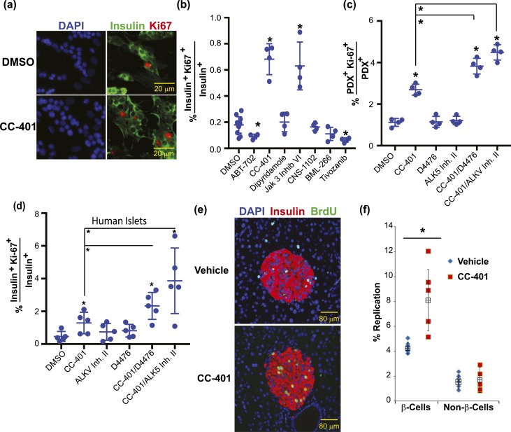
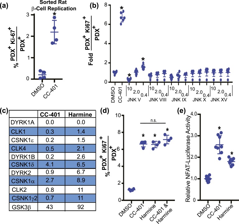
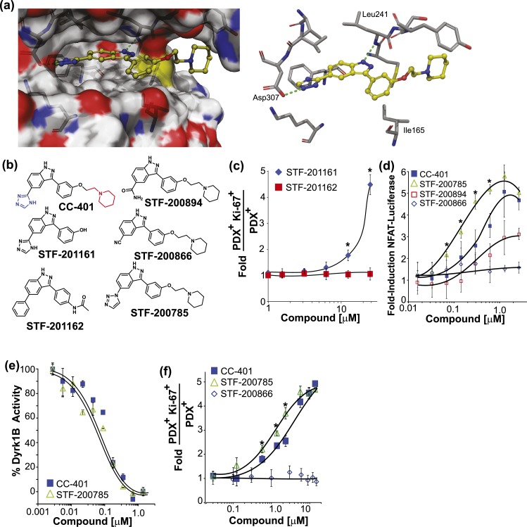
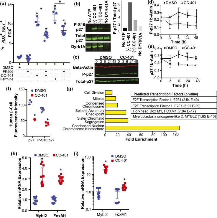
Pharmacologic expansion of endogenous β cells is a promising therapeutic strategy for diabetes. To elucidate the molecular pathways that control β-cell growth we screened ∼2400 bioactive compounds for rat β-cell replication-modulating activity. Numerous hit compounds impaired or promoted rat β-cell replication, including CC-401, an advanced clinical candidate previously characterized as a c-Jun N-terminal kinase inhibitor. Surprisingly, CC-401 induced rodent (in vitro and in vivo) and human (in vitro) β-cell replication via dual-specificity tyrosine phosphorylation-regulated kinase (DYRK) 1A and 1B inhibition. In contrast to rat β cells, which were broadly growth responsive to compound treatment, human β-cell replication was only consistently induced by DYRK1A/B inhibitors. This effect was enhanced by simultaneous glycogen synthase kinase-3β (GSK-3β) or activin A receptor type II-like kinase/transforming growth factor-β (ALK5/TGF-β) inhibition. Prior work emphasized DYRK1A/B inhibition-dependent activation of nuclear factor of activated T cells (NFAT) as the primary mechanism of human β-cell-replication induction. However, inhibition of NFAT activity had limited effect on CC-401-induced β-cell replication. Consequently, we investigated additional effects of CC-401-dependent DYRK1A/B inhibition. Indeed, CC-401 inhibited DYRK1A-dependent phosphorylation/stabilization of the β-cell-replication inhibitor p27Kip1. Additionally, CC-401 increased expression of numerous replication-promoting genes normally suppressed by the dimerization partner, RB-like, E2F and multivulval class B (DREAM) complex, which depends upon DYRK1A/B activity for integrity, including MYBL2 and FOXM1. In summary, we present a compendium of compounds as a valuable resource for manipulating the signaling pathways that control β-cell replication and leverage a DYRK1A/B inhibitor (CC-401) to expand our understanding of the molecular pathways that control β-cell growth.


Figures

Figure 1.
Figure 1.
Identification of small-molecule inducers of rat β-cell replication and their combinatorial use. (a) Summary of rat β-cell replication screening results. Compounds are clustered according to their annotated bioactivity. Supplemental Tables 1 and 2 list primary screening and confirmatory testing β-cell–replication results. (b) The β-cell replication–promoting profile of prioritized compounds. Data are presented as mean ± standard deviation (SD; n = 3 to 5 wells per treatment condition; results confirmed in n ≥ 3 independent experiments). (c) Representative images of rat islet cultures treated with vehicle (DMSO) or CC-401. (d) Representative images of compound-treated rat islet cultures. The percentage of replicating PDX1+ cells and compound treatments are indicated. (e) Rat islet β-cell replication indices Ki67 (blue) and PCNA (red). Compound concentrations were as indicated in (b) and was 1 μM for CHIR99021. Individual data points represent 2000 to 3000 β cells (n = 4 to 8 replicates per condition; mean ± SD shown). All compounds increased β-cell replication above DMSO (P < 0.01). All compound combinations increased β-cell replication above the relevant individual compound-treatment conditions (P < 0.01). (f) Rat islet β-cell replication index after compound treatment as indicated. Single compound treatment, CNS-1102 combinations, and BML-266 combinations are shown. All individual compound treatments increased replication (vs DMSO; P < 0.01); select combinations increased β-cell replication vs relevant individual compound-treated conditions. *P < 0.01. Individual data points represent 2000 to 3000 β cells (n = 4 to 8 replicates per condition; mean ± SD shown). Error bars represent the standard deviation of an experimental condition (n ≥ 3). cAMP, cyclic adenosine monophosphate; CAS#, Chemical Abstracts Service number; PDE, phosphodiesterase.
Figure 2.
Figure 2.
CC-401 stimulates human β-cell replication in vitro and mouse β-cell replication in vivo. (a) Representative immunofluorescence images of vehicle- (DMSO) and CC-401–treated human islet cultures [DAPI (blue); insulin (green)]. (b) The β-cell–replication index of dispersed human islet cultures after compound treatment (48 hours). Independent treatments along with mean ± standard deviation (SD) are shown. *P < 0.05. Similar data were obtained from at least five independent islet procurements. (c) Rat β-cell–replication index of compound-treated (CC-401, 10 µM; D4476, 5 µM; and ALK5 inhibitor II, 2 µM) primary islet cultures. Independent replicates (n = 4) are shown with mean ± SD. *P < 0.01; >1000 β cells counted per data point. (d) Human β-cell–replication index of compound-treated islet cultures. Independent replicates (n = 5) are shown with mean ± SD. *P < 0.05; >1000 β cells counted per data point. (e) Representative images of pancreatic sections from 8-week female vehicle- and CC-401–treated mice stained for insulin (red), BrdU (green), and nuclei (blue). See Supplemental Fig. 2 for determination of CC-401’s in vivo half-life and in vitro potency. (f) The BrdU incorporation index (percentage of replication) of β cells (insulin+) and non–β cells (insulin) after treatment with vehicle or CC-401 (25 mg/kg) for 1 week. Data from individual mice (n = 5) and mean ± SD shown. *P < 0.05. Error bars represent the standard deviation of an experimental condition (n ≥ 3). Two independent experiments were performed with similar results. See Supplemental Fig. 2 for in vitro replication effects on α cells, δ cells, and dermal fibroblasts. ALKV Inh. II, activin A receptor type II–like kinase inhibitor II.
Figure 3.
Figure 3.
CC-401 promotes β-cell replication via DYRK1A/B inhibition. (a) Replication index of vehicle- and CC-401–treated fluorescence-activated cell sorting–purified rat β cells (see also Supplemental Fig. 3). Individual data points and mean ± standard deviation (SD) are shown (n = 4 wells per treatment condition with >1000 β cells per well). *P < 0.05. (b) Rat β-cell–proliferation index of JNK inhibitor–treated islet cultures. Individual wells and mean ± SD shown (n = 6 wells per treatment condition). *P < 0.05 vs DMSO treatment. (c) Percent residual kinase activity after incubation with the specified compound (10 µM). See Supplemental Table 4 for complete kinome datasets and Supplemental Fig. 4(a)–4(c) for the replication-promoting activity of additional DYRK1A/B inhibitors. (d) In vitro rat β-cell–replication response to the indicated compound treatment(s) (48 hours). Data from individual wells with mean ± SD are shown (n = 4 wells per treatment condition; >1000 β cells counted per well). *P < 0.05. (e) Fold-induction of NFAT-firefly luciferase reporter activity from cells cotransfected with Renilla (used for normalization across transfections), NFATc1, and DYRK1A expression plasmids, and treated with DMSO, CC-401, or harmine. Data from individual wells (n = 8) are presented with mean ± SD. *P < 0.05. Error bars represent the standard deviation of an experimental condition (n ≥ 3).
Figure 4.
Figure 4.
Rationally designed CC-401 derivatives confirm DYRK1A/B as the relevant β-cell–replication target. (a) Space-filling (left) and stick (right) models of CC-401 binding to DYRK1A [4MQ2; Protein Data Bank (28)] generated with Schrodinger Glide software. Key hydrogen bond interactions are highlighted (green dashed line). (b) Chemical structures of CC-401 and synthesized derivatives. The piperidine group (red) and the triazole group (blue) are highlighted on CC-401. (c) Rat β-cell–proliferation response to STF-201161 and STF-201162. Data are presented as mean ± standard deviation (SD; n = 4 wells per treatment condition). *P < 0.05. (d) Fold-induction of NFAT-luciferase reporter activity in cells cotransfected with NFATc1, DYRK1A, and Renilla luciferase. Cells were compound treated as indicated. Data are presented as mean ± SD (n = 4 wells per experimental point). *P < 0.01 shown for STF-785 vs CC-401. A significant (P < 0.05) luciferase induction above DMSO occurred in response to CC-401 at a concentration >0.1 µM, STF-200785 (>0.05 µM), and STF-200894 (>2.0 µM). No luciferase induction occurred in response to STF-200866. (e) Recombinant DYRK1B activity measured in the presence of CC-401 or STF-200785 (n = 2 wells per experimental point). *P < 0.01. Half maximal inhibitory concentrations were not statistically different. (f) Fold increase in rat β-cell replication in response to the indicated compound treatment (n = 4 wells per treatment condition presented as mean ± SD). *P < 0.05 shown for STF-785 vs CC-401. No replication response occurred in response to STF-200866. Error bars represent the standard deviation of an experimental condition (n ≥ 3).
Figure 5.
Figure 5.
Inhibition of DYRK1A/B promotes β-cell replication via NFAT activation, p27kip1 destabilization, and DREAM-complex target gene derepression. (a) In vitro rat β-cell–replication indices after compound treatment as indicated. The results from individual wells (n = 4 to 7) are shown with mean ± standard deviation (SD). *P < 0.05 vs the DMSO-treated condition or as indicated. (b) Western blot analysis of phospho-S10p27, p27, and DYRK1A after in vitro incubation of recombinant p27 and DYRK1A with and without adenosine triphosphate, CC-401, or DYRK1A. Quantitation of S10 phosphorylated p27 intensity normalized to total p27 are shown in the right-side panel. (c) Representative western blot of R7T1 β-cell lysates probed for β-actin, p27, and phosphoS10-p27 after treatment of cultures with DMSO or CC-401 for the indicated time (hours). Quantitation of the relative levels of (d) phosphoS10-p27 and (e) total p27 obtained from western blots (n = 3) performed as in (c). Protein levels were normalized to β-actin levels. *P < 0.05 vs DMSO condition at the relevant time point. (f) The average fluorescence intensity of p27 and phospho-S10p27 measured in β cells (PDX1+) from human islet cultures treated with DMSO or CC-401. Individual wells (n = 4; >1500 β cells per well) are shown with mean ± SD. *P < 0.01. (g) Enriched gene ontology categories (left) and transcription factor networks (right) identified by MetaCore analysis of differentially expressed genes from RNA sequencing performed on fluorescence-activated cell–sorted β cells after vehicle- and CC-401–treatment (48 hours); see Supplemental Table 4 for complete results. (h) Relative gene expression levels determined by qPCR (normalized to Gapdh) after treatment of growth-arrested R7T1 β-cell cultures with DMSO or CC-401 for 48 hours. (i) Relative gene expression levels determined by qPCR (normalized to GAPDH) after treatment of human islet cultures with DMSO or CC-401 for 48 hours. Error bars represent the standard deviation of an experimental condition (n ≥ 3). ATP, adenosine triphosphate.

References

    1. Nichols RJ, New C, Annes JP. Adult tissue sources for new β cells. Transl Res. 2014;163(4):418–431. - PMC - PubMed
    1. Van Assche FA, Aerts L, De Prins F. A morphological study of the endocrine pancreas in human pregnancy. Br J Obstet Gynaecol. 1978;85(11):818–820. - PubMed
    1. Ritzel RA, Butler AE, Rizza RA, Veldhuis JD, Butler PC. Relationship between beta-cell mass and fasting blood glucose concentration in humans. Diabetes Care. 2006;29(3):717–718. - PubMed
    1. Morris AP, Voight BF, Teslovich TM, Ferreira T, Segrè AV, Steinthorsdottir V, Strawbridge RJ, Khan H, Grallert H, Mahajan A, Prokopenko I, Kang HM, Dina C, Esko T, Fraser RM, Kanoni S, Kumar A, Lagou V, Langenberg C, Luan J, Lindgren CM, Müller-Nurasyid M, Pechlivanis S, Rayner NW, Scott LJ, Wiltshire S, Yengo L, Kinnunen L, Rossin EJ, Raychaudhuri S, Johnson AD, Dimas AS, Loos RJ, Vedantam S, Chen H, Florez JC, Fox C, Liu CT, Rybin D, Couper DJ, Kao WH, Li M, Cornelis MC, Kraft P, Sun Q, van Dam RM, Stringham HM, Chines PS, Fischer K, Fontanillas P, Holmen OL, Hunt SE, Jackson AU, Kong A, Lawrence R, Meyer J, Perry JR, Platou CG, Potter S, Rehnberg E, Robertson N, Sivapalaratnam S, Stančáková A, Stirrups K, Thorleifsson G, Tikkanen E, Wood AR, Almgren P, Atalay M, Benediktsson R, Bonnycastle LL, Burtt N, Carey J, Charpentier G, Crenshaw AT, Doney AS, Dorkhan M, Edkins S, Emilsson V, Eury E, Forsen T, Gertow K, Gigante B, Grant GB, Groves CJ, Guiducci C, Herder C, Hreidarsson AB, Hui J, James A, Jonsson A, Rathmann W, Klopp N, Kravic J, Krjutškov K, Langford C, Leander K, Lindholm E, Lobbens S, Männistö S, Mirza G, Mühleisen TW, Musk B, Parkin M, Rallidis L, Saramies J, Sennblad B, Shah S, Sigurðsson G, Silveira A, Steinbach G, Thorand B, Trakalo J, Veglia F, Wennauer R, Winckler W, Zabaneh D, Campbell H, van Duijn C, Uitterlinden AG, Hofman A, Sijbrands E, Abecasis GR, Owen KR, Zeggini E, Trip MD, Forouhi NG, Syvänen AC, Eriksson JG, Peltonen L, Nöthen MM, Balkau B, Palmer CN, Lyssenko V, Tuomi T, Isomaa B, Hunter DJ, Qi L, Shuldiner AR, Roden M, Barroso I, Wilsgaard T, Beilby J, Hovingh K, Price JF, Wilson JF, Rauramaa R, Lakka TA, Lind L, Dedoussis G, Njølstad I, Pedersen NL, Khaw KT, Wareham NJ, Keinanen-Kiukaanniemi SM, Saaristo TE, Korpi-Hyövälti E, Saltevo J, Laakso M, Kuusisto J, Metspalu A, Collins FS, Mohlke KL, Bergman RN, Tuomilehto J, Boehm BO, Gieger C, Hveem K, Cauchi S, Froguel P, Baldassarre D, Tremoli E, Humphries SE, Saleheen D, Danesh J, Ingelsson E, Ripatti S, Salomaa V, Erbel R, Jöckel KH, Moebus S, Peters A, Illig T, de Faire U, Hamsten A, Morris AD, Donnelly PJ, Frayling TM, Hattersley AT, Boerwinkle E, Melander O, Kathiresan S, Nilsson PM, Deloukas P, Thorsteinsdottir U, Groop LC, Stefansson K, Hu F, Pankow JS, Dupuis J, Meigs JB, Altshuler D, Boehnke M, McCarthy MI;Wellcome Trust Case Control Consortium; Meta-Analyses of Glucose and Insulin-related traits Consortium (MAGIC) Investigators; Genetic Investigation of ANthropometric Traits (GIANT) Consortium; Asian Genetic Epidemiology Network–Type 2 Diabetes (AGEN-T2D) Consortium; South Asian Type 2 Diabetes (SAT2D) Consortium; DIAbetes Genetics Replication And Meta-analysis (DIAGRAM) Consortium . Large-scale association analysis provides insights into the genetic architecture and pathophysiology of type 2 diabetes. Nat Genet. 2012;44(9):981–990. - PMC - PubMed
    1. Steinthorsdottir V, Thorleifsson G, Sulem P, Helgason H, Grarup N, Sigurdsson A, Helgadottir HT, Johannsdottir H, Magnusson OT, Gudjonsson SA, Justesen JM, Harder MN, Jørgensen ME, Christensen C, Brandslund I, Sandbæk A, Lauritzen T, Vestergaard H, Linneberg A, Jørgensen T, Hansen T, Daneshpour MS, Fallah MS, Hreidarsson AB, Sigurdsson G, Azizi F, Benediktsson R, Masson G, Helgason A, Kong A, Gudbjartsson DF, Pedersen O, Thorsteinsdottir U, Stefansson K. Identification of low-frequency and rare sequence variants associated with elevated or reduced risk of type 2 diabetes. Nat Genet. 2014;46(3):294–298. - PubMed

Publication types

MeSH terms

Substances