

Brief electrical stimulation and synkinesis after facial nerve crush injury: a randomized prospective animal study
- PMID: 29514718
- PMCID: PMC5842591
- DOI: 10.1186/s40463-018-0264-0
Brief electrical stimulation and synkinesis after facial nerve crush injury: a randomized prospective animal study
Abstract
Background: Recent studies have examined the effects of brief electrical stimulation (BES) on nerve regeneration, with some suggesting that BES accelerates facial nerve recovery. However, the facial nerve outcome measurement in these studies has not been precise or accurate. Furthermore, no previous studies have been able to demonstrate the effect of BES on synkinesis. The objective of this study is to examine the effect of brief electrical stimulation (BES) on facial nerve function and synkinesis in a rat model.
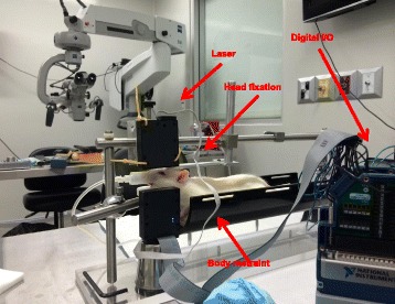
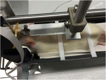
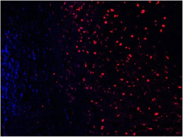
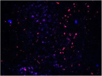
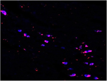
Methods: Four groups of six rats underwent a facial nerve injury procedure. Group 1 and 2 underwent a crush injury at the main trunk of the nerve, with group 2 additionally receiving BES for 1 h. Group 3 and 4 underwent a transection injury at the main trunk, with group 4 additionally receiving BES for 1 h. A laser curtain model was used to measure amplitude of whisking at 2, 4, and 6 weeks. Fluorogold and fluororuby neurotracers were additionally injected into each facial nerve to measure synkinesis. Buccal and marginal mandibular branches of the facial nerve were each injected with different neurotracers at 3 months following injury. Based on facial nucleus motoneuron labelling of untreated rats, comparison was made to post-treatment animals to deduce whether synkinesis had taken place. All animals underwent trans-cardiac perfusion with subsequent neural tissue sectioning.
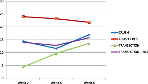
Results: At week two, the amplitude observed for group 1 and 2 was 14.4 and 24.0 degrees, respectively (p = 0.0004). Group 4 also demonstrated improved whisking compared to group 3. Fluorescent neuroimaging labelling appear to confirm improved pathway specific regeneration with BES following facial nerve injury.
Conclusions: This is the first study to use an implantable stimulator for serial BES following a crush injury in a validated animal model. Results suggest performing BES after facial nerve injury is associated with accelerated facial nerve function and improved facial nerve specific pathway regeneration in a rat model.
Keywords: Brief electrical stimulation; Electrical stimulation; Facial nerve; Peripheral nerve injury; Peripheral nerve regeneration; Regeneration; Synkinesis.
Conflict of interest statement
Ethics approval and consent to participate
Prior to commencement of this study ethics approval was obtained from the University of Alberta Health Research Ethics Board.
Consent for publication
Not applicable.
Competing interests
The authors declare that they have no competing interests.
Publisher’s Note
Springer Nature remains neutral with regard to jurisdictional claims in published maps and institutional affiliations.
Figures
References
-
- Hadlock T, Lindsay R, Edwards C, et al. The effect of electrical and mechanical stimulation on the regenerating rodent facial nerve. Laryngoscope. 2010;120(6):1094–1102. - PubMed
-
- Sunderland S. Nerve and nerve injuries. London: Churchill Livingstone; 1978.
Publication types
MeSH terms
Grants and funding
LinkOut - more resources
Full Text Sources
Other Literature Sources
