. 2018 Apr 27;92(10):e00088-18.
doi: 10.1128/JVI.00088-18. Print 2018 May 15.

Role of Microvesicles in the Spread of Herpes Simplex Virus 1 in Oligodendrocytic Cells

Affiliations

Role of Microvesicles in the Spread of Herpes Simplex Virus 1 in Oligodendrocytic Cells

Raquel Bello-Morales et al. J Virol. .

Abstract

Herpes simplex virus 1 (HSV-1) is a neurotropic pathogen that can infect many types of cells and establishes latent infections in the neurons of sensory ganglia. In some cases, the virus spreads into the central nervous system, causing encephalitis or meningitis. Cells infected with several different types of viruses may secrete microvesicles (MVs) containing viral proteins and RNAs. In some instances, extracellular microvesicles harboring infectious virus have been found. Here we describe the features of shedding microvesicles released by the human oligodendroglial HOG cell line infected with HSV-1 and their participation in the viral cycle. Using transmission electron microscopy, we detected for the first time microvesicles containing HSV-1 virions. Interestingly, the Chinese hamster ovary (CHO) cell line, which is resistant to infection by free HSV-1 virions, was susceptible to HSV-1 infection after being exposed to virus-containing microvesicles. Therefore, our results indicate for the first time that MVs released by infected cells contain virions, are endocytosed by naive cells, and lead to a productive infection. Furthermore, infection of CHO cells was not completely neutralized when virus-containing microvesicles were preincubated with neutralizing anti-HSV-1 antibodies. The lack of complete neutralization and the ability of MVs to infect nectin-1/HVEM-negative CHO-K1 cells suggest a novel way for HSV-1 to spread to and enter target cells. Taken together, our results suggest that HSV-1 could spread through microvesicles to expand its tropism and that microvesicles could shield the virus from neutralizing antibodies as a possible mechanism to escape the host immune response.IMPORTANCE Herpes simplex virus 1 (HSV-1) is a neurotropic pathogen that can infect many types of cells and establishes latent infections in neurons. Extracellular vesicles are a heterogeneous group of membrane vesicles secreted by most cell types. Microvesicles, which are extracellular vesicles which derive from the shedding of the plasma membrane, isolated from the supernatant of HSV-1-infected HOG cells were analyzed to find out whether they were involved in the viral cycle. The importance of our investigation lies in the detection, for the first time, of microvesicles containing HSV-1 virions. In addition, virus-containing microvesicles were endocytosed into CHO-K1 cells and were able to actively infect these otherwise nonpermissive cells. Finally, the infection of CHO cells with these virus-containing microvesicles was not completely neutralized by anti-HSV-1 antibodies, suggesting that these extracellular vesicles might shield the virus from neutralizing antibodies as a possible mechanism of immune evasion.

Keywords: extracellular vesicles; herpes simplex virus; microvesicles; oligodendrocytes; viral spread.


Figures

FIG 1
FIG 1
Isolation of MVs from the cell culture supernatants of HOG cells. (A) MVs were isolated by differential centrifugation from the supernatants of HOG cells that had been mock infected (Mock) and infected (Inf) with HSV-1 at an MOI of 1 for 24 h. MVs were adsorbed onto collodion-carbon-coated copper grids and negatively stained with aqueous uranyl acetate. Bars = 500 nm. (B) HOG cells were incubated at 4°C for 1 h with MVs isolated from HSV-1-infected HOG cells and then for 15 min at 37°C. After that the cells were fixed and processed for electron microscopy. The image shows the presence of virions and a heterogeneous group of MVs attached to the plasma membrane. (C) Four random images corresponding to the MV-untreated cell control, showing the plasma membrane free of attached vesicles. (D) The pellet containing the MVs obtained as described above was resuspended and kept at 4°C in serum-free DM before its analysis by nanoparticle tracking analysis (NTA). The plots and histogram show the average size of MVs. (E) Histograms representing the concentration of particles (×10E8) per milliliter show an increase in the concentrations of particles obtained from infected cells (dark bars) compared to mock-infected ones (light bars). To exclude the possibility of the presence of isolated virions, we also performed the analysis with particles with a size larger than 250 nm. (F) Immunoblots show the presence of integrin β-1, flotillin-1, LC3-II, and CD81 in MVs isolated from mock-infected (−) and infected (+) cells. CD63 was observed in the MV fraction isolated from infected cells. The myelin proteins CNPase and PLP were not detected in MVs. The bands corresponding to the anti-HSV-1 antibody are also shown.
FIG 2
FIG 2
Immunoelectron microscopy of MVs obtained from HOG cells infected with HSV-1. MVs from HOG cells infected with HSV-1 were fixed and processed for immunoelectron microscopy. (A) Vesicles labeled with an anti-HSV-1 antibody coupled to colloidal gold can be seen, although not all vesicles (arrows) are labeled with viral proteins. (B and C) In isolated virions (B) and MVs (C), viral proteins exposed outside the membrane (arrows) can be detected. (D to F) Images show the presence of MVs containing virions. (G to J) Four random images corresponding to the control of centrifugation, in which a mixture of virions with MVs was centrifuged to exclude the possibility that the presence of virions inside MVs was artifactual. Although the images corresponding to the centrifugation of virions with vesicles showed a compact group of vesicles and viral particles tightly situated, MVs containing virions were not detected. Bars = 90 nm (A to F) and 200 nm (G to J).
FIG 3
FIG 3
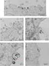
Electron microscopy of HOG cells incubated with MVs. MVs from HOG cells infected with HSV-1, isolated as described in the text, were layered onto HOG cells and incubated at 4°C for 1 h. After that, they were incubated for 15 min at 37°C, fixed, and processed for electron microscopy. (A and B) Although the majority of MVs lacked virions (A), occasionally, virion-containing MVs were observed (B). (C to E) Most of the virion-containing MVs enclosed only one viral particle, which lacked, in most cases, an organized envelope (black arrows). Occasionally, more virions or other vesicles (red arrow) were also found in the virion-containing MVs (D). MVs with inner virions attached to incipient cell membrane invaginations, resembling the initiation of an endocytic process, were also observed (D and E).
FIG 4
FIG 4
Immunofluorescence analysis of HOG cells incubated with MVs. MVs from HSV-1-infected HOG cells were stained with the red dye PKH26 and added to naive HOG cells. After 2 h of incubation at 37°C, the cells were fixed and stained with Alexa Fluor 647-phalloidin to visualize the contour of the cells. Images showed MVs colocalizing with the plasma membrane (double-headed arrow). In addition, MVs attached to the plasma membrane (arrow) and inside the cells (arrowhead) can be observed in the magnified region corresponding to the white square, suggesting a process of endocytosis of MVs. Images correspond to a 6-μm confocal plane.
FIG 5
FIG 5
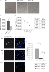
Infection of nonpermissive CHO-K1 cells with MVs obtained from HOG cells infected with HSV-1 K26-GFP. To evaluate the neutralization capacity of the anti-HSV-1 antibody, HOG cells were infected with K26-GFP incubated with or without an anti-HSV-1 polyclonal antibody. (A and B) The figure shows the lack of a cytopathic effect (A) and a significant decrease in viral production (B) in cells infected with antibody-incubated virus. (C) To quantify the neutralization capacity of the anti-HSV-1 antibody, we performed a titration assay as described in Materials and Methods. The figure shows that 100 μg of antibody can neutralize 1 × 10E8 TCID50. HOG cells were infected with K26-GFP. MVs from these HOG-infected cells, obtained as described above, were incubated with or without anti-HSV-1 antibody and subsequently added to receptor-deficient CHO-K1 cells, regarded as nonpermissive for HSV-1 infection, and incubated again for 1 h at 37°C. After adsorption, the cells were left for 24 h with DM. (D) Immunofluorescence images show the presence of infected CHO cells, suggesting that MVs from HOG-infected cells had transferred infectious virus to the nonpermissive CHO-K1 cell line. Infection of cells with antibody-incubated virus (MVs + Ab) showed a marked decrease, but despite that, infected CHO cells could be observed. A control for CHO cells infected directly with K26-GFP did not show a significant number of infected cells (Control). (E) Titration of viral production of CHO cells infected as described in the legend to panel D showed the same phenomenon: the susceptibility of CHO cells to MV-mediated infection (MVs), the existence of a significant viral production even in the presence of a neutralizing antibody (MVs + Ab), and the lack of significant viral production in CHO cells infected with the virus (Control). *, P < 0.05.
FIG 6
FIG 6
Analysis of MVs secreted by HeLa cells. MVs from HeLa cells infected with HSV-1 at an MOI of 1 for 24 h were isolated by differential centrifugation from the supernatants of HeLa cell cultures as previously described. (A to D) MVs were adsorbed onto collodion-carbon coated-copper grids and negatively stained and embedded with methylcellulose-uranyl acetate as described in Materials and Methods. (E and F) HeLa cells were incubated at 4°C for 1 h with MVs isolated from HSV-1-infected HeLa cell cultures and then for 15 min at 37°C. After that, the cells were fixed and processed for electron microscopy. Images show the presence of virions (solid arrows), MVs (dashed arrows), and virions enclosed in MVs (arrowheads). Bars = 200 nm. (G) Immunoblots show the presence of integrin β-1, flotillin-1, LC3-II, CD81, and CD63 in MVs isolated from mock-infected (−) and infected (+) cells. The bands corresponding to the anti-HSV-1 antibody are also shown.
FIG 7
FIG 7
Analysis of MVs secreted by MeWo cells. MVs from MeWo cells infected with HSV-1 at an MOI of 1 for 24 h were isolated by differential centrifugation from the supernatants of MeWo cell cultures as previously described. (A to D) MVs were adsorbed onto collodion-carbon-coated copper grids and negatively stained and embedded with methylcellulose-uranyl acetate as described in Materials and Methods. Images show the presence of virions (solid arrows), MVs (dashed arrows), and virions enclosed in MVs (arrowheads). Bars = 200 nm. (E) Immunoblots show the presence of integrin β-1, flotillin-1, LC3-II, CD81, and CD63 in MVs isolated from mock-infected (−) and infected (+) cells. The bands corresponding to the anti-HSV-1 antibody are also shown. In this cell line, two nonspecific bands can be observed in the mock-infected cell lysate lane.
FIG 8
FIG 8
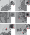
Electron microscopy of MeWo cells incubated with MVs. MVs from MeWo cells infected with HSV-1, isolated as described in the text, were layered onto MeWo cells and incubated at 4°C for 1 h. After that, they were incubated for 15 min at 37°C, fixed, and processed for electron microscopy. (A and B) Images show the presence of numerous MVs and virions (solid arrows). (B and C) MVs containing melanosomes (dashed arrows) were also found. (D to F) MVs containing virions (red dashed arrow) were occasionally observed. These virions contained a second envelope (red arrowhead), in addition to its viral envelope (e). Red solid arrows point to viral structural components: e, envelope; cp, capsid. Occasionally, in addition to virions, melanosomes were found in the same MV (F). Bars = 200 nm.

References

    1. Wald A, Corey L. 2007. Persistence in the population: epidemiology, transmission. In Arvin A, Campadelli-Fiume G, Mocarski E, Moore PS, Roizman B, Whitley R, Yamanishi K (ed), Human herpesviruses: biology, therapy, and immunoprophylaxis. Cambridge University Press, Cambridge, United Kingdom. - PubMed
    1. Roizman B, Zhou G, Du T. 2011. Checkpoints in productive and latent infections with herpes simplex virus 1: conceptualization of the issues. J Neurovirol 17:512–517. doi:10.1007/s13365-011-0058-x. - DOI - PubMed
    1. Whitley RJ. 2006. Herpes simplex encephalitis: adolescents and adults. Antiviral Res 71:141–148. doi:10.1016/j.antiviral.2006.04.002. - DOI - PubMed
    1. Bernstein DI, Bellamy AR, Hook EW III, Levin MJ, Wald A, Ewell MG, Wolff PA, Deal CD, Heineman TC, Dubin G, Belshe RB. 2013. Epidemiology, clinical presentation, and antibody response to primary infection with herpes simplex virus type 1 and type 2 in young women. Clin Infect Dis 56:344–351. doi:10.1093/cid/cis891. - DOI - PMC - PubMed
    1. Horowitz R, Aierstuck S, Williams EA, Melby B. 2010. Herpes simplex virus infection in a university health population: clinical manifestations, epidemiology, and implications. J Am College Health 59:69–74. doi:10.1080/07448481.2010.483711. - DOI - PubMed

Publication types

MeSH terms

Substances