. 2018 Apr 27;92(10):e02062-17.
doi: 10.1128/JVI.02062-17. Print 2018 May 15.

Reovirus-Induced Apoptosis in the Intestine Limits Establishment of Enteric Infection

Affiliations

Reovirus-Induced Apoptosis in the Intestine Limits Establishment of Enteric Infection

Judy J Brown et al. J Virol. .

Abstract

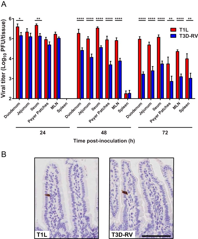
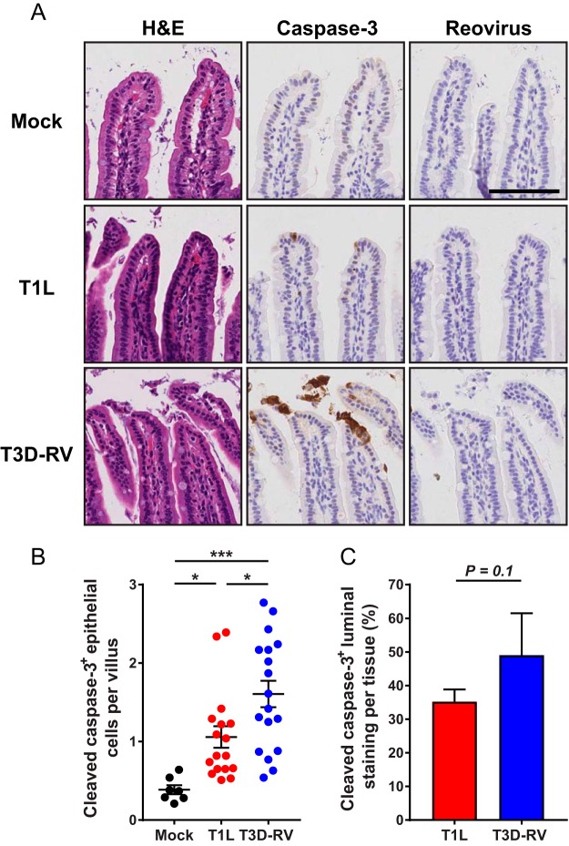
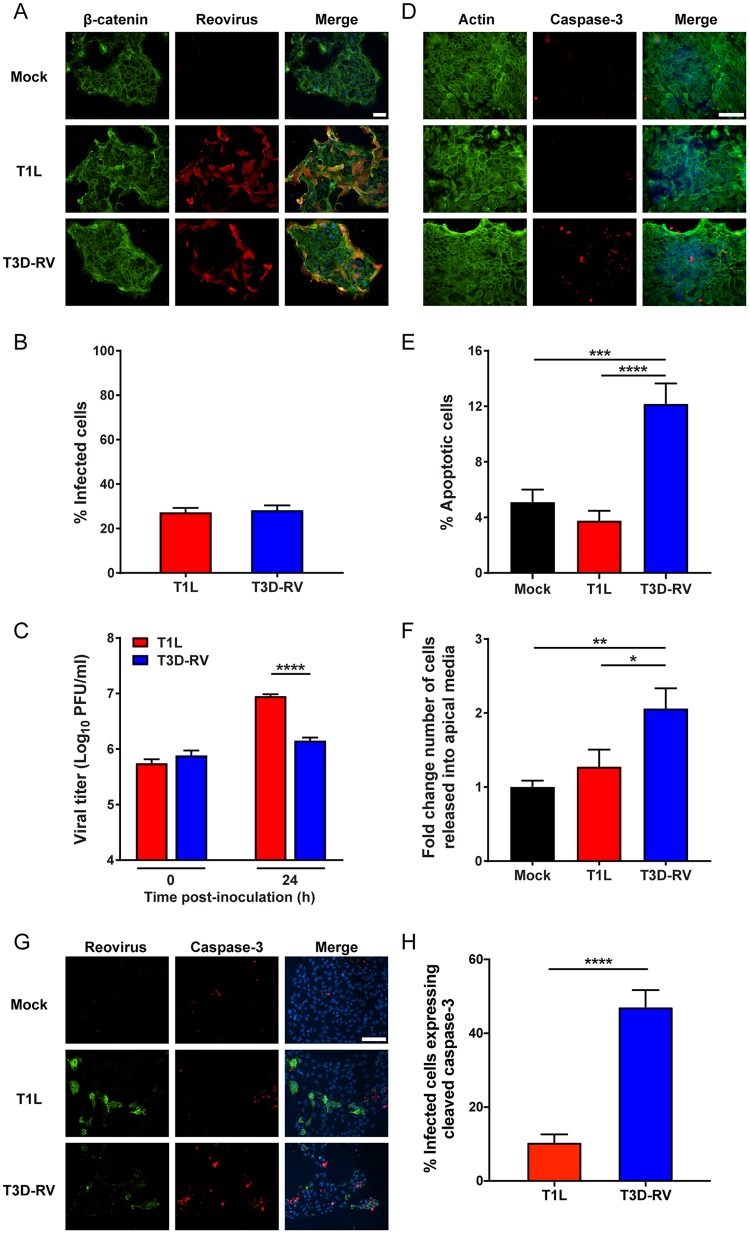
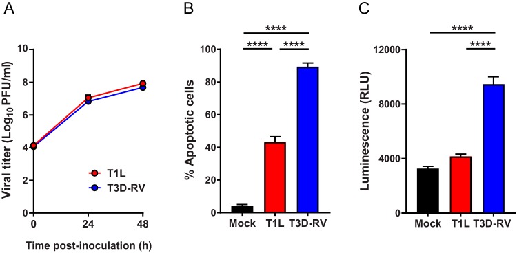
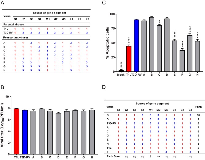
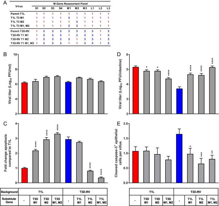
Several viruses induce intestinal epithelial cell death during enteric infection. However, it is unclear whether proapoptotic capacity promotes or inhibits replication in this tissue. We infected mice with two reovirus strains that infect the intestine but differ in the capacity to alter immunological tolerance to new food antigen. Infection with reovirus strain T1L, which induces an inflammatory immune response to fed antigen, is prolonged in the intestine, whereas T3D-RV, which does not induce this response, is rapidly cleared from the intestine. Compared with T1L, T3D-RV infection triggered apoptosis of intestinal epithelial cells and subsequent sloughing of dead cells into the intestinal lumen. We conclude that the infection advantage of T1L derives from its capacity to subvert host restriction by epithelial cell apoptosis, providing a possible mechanism by which T1L enhances inflammatory signals during antigen feeding. Using a panel of T1L × T3D-RV reassortant viruses, we identified the viral M1 and M2 gene segments as determinants of reovirus-induced apoptosis in the intestine. Expression of the T1L M1 and M2 genes in a T3D-RV background was sufficient to limit epithelial cell apoptosis and enhance viral infection to levels displayed by T1L. These findings define additional reovirus gene segments required for enteric infection of mice and illuminate the antiviral effect of intestinal epithelial cell apoptosis in limiting enteric viral infection. Viral strain-specific differences in the capacity to infect the intestine may be useful in identifying viruses capable of ameliorating tolerance to fed antigen in autoimmune conditions like celiac disease.IMPORTANCE Acute viral infections are thought to be cleared by the host with few lasting consequences. However, there may be much broader and long-lasting effects of viruses on immune homeostasis. Infection with reovirus, a common, nonpathogenic virus, triggers inflammation against innocuous food antigens, implicating this virus in the development of celiac disease, an autoimmune intestinal disorder triggered by exposure to dietary gluten. Using two reovirus strains that differ in the capacity to abrogate oral tolerance, we found that strain-specific differences in the capacity to replicate in the intestine inversely correlate with the capacity to induce apoptotic death of intestinal epithelial cells, providing a host-mediated process to restrict intestinal infection. This work contributes new knowledge about virus-host interactions in the intestine and establishes a foundation for future studies to define mechanisms by which viruses break oral tolerance in celiac disease.

Keywords: apoptosis; cell death; enteroid; gastrointestinal infection; mucosal immunity; reovirus.


Figures

FIG 1
FIG 1
Viral infection in murine tissues following inoculation with reoviruses T1L and T3D-RV. Mice were inoculated perorally with 1010 PFU of T1L or T3D-RV (n = 7 to 10 mice per virus strain). (A) Titers of T1L and T3D-RV in different regions of the intestine and secondary lymphoid organs were determined at the times shown by plaque assay. The small intestine was sectioned into thirds, approximating the duodenum, jejunum, and ileum. Viral titers are expressed as PFU per tissue. The 24-hpi titer values were previously published in reference ; data are used with permission of the publisher. (B) One day after inoculation, intestines were resected, and the distal half was flushed, Swiss rolled, and processed for histology. Sections were stained with a polyclonal antiserum specific for reovirus. Representative sections of jejunum are shown (scale bar, 100 μm). Error bars indicate SEMs. *, P < 0.05; **, P < 0.01; ****, P < 0.0001; one-way ANOVA and Sidak's multiple-comparison test.
FIG 2
FIG 2
Cleaved caspase-3 in the intestines of mice following infection with reovirus T1L or T3D-RV. Mice were inoculated perorally with 108 PFU of T1L or T3D-RV or PBS (mock). One day after inoculation, intestines were resected. The distal half was flushed, Swiss rolled, and processed for histology. (A) Sections were stained with H&E, reovirus polyclonal antiserum, or antibody against cleaved caspase-3. Representative sections of jejunum are shown (scale bar, 100 μm). (B) Cells positive for cleaved caspase-3 were enumerated manually and normalized per villus. Each symbol represents an individual mouse (n = 5 to 18 mice per group). (C) Cleaved-caspase-3 staining in the lumen was quantified by outlining the luminal region using the Digital Histology Shared Resource tool (n = 3 mice per virus). The percent luminal staining was determined as follows: (area in the lumen positive for cleaved-caspase-3 staining/area in the whole tissue positive for cleaved-caspase-3 staining) × 100. (B) Error bars indicate SEMs. (C) Error bars indicate SDs. *, P < 0.05; ***, P < 0.001. P values were determined by one-way ANOVA and Tukey's multiple-comparison test (B) and Mann-Whitney test (C).
FIG 3
FIG 3
Viral infectivity and apoptosis in murine-derived enteroids following reovirus T1L and T3D-RV infection. Intestinal crypts were harvested from mice and established as enteroids in Matrigel. The enteroids were dissociated, seeded onto Transwell plates, and incubated at 37°C for 4 days. Enteroids were adsorbed with T1L or T3D-RV at an MOI of 100 PFU/cell or with PBS as a mock control. At 24 hpi, cells were fixed and stained with antibodies specific for β-catenin and reovirus polyclonal antiserum (A and B) or actin and cleaved caspase-3 (D and E). Nuclei were labeled with ProLong Gold antifade mountant containing DAPI (scale bar, 50 μm). (B) The percentage of infected cells was determined by enumeration of reovirus-positive cells from immunofluorescence images. (C) Viral titers were determined at the intervals shown by plaque assay and expressed as PFU per milliliter of cell homogenate. (E) The percentage of apoptotic cells was determined using the Nikon Elements Basic Research analysis software and represents the number of cleaved caspase-3-positive cells per total number of cells in the selected field. (F) The number of cells released into the apical medium was quantified using an automated cell counter. (G and H) At 24 hpi, cells were fixed and stained with an antibody specific for cleaved caspase-3 and a polyclonal antiserum specific for reovirus nonstructural protein μNS. Nuclei were labeled with ProLong Gold antifade mountant containing DAPI (scale bar, 50 μm). (H) The percentage of infected cells expressing cleaved caspase-3 was determined by dividing the number of costained reovirus-positive and cleaved-caspase-3-positive cells by the number of reovirus-positive cells and then multiplying the quotient by 100. Data represent results from two to four independent experiments performed in triplicate. Error bars indicate SEMs. *, P < 0.05; **, P < 0.01; ***, P < 0.001; ****, P < 0.0001. P values were determined by Mann-Whitney test (C and H) and one-way ANOVA and Tukey's multiple-comparison test (E and F).
FIG 4
FIG 4
Viral titers and apoptosis in L cells following reovirus T1L and T3D-RV infection. (A) L cells were adsorbed with T1L or T3D-RV at an MOI of 1 PFU/cell, and viral titers were determined at the intervals shown by plaque assay. Viral titers are expressed as PFU per milliliter of cell homogenate. (B and C) L cells were adsorbed with T1L or T3D-RV at an MOI of 100 PFU/cell. (B) Cells were evaluated by AO assay at 38 hpi. The results are expressed as percentage of apoptotic cells per field of view. (C) Cell lysates were subjected to a Caspase-Glo 3/7 assay at 24 hpi. Caspase-3 activity is expressed in relative luminescence units. Data represent results from three independent experiments performed in triplicate. Error bars indicate SEMs. ****, P < 0.0001; one-way ANOVA and Tukey's multiple-comparison test.
FIG 5
FIG 5
Viral replication and apoptosis in L cells following infection with reovirus reassortant viruses. (A) T1L × T3D-RV reassortant viruses used to identify genes that segregate with strain-specific differences in the capacity of reovirus to induce apoptosis in the intestine. Gene segments labeled “1” in red represent those derived from T1L; gene segments labeled “3” in blue represent those derived from T3D. (B) L cells were adsorbed with T1L, T3D-RV, or one of eight T1L × T3D-RV reassortants at an MOI of 1 PFU/cell, and viral titers were determined at 48 hpi by plaque assay. Viral titers are expressed as PFU per milliliter of cell homogenate. (C) L cells were adsorbed with T1L, T3D-RV, or one of eight T1L × T3D-RV reassortants at an MOI of 100 PFU/cell. PBS-inoculated samples (mock) were used as controls. Cells were evaluated by AO assay at 38 hpi. The results are expressed as the percentage of apoptotic cells per field of view. (D) Rank sum analysis of reassortants used to identify genes that segregate with the capacity to induce apoptosis. The rank number of each reassortant and the significance levels of each gene segment are shown. Data represent results from two or three experiments performed in triplicate. Error bars indicate SEMs. ns, nonsignificant. #, P = 0.06; **, P < 0.01; ****, P < 0.0001. P values were determined by one-way ANOVA and Dunnett's multiple-comparison test (compared with T3D-RV) (C) and Wilcoxon rank sum distribution test (D).
FIG 6
FIG 6
Viral replication and apoptosis in cultured cells and murine intestine following infection with M gene reassortant viruses. (A) T1L × T3D-RV M gene reassortant viruses used to define genes that segregate with the apoptosis-inducing capacity of reovirus. Gene segments labeled “1” in red represent those derived from T1L; gene segments labeled “3” in blue represent those derived from T3D. (B) L cells were adsorbed with T1L, T3D-RV, or one of six T1L × T3D-RV M gene reassortants at an MOI of 1 PFU/cell, and viral titers were determined at 24 hpi by plaque assay. Viral titers are expressed as PFU per milliliter of cell homogenate. (C) L cells were adsorbed with T1L, T3D-RV, or one of six T1L × T3D-RV M gene reassortants at an MOI of 100 PFU/cell. Cells were evaluated by AO assay at 38 hpi. The results are expressed as fold change in apoptosis compared with the value for T1L. Data from two experiments performed in triplicate are presented. (D and E) Mice were inoculated perorally with 108 PFU of T1L, T3D-RV, or one of six T1L × T3D-RV M gene reassortants. The proximal half of the intestine was processed for viral titer, and the distal half was flushed, Swiss rolled, and processed for histology. (D) Titers of reovirus in the intestine were determined at 72 hpi by plaque assay (n ≥ 10 mice per virus). (E) At 24 hpi, histological sections were stained with an antibody against cleaved caspase-3. Cells positive for cleaved caspase-3 were enumerated manually and normalized per villus (n ≥ 8 mice per virus). Error bars indicate SEMs. *, P < 0.05; **, P < 0.01; ***, P < 0.001; ****, P < 0.0001. P values in panels C to E were determined by one-way ANOVA and Dunnett's multiple-comparison test (compared with the parental strain).

Similar articles

Cited by

References

    1. Cuconati A, Degenhardt K, Sundararajan R, Anschel A, White E. 2002. Bak and Bax function to limit adenovirus replication through apoptosis induction. J Virol 76:4547–4558. doi:10.1128/JVI.76.9.4547-4558.2002. - DOI - PMC - PubMed
    1. Handke W, Luig C, Popovic B, Krmpotic A, Jonjic S, Brune W. 2013. Viral inhibition of BAK promotes murine cytomegalovirus dissemination to salivary glands. J Virol 87:3592–3596. doi:10.1128/JVI.02657-12. - DOI - PMC - PubMed
    1. Bouziat R, Hinterleitner R, Brown JJ, Stencel-Baerenwald JE, Ikizler M, Mayassi T, Meisel M, Kim SM, Discepolo V, Pruijssers AJ, Ernest JD, Iskarpatyoti JA, Costes LM, Lawrence I, Palanski BA, Varma M, Zurenski MA, Khomandiak S, McAllister N, Aravamudhan P, Boehme KW, Hu F, Samsom JN, Reinecker HC, Kupfer SS, Guandalini S, Semrad CE, Abadie V, Khosla C, Barreiro LB, Xavier RJ, Ng A, Dermody TS, Jabri B. 2017. Reovirus infection triggers inflammatory responses to dietary antigens and development of celiac disease. Science 356:44–50. doi:10.1126/science.aah5298. - DOI - PMC - PubMed
    1. Hengartner MO. 2000. The biochemistry of apoptosis. Nature 407:770–776. doi:10.1038/35037710. - DOI - PubMed
    1. Elmore S. 2007. Apoptosis: a review of programmed cell death. Toxicol Pathol 35:495–516. doi:10.1080/01926230701320337. - DOI - PMC - PubMed

Publication types

Substances

LinkOut - more resources