. 2018 May 14;92(11):e00100-18.
doi: 10.1128/JVI.00100-18. Print 2018 Jun 1.

Porcine MKRN1 Modulates the Replication and Pathogenesis of Porcine Circovirus Type 2 by Inducing Capsid Protein Ubiquitination and Degradation

Affiliations

Porcine MKRN1 Modulates the Replication and Pathogenesis of Porcine Circovirus Type 2 by Inducing Capsid Protein Ubiquitination and Degradation

Tongtong Wang et al. J Virol. .

Erratum in

Abstract

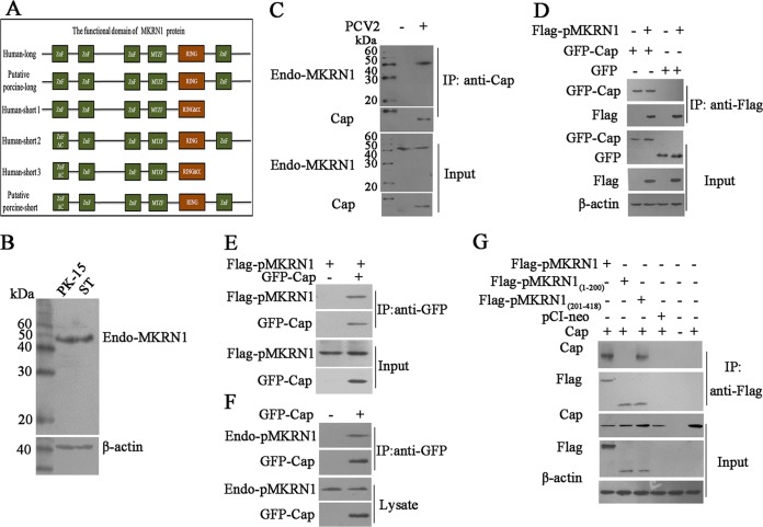
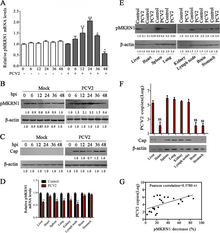
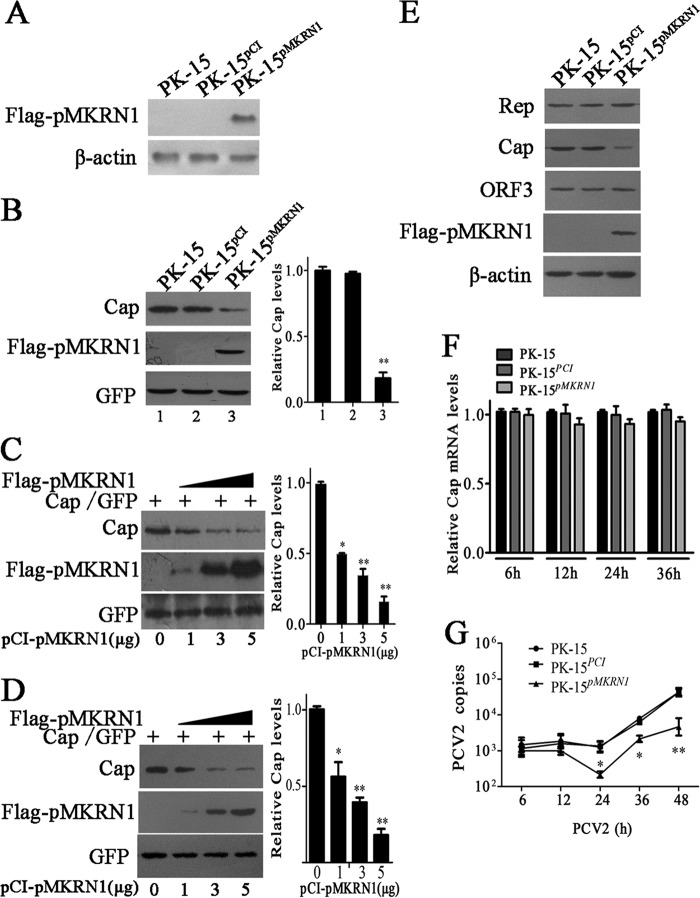
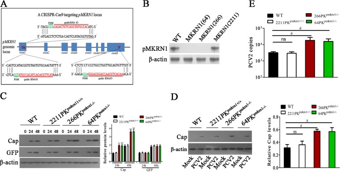
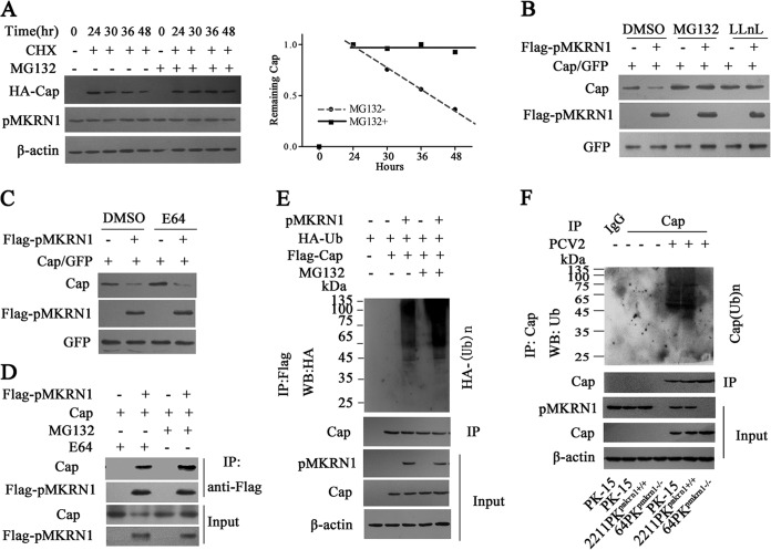
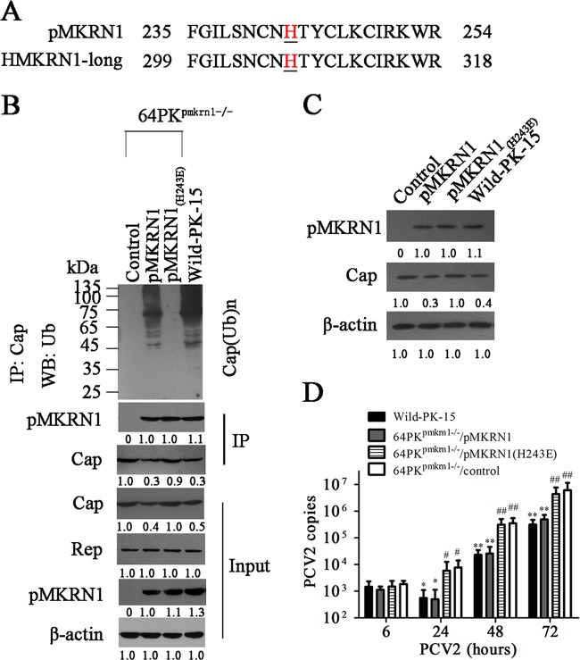
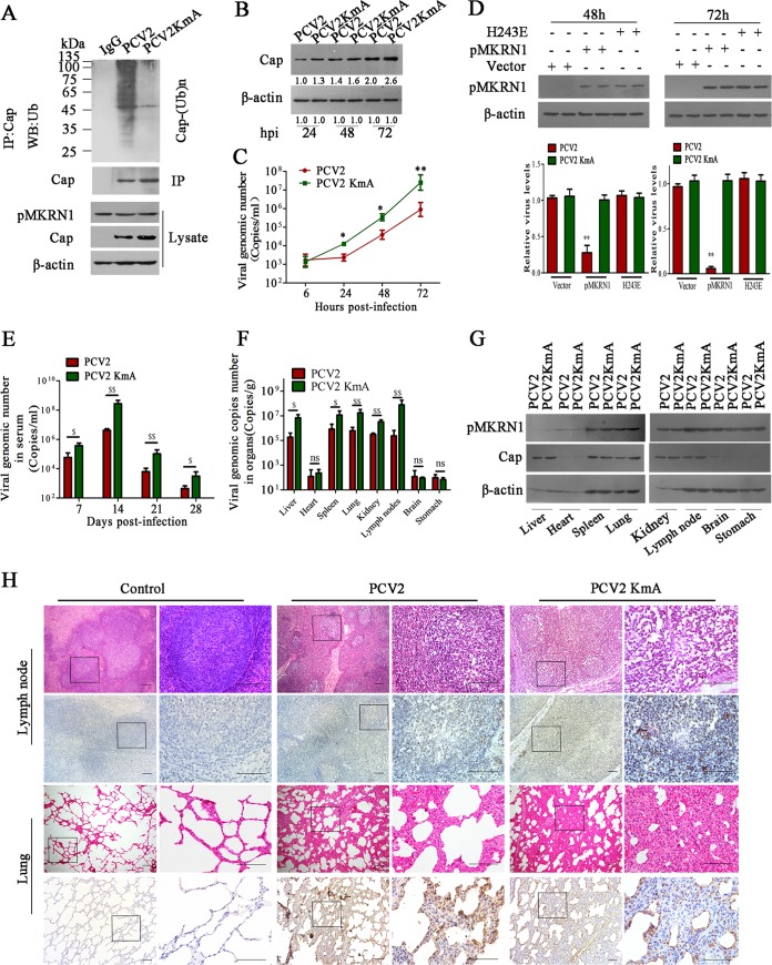
Porcine circovirus type 2 (PCV2) capsid protein (Cap) is a unique structure protein that plays pivotal roles in the process of viral replication and pathogenesis. Herein, we characterized a putative porcine Makorin RING finger protein 1 (pMKRN1) variant, an N-terminal-truncated variant of putative full-size porcine MKRN1 which has a unique expression pattern resulting from the porcine mkrn1 gene and which interacts with PCV2 Cap. A domain mapping assay showed that the C terminus of pMKRN1 and fragments (amino acids 108 to 198) of Cap are required for this interaction. PCV2 transiently upregulated pMKRN1 in PK-15 cells, but persistent viral infection downregulated pMKRN1 in major pathological tissues of PCV2-infected piglets. Overexpression of pMKRN1 significantly inhibited the generation of progeny PCV2 via ubiquitination and degradation of Cap, whereas knockout of pMKRN1 blocked Cap degradation and promoted progeny virus replication. pMKRN1 specifically targeted PCV2 Cap lysine residues 164, 179, and 191 to induce polyubiquitination and subsequent degradation. Mutation of either of the three lysine residues in the Cap protein or mutation of the histidine at residue 243 within the RING finger domain of pMKRN1 abrogated the E3 ligase activity of pMKRN1, rendering cells incapable of inducing Cap ubiquitination and degradation. Consistent with this finding, a Cap ubiquitination-deficient PCV2 strain showed enhanced virus replication and produced severe histological lesions in the lung and lymph node tissues compared with wild-type PCV2. Taken together, the results presented here suggest that PCV2 downregulates the pMKRN1 variant to avoid pMKRN1-mediated Cap ubiquitination and degradation, thus promoting viral replication and pathogenesis in its targeted tissues.IMPORTANCE Porcine circovirus type 2 is the pathogen to which pigs are the most susceptible, causing immense economic losses in the global swine industry, but whether host cells have developed some strategies to prevent viral replication is still unclear. Here, we found that porcine MKRN1 (pMKRN1) was upregulated in the early stage of PCV2 infection and mediated the polyubiquitination and degradation of Cap protein to block PCV2 replication, yet persistent PCV2 infection downregulated pMKRN1 levels to avoid degradation, promoting viral replication and pathogenesis in its targeted tissues. These data present new insight into the molecular mechanisms underlying the antiviral effects of pMKRN1 E3 ligase during PCV2 infection and also suggest potential new control measures for PCV2 outbreaks.

Keywords: PCV2; pMKRN1; replication; ubiquitination.


Figures

FIG 1
FIG 1
Porcine MKRN1 interaction with PCV2 Cap. (A) Schematic comparison of the functional domains of MKRN1-long (Human-long; GenBank accession no. NP_038474), putative porcine full-size MKRN1 protein (Porcine-long; GenBank accession no. XM_003134608.4), the human MKRN1 variants human-MKRN1-short 1 (Human-short1; GenBank accession no. NP_001138597.1), human MKRN1-short 2 (Human-short2; GenBank accession no. XP_011514299.1, XP_011514300.1), and human MKRN1-short 3 (Human-short3; GenBank accession no. NP_001278592.1), and the putative porcine MKRN1 variant pMKRN1-short (Putative porcine-short; UniProtKB accession no. A0A287ALB7). (B) Molecular expression patterns of MKRN1 in PK-15 cells and ST cells. PK-15 cells and ST cells were lysed by RIPA, and the lysates were subjected to Western blotting with MKRN1 antibodies. (C) The putative porcine MKRN1 variant (pMKRN1-short) interacts with PCV2 Cap protein. PK-15 cells were infected with PCV2 (MOI = 1) for 24 h, and lysates were immunoprecipitated (IP) using an anti-Cap antibody; an anti-MKRN1 antibody was used for immunoblotting. (D, E) Cap interacts with pMKRN1 in transfected cells. HEK293T cells were cotransfected with GFP-Cap and Flag-pMKRN1 expression plasmids or cotransfected with the pEGFP control vector and the Flag-pMKRN1 expression plasmid. The cells transfected with the pEGFP control vector, the GFP-Cap expression plasmid, or the Flag-pMKRN1 expression plasmid alone served as controls. Immunoprecipitation was performed to detect the GFP-Cap interaction with Flag-pMKRN1 using anti-Flag antibodies or anti-GFP antibodies. (F) Endogenous MKRN1 interacts with GFP-Cap. PK-15 cells were transfected with GFP-Cap, cells were lysed for immunoprecipitation with GFP monoclonal antibodies, and anti-MKRN1 antibodies were used for immunoblotting. The cells untransfected with GFP-Cap served as a control. (G) The C terminus (aa 201 to 418) of pMKRN1 is responsible for interacting with Cap. HEK293T cells were transfected with plasmids to express the indicated proteins, an immunoprecipitation assay was performed using anti-Flag monoclonal antibodies, and anti-Cap antibodies were used for immunoblotting. These results were confirmed by three independent experiments.
FIG 2
FIG 2
The expression of pMKRN1 is regulated by PCV2 and inversely associated with the Cap level in PCV2-infected cells and tissues. (A to C) PK-15 cells were infected with PCV2 at an MOI of 1 or mock infected for the indicated times, and the mRNA levels of pMKRN1 (A), the protein levels of pMKRN1 (B), and the levels of Cap (C) were measured. The results are the means ± SEMs from 3 independent experiments. *, P < 0.05 versus mock-infected cells at 0 h; **, P < 0.01 versus mock-infected cells at 0 h. (D to F) Piglets were infected with PCV2 or mock infected for 14 or 28 days. Different tissues were used for detecting pMKRN1 mRNA levels in PCV2-infected or control piglets at 14 days postinfection (D), the pMKRN1 levels (E) and Cap levels (F) were compared at 28 days postinfection by immunoblotting, and quantitative PCR was used to analyze PCV2 copies number (F). The data shown are representatives from three independent experiments. *, P < 0.05 versus the same mock-infected tissues; **, P < 0.01 versus the same mock-infected tissues; $$, P < 0.01 versus other tissues of the PCV2-infected group. (G) The levels of the pMKRN1 protein and the number of PCV2 copies (log) in different tissues of PCV2-infected piglets were measured, the decreased percentages of pMKRN1 in each detected tissue (100% minus the percentage of pMKRN1 in the PCV2-infected group relative to the amount in the mock-infected group [quantity for the PCV2-infected group/quantity for the mock-infected group]) were calculated, and then their associations were evaluated using Pearson's coefficient of correlation analysis, as shown by positive trend lines. The numbers immediately beneath the lanes in panels B, C, and E represent the quantity of the indicated bands normalized to the content in the control lane in each blot (lane 1). ##, P < 0.01, which demonstrates that the reduction of pMKRN1 has a significant positive association with PCV2 copy numbers.
FIG 3
FIG 3
Overexpression of pMKRN1 decreases ORF2 protein levels and inhibits the replication of PCV2. (A) A stable cell line, PK-15pMKRN1, expresses the pMKRN1 protein. PK-15 cells were transfected with linearized pCI-Flag-pMKRN1 or the control vector, pCI-neo, to establish PK-15pMKRN1 and PK-15PCI cells selected by G418, respectively. The cells were analyzed by immunoblotting using an anti-Flag antibody. β-Actin was detected as an internal control. (B) The level of Cap was decreased in cells overexpressing pMKRN1. (Left) The pcDNA-Cap and pEGFP vectors were cotransfected into PK-15, PK-15PCI, or PK-15pMKRN1 cells, and Cap, pMKRN1, and GFP levels were detected by immunoblotting. (Right) The ImageJ program was used to quantify the relative intensities of the bands. **, P < 0.01 versus the Cap level of PK-15 cells. (C and D) Increased pMKRN1 expression is correlated with decreased Cap. (Left) Different doses of pCI-Flag-pMKRN1 plasmids (0, 1, 3, 5 μg) were transfected with a fixed amount of Cap expression vectors into PK-15 cells (C) or ST cells (D), and Western blotting was used to examine Cap expression. GFP was used as a control. (Right) Relative intensities of the bands. *, P < 0.05 versus Cap levels in PK-15 cells or ST cells with 0 μg of pCI-Flag-pMKRN1 transfection; **, P < 0.01 versus Cap levels in PK-15 cells or ST cells with 0 μg of pCI-Flag-pMKRN1 transfection. (E to G) PK-15, PK-15PCI, and PK-15pMKRN1 cells were infected with PCV2 at an MOI of 1 for the indicated periods, and PCV2 Rep, Cap, and ORF3 protein levels were determined by Western blotting (E); the mRNA levels of Cap (F) and PCV2 copy numbers (G) were determined by quantitative PCR. The Cap mRNA levels of PK-15 cells at each time point were defined as 1 in panel F. The data shown are representatives from three independent experiments. *, P < 0.05 versus PCV2-infected PK-15 cells at the same time points; **, P < 0.01 versus PCV2-infected PK-15 cells at the same time points.
FIG 4
FIG 4
Knockout of pMKRN1 by CRISPR-Cas9 increases Cap protein and promotes progeny virus replication. (A) Schematic diagram of gRNA targeting sites at the pMKRN1 genomic region. The gRNA-targeting sites (sites 1, 2, and 3) located on exon 1, exon 1, and exon 8 are shown. Red arrows indicate gRNA-targeting sites, the gRNA-targeting sequences are in red, and the protospacer adjacent motif (PAM) sequences are shown in green. (B) Examination of pMKRN1 expression in PK-15 cells with a CRISPR-Cas9 system targeting the pMKRN1 locus. Three single-cell clones [MKRN1(64), MKRN1(266), MKRN1(2211)] were derived from cells infected with lentiviral pseudotypes expressing gRNAs 1, 2, and 3, respectively. (C) Knockout of pMKRN1 increases Cap expression. Wild-type PK-15, 2211PKpmkrn1+/+, 266PKpmkrn1−/−, and 64PKpmkrn1−/− were transfected with Cap expression vectors or pEGFP control vectors, and Cap and GFP expression levels were detected by Western blotting at the indicated times (in hours). **, P < 0.01 versus the indicated protein levels in wild-type PK-15 cells at the same time. (D, E) Knockout of pMKRN1 promotes PCV2 progeny virion production. Wild-type PK-15, 2211PKpmkrn1+/+, 266PKpmkrn1−/−, and 64PKpmkrn1−/− cells were infected with PCV2 at an MOI of 1 for 24 h, and Western blotting was performed to detect PCV2 Cap (D) and quantitative PCR analysis was used to determine the number of PCV2 copies (E). #, P < 0.05; ns, not significant.
FIG 5
FIG 5
pMKRN1 induces Cap ubiquitination, resulting in its degradation via the proteasome pathway. (A to D) pMKRN1 promotes the degradation of Cap via the proteasome pathway. PK-15 cells were transfected with HA-Cap; at 24 h posttransfection, the cells were treated with CHX (100 μg/ml) for another 24 h; and the expression of HA-Cap was tested at the indicated times by immunoblotting (A). 64PKpmkrn1−/− cells were transfected with pCI-Flag-pMKRN1, pcDNA-Cap, and pEGFP, as indicated; the cells were treated with 10 μM MG132 (a proteasome inhibitor), 10 μM LLnL (an MG132 analogue) (B), or 10 μM E64 (a lysosome inhibitor) (C); and the expression of Flag-pMKRN1, Cap, or GFP was analyzed by Western blotting (WB). The lysates were immunoprecipitated with anti-Flag antibodies, and Western blotting was used to detect Cap (D). (E) pMKRN1 induces Cap ubiquitination. 64PKpmkrn1−/− cells were transfected with the indicated expression plasmids, followed by treatment of the cells with MG132 (10 μM) for 6 h. Cap proteins were immunoprecipitated using an anti-Flag antibody, and ubiquitinated proteins were immunoblotted using an anti-HA antibody. (F) The ubiquitination of Cap is lost in pMKRN1 knockout cells. Wild-type PK-15, 2211PKpmkrn1+/+, or 64PKpmkrn1−/− cells were infected with PCV2, cell lysates were immunoprecipitated using anti-Cap antibodies, and polyubiquitinated Cap [Cap(Ub)n] was detected by immunoblotting using anti-ubiquitin antibodies. These results were confirmed in three independent experiments.
FIG 6
FIG 6
The histidine 243 residue is critical for the E3 ligase activity of pMKRN1. (A) The histidine residue is conserved between pMKRN1 and human MKRN1-long. (B to D) Cap ubiquitination can be restored by wild-type pMKRN1 but not mutant pMKRN1(H243E) in pMKRN1 knockout cells. 64PKpmkrn1−/− cells were transfected with plasmids to express wild-type pMKRN1 (64PKpmkrn1−/−/pMKRN1) or mutated pMKRN1 [64PKpmkrn1−/−/pMKRN1(H243E)] or transfected with control plasmids (64PKpmkrn1−/−/control); the cells were then infected with PCV2 (MOI = 1), and ubiquitination of Cap (B), the expression of Cap and pMKRN1 (C), and the production of progeny PCV2 were measured (D). The data shown are representatives from three independent experiments. *, P < 0.05 versus PCV2-infected 64PKpmkrn1−/−/control cells; **, P < 0.01 versus PCV2-infected 64PKpmkrn1−/−/control cells; #, P < 0.05 versus PCV2-infected 64PKpmkrn1−/−/pMKRN1 cells; ##, P < 0.01 versus PCV2-infected 64PKpmkrn1−/−/pMKRN1 cells. The numbers under the gels represent quantification of indicated bands, normalized to the first visible band in each blot (C and D). DMSO, dimethyl sulfoxide.
FIG 7
FIG 7
PCV2 Cap lysine residues 164, 179, and 191 are the sites of ubiquitination by pMKRN1. (A) Schematic diagram of N- and C-terminal truncation mutants of PCV2 Cap. (B) Mapping the regions of PCV2 Cap that interact with pMKRN1. HEK293T cells were transfected with plasmids to express Flag-pMKRN1 and Cap truncation mutants fused with the GFP tag, and lysates were immunoprecipitated with an anti-GFP antibody, followed by immunoblotting using an anti-MKRN1 antibody, anti-GFP antibody, or anti-β-actin antibody. The middle white space divides the samples that were resolved by two SDS-PAGEs due to the limit of the gel lanes. (C) PCV2 Cap containing amino acid residues 162 to 198 is degraded by pMKRN1. HEK293T cells were transfected with plasmids to express Flag-pMKRN1 and the indicated Cap mutants, and Western blotting was used to detect the levels of Cap mutant expression with anti-HA antibodies. (D to F) K164, K179, and K191 were the ubiquitination sites of the PCV2 Cap protein. The Cap lysine residues at residues 164, 179, and 191 were replaced with alanine (D). HEK293T cells were transfected to express Flag-pMKRN1 and the indicated Cap lysine mutants, Western blotting was used to detect the expression levels of Cap or mutants using anti-Cap antibodies (E), and the ubiquitination of Cap mutants was detected (F). These results were confirmed in three independent experiments.
FIG 8
FIG 8
Mutation of ubiquitination sites of Cap protein enhances the replication and pathogenesis of PCV2. (A to D) Mutant PCV2 (PCV2KmA) containing the three lysine residues (164, 179, and 191) replaced by alanine shows increased Cap levels and viral reproduction due to the loss of ubiquitination. PK-15 cells were infected with PCV2 or PCV2KmA, and Cap ubiquitination was examined (A), Cap expression levels were determined (B), and viral copy numbers were measured by quantitative PCR (C). *, P < 0.05 versus PCV2-infected cells; **, P < 0.01 versus PCV2-infected cells. (D) 64PKpmkrn1−/− cells were transfected with wild-type pMKRN1, the pMKRN1(H243E) mutant, or the pCI-neo vector, and then the cells were infected by PCV2 or the PCV2KmA mutant for 48 h and 72 h. Relative PCV2 or PCV2KmA levels were measured by quantitative PCR. **, P < 0.01 versus pCI-neo vector-transfected cells. (E to G) Piglets were infected with PCV2 or PCV2KmA (5 × 10⁵ TCID50) by intranasal injection. The numbers of PCV2 copies in serum at the indicated times (E) and at 28 days postinfection of tissues (F) were measured by quantitative PCR. The pMKRN1 expression levels in tissues at 28 days postinfection were measured by Western blotting (G). $, P < 0.05; $$, P < 0.01; ns, not significant. Results were confirmed in three independent experiments. (H) Hematoxylin and eosin staining of lung and lymph node tissues and immunohistochemical staining with anti-ORF1 antibody of lung and lymph node tissues derived from PCV2- or PCV2KmA-infected piglets at 28 days postinfection. The streptavidin-biotin-peroxidase complex method with counterstaining with hematoxylin was used. The regions with black rectangles were amplified, and the images are shown on the right. Bars, 100 μm.

Similar articles

Cited by

References

    1. Opriessnig T, Meng XJ, Halbur PG. 2007. Porcine circovirus type 2 associated disease: update on current terminology, clinical manifestations, pathogenesis, diagnosis, and intervention strategies. J Vet Diagn Invest 19:591–615. doi: 10.1177/104063870701900601. - DOI - PubMed
    1. Thomson J, Smith B, Allan G, McNeilly F, McVicar C. 2000. PDNS, PMWS and porcine circovirus type 2 in Scotland. Porcine dermatitis and nephropathy syndrome. Post-weaning multisystemic wasting syndrome. Vet Rec 146:651–652. - PubMed
    1. Pineyro PE, Kenney SP, Gimenez-Lirola LG, Opriessnig T, Tian D, Heffron CL, Meng XJ. 2016. Evaluation of the use of non-pathogenic porcine circovirus type 1 as a vaccine delivery virus vector to express antigenic epitopes of porcine reproductive and respiratory syndrome virus. Virus Res 213:100–108. doi: 10.1016/j.virusres.2015.11.005. - DOI - PubMed
    1. Nawagitgul P, Morozov I, Bolin SR, Harms PA, Sorden SD, Paul PS. 2000. Open reading frame 2 of porcine circovirus type 2 encodes a major capsid protein. J Gen Virol 81:2281–2287. doi: 10.1099/0022-1317-81-9-2281. - DOI - PubMed
    1. Mankertz A, Mankertz J, Wolf K, Buhk HJ. 1998. Identification of a protein essential for replication of porcine circovirus. J Gen Virol 79(Pt 2):381–384. - PubMed

Publication types

MeSH terms