. 2018 May 14;92(11):e00196-18.
doi: 10.1128/JVI.00196-18. Print 2018 Jun 1.

HIV-1 Nef Antagonizes SERINC5 Restriction by Downregulation of SERINC5 via the Endosome/Lysosome System

Affiliations

HIV-1 Nef Antagonizes SERINC5 Restriction by Downregulation of SERINC5 via the Endosome/Lysosome System

Jing Shi et al. J Virol. .

Abstract

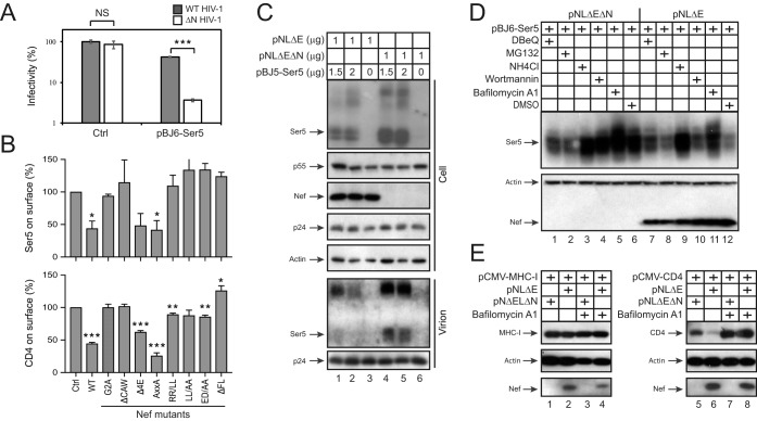
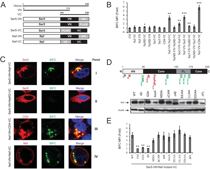
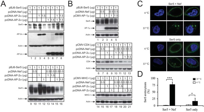
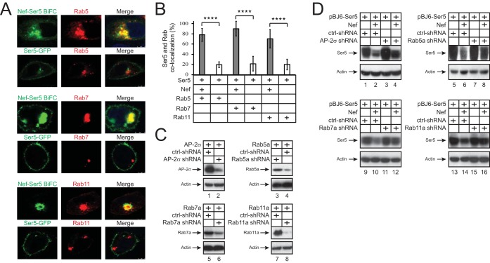
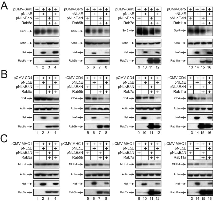
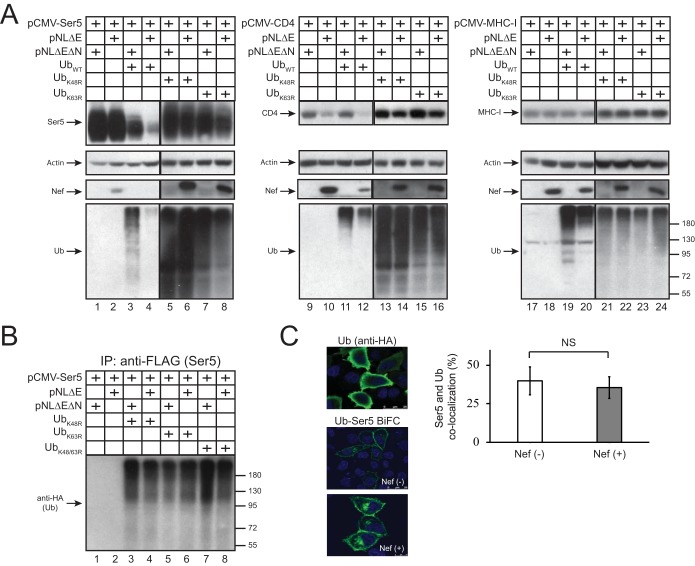
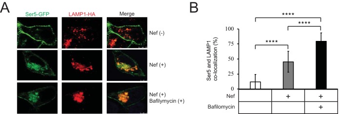
The primate lentiviral accessory protein Nef downregulates CD4 and major histocompatibility complex class I (MHC-I) from the cell surface via independent endosomal trafficking pathways to promote viral pathogenesis. In addition, Nef antagonizes a novel restriction factor, SERINC5 (Ser5), to increase viral infectivity. To explore the molecular mechanism of Ser5 antagonism by Nef, we determined how Nef affects Ser5 expression and intracellular trafficking in comparison to CD4 and MHC-I. We confirm that Nef excludes Ser5 from human immunodeficiency virus type 1 (HIV-1) virions by downregulating its cell surface expression via similar functional motifs required for CD4 downregulation. We find that Nef decreases both Ser5 and CD4 expression at steady-state levels, which are rescued by NH4Cl or bafilomycin A1 treatment. Nef binding to Ser5 was detected in living cells using a bimolecular fluorescence complementation assay, where Nef membrane association is required for interaction. In addition, Nef triggers rapid Ser5 internalization via receptor-mediated endocytosis and relocalizes Ser5 to Rab5+ early, Rab7+ late, and Rab11+ recycling endosomes. Manipulation of AP-2, Rab5, Rab7, and Rab11 expression levels affects the Nef-dependent Ser5 and CD4 downregulation. Moreover, although Nef does not promote Ser5 polyubiquitination, Ser5 downregulation relies on the ubiquitination pathway, and both K48- and K63-specific ubiquitin linkages are required for the downregulation. Finally, Nef promotes Ser5 colocalization with LAMP1, which is enhanced by bafilomycin A1 treatment, suggesting that Ser5 is targeted to lysosomes for destruction. We conclude that Nef uses a similar mechanism to downregulate Ser5 and CD4, which sorts Ser5 into a point-of-no-return degradative pathway to counteract its restriction.IMPORTANCE Human immunodeficiency virus (HIV) and simian immunodeficiency virus (SIV) express an accessory protein called Nef to promote viral pathogenesis. Nef drives immune escape in vivo through downregulation of CD4 and MHC-I from the host cell surface. Recently, Nef was reported to counteract a novel host restriction factor, Ser5, to increase viral infectivity. Nef downregulates cell surface Ser5, thus preventing its incorporation into virus particles, resulting in disruption of its antiviral activity. Here, we report mechanistic studies of Nef-mediated Ser5 downregulation in comparison to CD4 and MHC-I. We demonstrate that Nef binds directly to Ser5 in living cells and that Nef-Ser5 interaction requires Nef association with the plasma membrane. Subsequently, Nef internalizes Ser5 from the plasma membrane via receptor-mediated endocytosis, and targets ubiquitinated Ser5 to endosomes and lysosomes for destruction. Collectively, these results provide new insights into our ongoing understanding of the Nef-Ser5 arms race in HIV-1 infection.

Keywords: CD4; MHC-I; Nef; SERINC5; downregulation; endocytosis; restriction factor.


Figures

FIG 1
FIG 1
Nef decreases Ser5 expression at steady-state levels. (A) Wild-type (WT) or Nef-deficient (ΔN) HIV-1 pseudoviruses were produced from 293T cells in the presence of pBJ6-Ser5 or a control (Ctrl) vector, and viral infectivity was determined in TZM-bI cells. Error bars indicate standard deviations (SD) from three independent experiments. (B) pBJ5-iHA-Ser5 and pMSCV-CD4-FLAG were expressed in 293T cells in the presence of a WT or indicated Nef mutant expression vector, and the Ser5 and CD4 expression on cell surface were determined by flow cytometry. Error bars indicate the standard errors of the mean (SEM) from three independent experiments. (C) 293T cells were transfected with pNLΔE or pNLΔEΔN in the presence of increasing amounts of pBJ5-iHA-Ser5. Virions were purified from culture supernatants via ultracentrifugation. Protein expression in cells and virions was analyzed by Western blotting. (D) Ser5 was expressed in 293T cells in the presence or absence of Nef. Cells were treated with DBeQ (15 μM), MG132 (10 μM), NH4Cl (20 μM), wortmannin (100 nM), bafilomycin A1 (100 nM), or dimethyl sulfoxide for 24 h, and protein expression was analyzed by Western blotting. (E) MHC-I and CD4 were expressed in 293T cells in the presence or absence of Nef and treated with bafilomycin A1 (100 nM) for 24 h. Protein expression was analyzed by Western blotting.
FIG 2
FIG 2
Nef binds to Ser5. (A) Schematic of the BiFC fusion proteins is presented. Venus is divided into an N-terminal region from residues 2 to 173 (VN) and a C-terminal region from residues 154 to 238 (VC). VN and VC were fused to the C terminus of Ser5 or Nef, and each fusion protein also contains an HA, V5, or FLAG epitope. (B) pcDNA3.1-Nef-VN-HA, pcDNA3.1-Nef4D-VN-HA, pcDNA3.1-Ser5-VN-HA, pcDNA3.1-Nef-V5-VC, pcDNA3.1-Nef4D-V5-VC, pcDNA3.1-CD4-V5-VC, and pcDNA3.1-Ser5-FLAG-VC were expressed individually or pairwise in 293T cells as indicated. The mean fluorescence intensities (MFI) of BiFC fluorescent signals were determined by flow cytometry and are presented as relative values to the signal from untransfected cells. Error bars represent the SD from three independent experiments. (C) The vectors in panel B are expressed pairwise in HeLa cells as indicated. Ser5-VN-HA (panels I and II) and Nef-VN-HA (panel IV) fusion proteins were stained with a rabbit anti-HA monoclonal antibody at a dilution of 1:500, followed by an Alexa Fluor 647-conjuated donkey anti-rabbit antibody at a 1:1,000 dilution. CD4-V5-VC (panel III) fusion was stained with a mouse anti-V5 antibody at a 1:250 dilution, followed by an Alexa Fluor 647-conjugated goat anti-mouse antibody at a 1:1,000 dilution. Colocalization of the red and the BiFC green fluorescent signals were visualized by confocal microscopy. (D) Schematic of the HIV-1 NL4-3 Nef protein. The N-terminal flexible anchor domain (AN), core domains, internal flexible loop (FL), and critical residues and motifs for MHC-I (red) and CD4 (green) downregulation are shown. The indicated Nef mutations were created in the pcDNA3.1-Nef-V5-VC vector, and Nef expression was determined by Western blotting with an anti-V5 antibody. (E) pMSCV-Ser5-VN-HA was expressed with pcDNA3.1-Nef-V5-VC vectors expressing the indicated Nef mutants in 293T cells. The BiFC MFI was determined by flow cytometry and are presented as values relative to the signal from untransfected cells. Error bars represent the SD from three independent experiments.
FIG 3
FIG 3
Nef internalizes Ser5 via receptor-mediated endocytosis. (A) Ser5 was expressed with Nef and/or indicated AP-2 proteins in 293T cells, and protein expression was determined by Western blotting. (B) Ser5, CD4, or MHC-I was expressed with Nef and/or indicated AP-1 or AP-2 proteins in 293T cells, and the protein expression was determined by Western blotting. (C) pBJ5-iHA-Ser5 was expressed with pNLΔE or pNLΔEΔN in 293T cells. The Ser5 endocytosis was determined by an antibody uptake experiment as described in Materials and Methods. Cell nuclei were stained with DAPI. (D) The levels of Ser5 endocytosis were calculated as the detection frequency of cells where Ser5 was internalized from the plasma membrane. Error bars indicate the SD from three independent experiments.
FIG 4
FIG 4
Intracellular trafficking of the Nef-Ser5 complex. (A) pCMV-DsRed-HA-Rab5b, pCMV-DsRed-2xHA-Rab7a, or pCMV-DsRed-2xHA-Rab11a was expressed in HeLa cells with the pcDNA3.1-Ser5-VN-FLAG/pcDNA3.1-Nef-V5-VC pair or with pCMV-Ser5-eGFP alone. Ectopic Rab proteins were stained with a mouse anti-HA antibody, followed by Alexa Fluor 647-conjugated secondary antibodies. Colocalization of these fluorescent signals was visualized by confocal microscopy. (B) The colocalization of Ser5 with Rab small GTPases in the presence or absence of Nef in panel A was statistically analyzed. Error bars indicate the SD from three independent experiments. (C) The indicated AP-2 or Rab proteins were expressed with their specific short hairpin RNAs (shRNAs) or scrambled shRNAs as a control (ctrl). Protein expression was determined by Western blotting. (D) Ser5 was expressed with Nef in the presence of indicated shRNAs, and protein expression was determined by Western blotting.
FIG 5
FIG 5
Role of Rab GTPases in Nef-mediated Ser5 downregulation. Ser5 (A), CD4 (B), and MHC-I (C) were expressed with Rab5, Rab7, or Rab11 in the presence or absence of Nef in 293T cells, and protein expression was analyzed by Western blotting.
FIG 6
FIG 6
Role of ubiquitination in Nef-mediated Ser5 downregulation. (A) Ser5, CD4, and MHC-I were expressed with WT ubiquitin (UbWT) or its mutants (UbK48R and UbK63R) in the presence or absence of Nef in 293T cells, and protein expression was determined by Western blotting. (B) Ser5 was expressed with the indicated ubiquitin expression vector in the presence or absence of Nef expression in 293T cells. Proteins were immunoprecipitated (IP) by an anti-FLAG antibody that targets Ser5 and analyzed by an anti-HA antibody that targets ubiquitin via Western blotting. (C) pcDNA3.1-Ub-VN-HA was expressed alone or with pcDNA3.1-Ser5-FLAG-VC in HeLa cells in the presence of pNLΔE or pNLΔEΔN. The single Ub expression was detected with an anti-HA antibody, followed by Alexa Fluor 488-conjugated secondary antibodies. Nuclei were stained with DAPI. Fluorescent signals were visualized by confocal microscopy. The colocalization of Ser5 and ubiquitin in the presence or absence of Nef was statistically analyzed. Error bars indicate the SD from three independent experiments.
FIG 7
FIG 7
Role of lysosomes in Nef-mediated Ser5 downregulation. (A) pCMV-Ser5-eGFP and pCMV-DsRed-HA-LAMP1 were expressed in HeLa cells in the presence or absence of pcDNA3.1-Nef-V5-VC. Cells were further treated with bafilomycin A1. LAMP1 was stained with rabbit anti-LAMP1, followed by Alexa Fluor 647-conjugated secondary antibodies. Colocalization of these fluorescent signals was visualized by confocal microscopy. (B) The colocalization of Ser5 and LAMP1 in the presence of Nef expression and bafilomycin A1 treatment was statistically analyzed. Error bars indicate the SD from three independent experiments.
FIG 8
FIG 8
Nef downregulation of Ser5 in human T cells. (A) Jurkat-TAg cells were transfected with pLPCX-iHA-Ser5 and an HIV-1 proviral vector pH22 or its Nef-defective control pH22ΔN. In addition, cells were transfected with an eGFP expression vector. The cell surface Ser5 proteins were stained with an Alexa Fluor 647-conjugated anti-HA antibody (BioLegend). Ser5 and eGFP expression were analyzed by flow cytometry. (B) Jurkat-TAg-KO cells were transfected with the indicated amounts of expression vectors, and protein expression was analyzed by Western blotting.

References

    1. Deacon NJ, Tsykin A, Solomon A, Smith K, Ludford-Menting M, Hooker DJ, McPhee DA, Greenway AL, Ellett A, Chatfield C, Lawson VA, Crowe S, Maerz A, Sonza S, Learmont J, Sullivan JS, Cunningham A, Dwyer D, Dowton D, Mills J. 1995. Genomic structure of an attenuated quasispecies of HIV-1 from a blood transfusion donor and recipients. Science 270:988–991. doi: 10.1126/science.270.5238.988. - DOI - PubMed
    1. Kestler HW III, Ringler DJ, Mori K, Panicali DL, Sehgal PK, Daniel MD, Desrosiers RC. 1991. Importance of the nef gene for maintenance of high virus loads and for development of AIDS. Cell 65:651–662. doi: 10.1016/0092-8674(91)90097-I. - DOI - PubMed
    1. Kirchhoff F, Greenough TC, Brettler DB, Sullivan JL, Desrosiers RC. 1995. Brief report: absence of intact nef sequences in a long-term survivor with nonprogressive HIV-1 infection. N Engl J Med 332:228–232. doi: 10.1056/NEJM199501263320405. - DOI - PubMed
    1. Robert-Guroff M, Popovic M, Gartner S, Markham P, Gallo RC, Reitz MS. 1990. Structure and expression of tat-, rev-, and nef-specific transcripts of human immunodeficiency virus type 1 in infected lymphocytes and macrophages. J Virol 64:3391–3398. - PMC - PubMed
    1. Garcia JV, Miller AD. 1991. Serine phosphorylation-independent downregulation of cell surface CD4 by nef. Nature 350:508–511. doi: 10.1038/350508a0. - DOI - PubMed

Publication types

MeSH terms