. 2018 May 14;92(11):e01544-17.
doi: 10.1128/JVI.01544-17. Print 2018 Jun 1.

A Stem-Loop Structure in Potato Leafroll Virus Open Reading Frame 5 (ORF5) Is Essential for Readthrough Translation of the Coat Protein ORF Stop Codon 700 Bases Upstream

Affiliations

A Stem-Loop Structure in Potato Leafroll Virus Open Reading Frame 5 (ORF5) Is Essential for Readthrough Translation of the Coat Protein ORF Stop Codon 700 Bases Upstream

Yi Xu et al. J Virol. .

Abstract

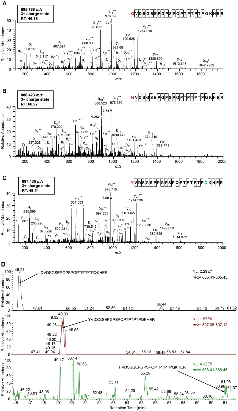
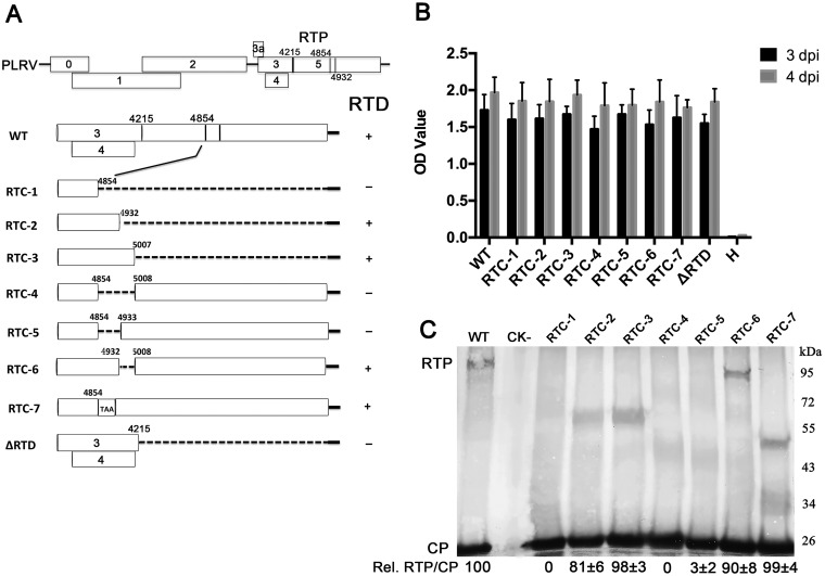
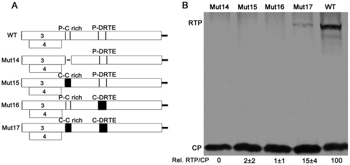
Translational readthrough of the stop codon of the capsid protein (CP) open reading frame (ORF) is used by members of the Luteoviridae to produce their minor capsid protein as a readthrough protein (RTP). The elements regulating RTP expression are not well understood, but they involve long-distance interactions between RNA domains. Using high-resolution mass spectrometry, glutamine and tyrosine were identified as the primary amino acids inserted at the stop codon of Potato leafroll virus (PLRV) CP ORF. We characterized the contributions of a cytidine-rich domain immediately downstream and a branched stem-loop structure 600 to 700 nucleotides downstream of the CP stop codon. Mutations predicted to disrupt and restore the base of the distal stem-loop structure prevented and restored stop codon readthrough. Motifs in the downstream readthrough element (DRTE) are predicted to base pair to a site within 27 nucleotides (nt) of the CP ORF stop codon. Consistent with a requirement for this base pairing, the DRTE of Cereal yellow dwarf virus was not compatible with the stop codon-proximal element of PLRV in facilitating readthrough. Moreover, deletion of the complementary tract of bases from the stop codon-proximal region or the DRTE of PLRV prevented readthrough. In contrast, the distance and sequence composition between the two domains was flexible. Mutants deficient in RTP translation moved long distances in plants, but fewer infection foci developed in systemically infected leaves. Selective 2'-hydroxyl acylation and primer extension (SHAPE) probing to determine the secondary structure of the mutant DRTEs revealed that the functional mutants were more likely to have bases accessible for long-distance base pairing than the nonfunctional mutants. This study reveals a heretofore unknown combination of RNA structure and sequence that reduces stop codon efficiency, allowing translation of a key viral protein.IMPORTANCE Programmed stop codon readthrough is used by many animal and plant viruses to produce key viral proteins. Moreover, such "leaky" stop codons are used in host mRNAs or can arise from mutations that cause genetic disease. Thus, it is important to understand the mechanism(s) of stop codon readthrough. Here, we shed light on the mechanism of readthrough of the stop codon of the coat protein ORFs of viruses in the Luteoviridae by identifying the amino acids inserted at the stop codon and RNA structures that facilitate this "leakiness" of the stop codon. Members of the Luteoviridae encode a C-terminal extension to the capsid protein known as the readthrough protein (RTP). We characterized two RNA domains in Potato leafroll virus (PLRV), located 600 to 700 nucleotides apart, that are essential for efficient RTP translation. We further determined that the PLRV readthrough process involves both local structures and long-range RNA-RNA interactions. Genetic manipulation of the RNA structure altered the ability of PLRV to translate RTP and systemically infect the plant. This demonstrates that plant virus RNA contains multiple layers of information beyond the primary sequence and extends our understanding of stop codon readthrough. Strategic targets that can be exploited to disrupt the virus life cycle and reduce its ability to move within and between plant hosts were revealed.

Keywords: RNA structure; polerovirus; readthrough; systemic infection; translational control.


Figures

FIG 1
FIG 1
High-resolution mass spectrometry identifies the amino acid residue incorporated at the CP amber stop codon position during readthrough. (A to C) Tandem mass spectra (MS2) of tryptic peptides spanning residues 209 to 233 in the PLRV RTP identified by affinity purification-MS analysis: K.Q209VDSGSEPGPSPQPTPTPTPQKHER.F (m/z 885.768) (A), K.H209VDSGSEPGPSPQPTPTPTPQKHER.F (m/z 888.423) (B), and K.Y209VDSGSEPGPSPQPTPTPTPQKHER.F (m/z 897.435) (C). Sequences show fragmentation along the peptide backbone and indicate the identity of residue 209 (red) and other ions that were used in peptide identification. For simplicity, only a representative amount of fragment ions in spectra are labeled. The ion peak highlighted in red indicates an immonium ion corresponding to the identity of a tyrosine residue at position 209. The residue highlighted in green indicates deamidation. RT, peptide retention time; ++, doubly charged ion; o, loss of H2O; *, loss of NH3; ?, contaminating fragment ions most likely from a coeluting peptide. (D) Relative abundances of the K.X209VDSGSEPGPSPQPTPTPTPQKHER.F peptide isoforms identified in the same PLRV affinity purification from locally infected N. benthamiana. Extracted MS1 chromatograms for precursor ions with m/z values of 885.41 to 885.45 (top), 897.08 to 897.12 (middle), and 888.41 to 888.45 (low) detected between 45.80 and 61.80 min (retention time) are shown. Arrows indicate the MS1 peak corresponding to peptide K.Q209VDSGSEPGPSPQPTPTPTPQKHER.F (retention time, 46.27 min) (A), K.Y209VDSGSEPGPSPQPTPTPTPQKHER.F (retention time, 49.39 min) (B), and K.H209VDSGSEPGPSPQPTPTPTPQKHER.F (retention time, 60.96 min) (C). The area under each peak is equal to the relative abundance of each peptide ion, with 100 on the y axis equaling the normalization (NL) value given in each panel.
FIG 2
FIG 2
Identification of the PLRV DRTE responsible for RTP translation. (A) Schematic representation of the wild-type PLRV subgenomic RNA1 ORFs and the deletion or insertion mutations that were used to define the DRTE regulating readthrough. Detection of RTP translation based on the Western blots represented in panel C is indicated to the right of the schematic. (B) Accumulation of PLRV antigen, measured by DAS-ELISA, in N. benthamiana leaves 3 and 4 days after agroinfiltration with wild-type virus (WT) or the RTD deletion mutants. Healthy controls were agroinfiltrated with bacteria that do not contain the PLRV genome insert. H, noninfiltrated plant leaves. (C) Western blot analysis of PLRV proteins in N. benthamiana tissue 3 to 5 days following agroinoculation with wild-type PLRV or the RTP mutants. Relative readthrough (Rel. RTP/CP) was calculated as the RTP/CP ratio, with that for wild-type PLRV set as 100%. Values represent the means (± standard error) determined from three independent experiments.
FIG 3
FIG 3
Stem-loop RNA structure of the distal element is required for RTP translation. (A) Predicted RNA secondary structure of the PLRV distal element and the mutants constructed for detecting RTP translation. (B) Western blot analysis of the role of stem-loop 1 in mediating RTP translation. (C) Western blot analysis of role of stem loops 3 and 4 in mediating RTP translation and the functional effects of the size and position of stem loop 1. Relative readthrough (Rel. RTP/CP) was calculated as the RTP/CP ratio, with that for wild-type PLRV set as 100%. Values represent the means (± standard error) determined from three independent experiments.
FIG 4
FIG 4
Time course of PLRV antigen accumulation in hairy nightshade plants systemically infected with wild-type PLRV and the various stem-loop mutants. Antigen was measured by DAS-ELISA in three randomly selected developing leaves from each of 40 plants. Data are displayed as average absorbance values (A405 minus A490). Detection of RTP translation based on the Western blot data (Fig. 2) is indicated to the right of the graph. Only plants with an absorbance value above background at 3 weeks after agroinoculation were considered to be successfully agroinfected and were included in the calculations.
FIG 5
FIG 5
Secondary structures of the distal translation elements from wild-type virus (WT) and from stem-loop mutants Mut1, Mut3, and Mut5. The blot shows RTP translation by Mut1R, Mut3R, and Mut5R in agroinfiltrated N. benthamiana tissues at 3 dpi. Relative readthrough (Rel. RTP/CP) was calculated as the RTP/CP ratio, with that for wild-type PLRV set as 100%. Values represent the means (± standard error) determined from three independent experiments. Secondary structures of wild-type, mutant, and second-site mutant (“revertant”) DRTEs that were recovered from plants that were symptomatic at 7 to 8 wpi are also shown. Each RNA was subjected to SHAPE probing, from which structure was predicted using SAFA and MFOLD software (see Materials and Methods). The degree of chemical modification (“single-strandedness”) of each base is indicated by color-coded circles, with red indicating the most modified and blue the least. Uncolored bases showed no modification. Mutations are boxed, with constructed mutations in red and revertants in gray. Magenta boxes on wild-type RNA indicate bases predicted to base pair to the C-rich region adjacent to the leaky stop codon (Fig. 7).
FIG 6
FIG 6
Communication between the DRTE and the C-rich domain is virus specific and indispensable for RTP translation. (A) Schematic of the chimeric ORF5 constructs, including a deletion of the PLRV C-rich domain (Mut14), a substitution with the homologous domain from CYDV-RPV (Mut15), a substitution of the PLRV DRTE with the homologous domain from CYDV-RPV (Mut16), and substitution of both PLRV domains with the homologous domains from CYDV-RPV (Mut17). (B) Western blot analysis of proteins in agroinfiltrated N. benthamiana tissue. Relative readthrough (Rel. RTP/CP) was calculated as the RTP/CP ratio, with that for wild-type PLRV set as 100%. Values represent the means (± standard error) determined from three independent experiments.
FIG 7
FIG 7
Long-range base pairing between the DRTE and 5′ end of C-rich domain. (A) Alignment of potentially complementary regions of the C-rich motif adjacent to the leaky stop codon (lightface) and the portion of the DRTE adjacent to the conserved EXG[X]6DE motif. The bases coding for DE (GAYGAR) are in lightface, with exceptions in lowercase. Complementary bases are in underlined magenta, base positions are indicated at ends, and the gap length between the two regions is in parentheses. Regions in the PLRV sequence highlighted in yellow indicate the bases deleted in the Δ4222-4236 and Δ4897-4911 mutants. Abbreviations not defined elsewhere: TobV2, Tobacco virus 2; CRLV, Carrot red leaf virus; TVDV, Tobacco vein distortion virus; BMYV, Beet mild yellowing virus; TuYV, Turnip yellows virus; BCV, Beet chlorosis virus; BrYV, Brassica yellows virus; WYDV, Wheat yellow dwarf virus; MABYV, Melon aphid-borne yellows virus; CLRDV, Cotton leafroll dwarf virus; CABYV, Cucurbit aphid-borne yellows virus; PeVYV, Pepper vein yellows virus; CpCSV, Chickpea chlorotic stunt virus; LABYV, Luffa aphid-borne yellows virus; PABYV, Pepo aphid-borne yellows virus; SCYLV, Sugarcane yellow leaf virus; MYDV-RMV, Maize yellow dwarf virus-RMV; WLYaV, Wheat leaf yellowing-associated virus; WCMV, White clover mottle virus; CpPV, Chickpea polerovirus; SPV1, Strawberry polerovirus 1; IxYMV1, Ixeridium yellow mottle virus 1; AEYV, African eggplant yellows virus; SYV, Sauropus yellowing virus; BLRV, Bean leafroll virus; SbDV, Soybean dwarf virus; ChaLV, Cherry-associated luteovirus; PaLV, Peach-associated luteovirus; AaLV, Apple-associated luteovirus; NSPaV, Nectarine stem-pitting-associated virus; RSDaV, Rose spring dwarf-associated virus; PEMV1, Pea enation mosaic virus 1; AEV1, Alfalfa enamovirus 1; CVEV, Citrus vein enation virus 1; GEV1, Grapevine enamovirus 1. (B) Diagram of the wild-type PLRV and mutants used for testing the effect of the complementary long-range base-pairing. Mut4222–4236-AA, replacement of nine out of 15 nucleotides in the 4222–4236 domain and no changes of original amino acids. Mut4222–4236-AA-C, complementary mutant of Mut4222–4236-AA in the DRTE; Mut4233–4236-AGGA: replacement of GCCU by AGGA in the domain of 4233–4236. Mut4233–4236-AGGA-C, complementary mutant of Mut4233–4236-AGGA. Arrows indicated the amino acid changes. (C) Western blot analysis of proteins in agroinfiltrated N. benthamiana tissue. Δ4222–4236, deletion of nt 4222 to 4236 in PLRV C-rich domain; Δ4897–4911, deletion of nt 4897 to 4911 in the DRTE; Δ4222–4233, deletion of nt 4222 to 4233. All the deletion mutations also did not change the ORF. Relative readthrough (Rel. RTP/CP) was calculated as the RTP/CP ratio, with that for WT PLRV set as 100%. Values represent the means (± standard error) determined from three independent experiments.
FIG 8
FIG 8
(A and C) Tissue prints showing PLRV infection foci in HNS stem and leaf tissues systemically infected with Mut1, PLRV-ΔRTD (which does not translate RTP), and Mut7 (which translates RTP at near-wild-type levels). Tissue was collected at 3 wpi (A) and 9 wpi (C). By 9 wpi Mut1 has accumulated compensatory mutations that restored RTP translation. (B and D) Actual counts of infection foci. Tissue prints were developed with antibodies to PLRV, and virus was visualized as blue-stained foci of indoxyl precipitate. The prints were photographed under a microscope at a magnification of ×10 to ×35. Graphs show the average of 15 infection foci and represent the number of virus infection foci in the immunoprint cross sections photographed under a microscope. **, significant differences in the number of stained foci (P < 0.01 by one-way analysis of variance [ANOVA]); n.s., no significant difference. (A) Bars in panels a, c, d, f, g, i, j, and l represent 1 mm, and those in panels b, e, h, and k represent 100 μm. (C) Bars in panels a, c, e, and g represent 1 mm, and those in panels b, d, f, and h represent 100 μm.

Similar articles

Cited by

References

    1. Beier H, Grimm M. 2001. Misreading of termination codons in eukaryotes by natural nonsense suppressor tRNAs. Nucleic Acids Res 29:4767–4782. doi:10.1093/nar/29.23.4767. - DOI - PMC - PubMed
    1. Dreher TW, Miller WA. 2006. Translational control in positive strand RNA plant viruses. Virology 344:185–197. doi:10.1016/j.virol.2005.09.031. - DOI - PMC - PubMed
    1. Firth AE, Brierley I. 2012. Non-canonical translation in RNA viruses. J Gen Virol 93:1385–1409. doi:10.1099/vir.0.042499-0. - DOI - PMC - PubMed
    1. Newburn LR, White KA. 2015. Cis-acting RNA elements in positive-strand RNA plant virus genomes. Virology 479–480:434–443. - PubMed
    1. Jungreis I, Lin MF, Spokony R, Chan CS, Negre N, Victorsen A, White KP, Kellis M. 2011. Evidence of abundant stop codon readthrough in Drosophila and other metazoa. Genome Res 21:2096–2113. doi:10.1101/gr.119974.110. - DOI - PMC - PubMed

Publication types

MeSH terms

Supplementary concepts