. 2018 May 14;92(11):e02103-17.
doi: 10.1128/JVI.02103-17. Print 2018 Jun 1.

Adenovirus Vector Vaccination Impacts NK Cell Rheostat Function following Lymphocytic Choriomeningitis Virus Infection

Affiliations

Adenovirus Vector Vaccination Impacts NK Cell Rheostat Function following Lymphocytic Choriomeningitis Virus Infection

Eryn Blass et al. J Virol. .

Abstract

Natural killer (NK) cells respond rapidly as a first line of defense against infectious pathogens. In addition, NK cells may provide a "rheostat" function and have been shown to reduce the magnitude of antigen-specific T cell responses following infection to avoid immunopathology. However, it remains unknown whether NK cells similarly modulate vaccine-elicited T cell responses following virus challenge. We used the lymphocytic choriomeningitis virus (LCMV) clone 13 infection model to address whether NK cells regulate T cell responses in adenovirus vector-vaccinated mice following challenge. As expected, NK cell depletion in unvaccinated mice resulted in increased virus-specific CD4+ and CD8+ T cell responses and immunopathology following LCMV challenge. In contrast, NK cell depletion had minimal to no impact on antigen-specific T cell responses in mice that were vaccinated with an adenovirus serotype 5 (Ad5)-GP vector prior to LCMV challenge. Moreover, NK cell depletion in vaccinated mice prior to challenge did not result in immunopathology and did not compromise protective efficacy. These data suggest that adenovirus vaccine-elicited T cells may be less sensitive to NK cell rheostat regulation than T cells primed by LCMV infection.IMPORTANCE Recent data have shown that NK cell depletion leads to enhanced virus-elicited T cell responses that can result in severe immunopathology following LCMV infection in mice. In this study, we observed that NK cells exerted minimal to no impact on vaccine-elicited T cells following LCMV challenge, suggesting that adenovirus vaccine-elicited T cells may be less subject to NK cell regulation. These data contribute to our understanding of NK cell regulatory functions and T cell-based vaccines.

Keywords: NK cells; adenoviruses; lymphocytic choriomeningitis virus; vaccines.


Figures

FIG 1
FIG 1
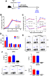
NK cell depletion has a minimal impact on CD4+ and CD8+ T cell responses elicited by Ad5-GP. Naive C57BL/6 mice received 500 μg of anti-NK1.1 or isotype control antibody prior to immunization with Ad5-GP. (A) Schematic of the experimental setup. (B) GP33-specific CD8+ T cell responses measured via Db/AL11 tetramer binding assays. (C) Phenotypic differentiation of GP33-specific CD8+ T cells. (D) PD-1 (mean fluorescence intensity [MFI]) expression on GP33 tetramer-positive CD8+ T cells. (E) Intracellular cytokine staining of GP33- and GP276-specific CD8+ T cells. (F) Intracellular cytokine staining of GP61-specific CD4+ T cells. Error bars represent standard errors of the means for 5 mice per group with 1 sham-vaccinated control. Statistically significant values are indicated (**, P < 0.01 by a Mann-Whitney U test).
FIG 2
FIG 2
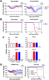
NK cell modulation of T cell responses in vaccinated and unvaccinated mice following LCMV Cl-13 challenge. (A) Schematic outlining the experimental setup. (B) Vaccinated and unvaccinated mice were challenged with LCMV Cl-13. At day 3 following infection, animals were sacrificed, and NK cell responses in blood, spleen, and lymph node were evaluated as the percentage of activated NK cells, as marked by the upregulation of CD69, NKG2D expression on NK cells, and 2B4 expression on NK cells. Error bars represent standard errors of the means for 5 five mice per group. Statistically significant values are indicated (bold line, P < 0.01 [determined by a Mann-Whitney U test]). (C and D) Vaccinated or unvaccinated C57BL/6 mice received anti-NK1.1 or isotype control antibody prior to infection with LCMV Cl-13. Animals were sacrificed at day 5 postinfection, and GP33 and GP276 (C) and NP396 (D) CD8+ T cell responses in the spleen were evaluated by intracellular cytokine staining. (E and F) GP61 CD4+ T cell responses in the spleen were evaluated by intracellular cytokine staining. Error bars represent standard errors of the means for 10 mice per group with 2 naive controls. Statistically significant values are indicated (**, P < 0.01; ***, P < 0.005; ****, P < 0.0001 [by a Mann-Whitney U test]).
FIG 3
FIG 3
Vaccination prevents immunopathology associated with NK cell depletion following LCMV Cl-13 challenge. Naive or Ad5-GP-vaccinated C57BL/6 mice received 500 μg anti-NK1.1 or isotype control antibody prior to infection with LCMV Cl-13. (A) Body weights were evaluated daily. (B) Survival of animals following LCMV Cl-13 challenge. (C) LCMV titers in serum as measured by a plaque assay. (D) Weight loss and survival in unvaccinated animals following depletion of NK cells, CD8+ T cells, or both. Error bars represent standard errors of the means for 5 to 15 mice per group with 1 to 3 naive controls. B represents baseline body weight. ND, not detectable. The dotted line represents weight loss euthanasia criteria. Statistically significant values (P < 0.005 by a Mantel-Cox test) are indicated by arrows.
FIG 4
FIG 4
Prior vaccination prevents NK cell-mediated augmentation of pathology. Vaccinated and unvaccinated mice were given anti-NK1.1 or isotype control antibody and then subsequently challenged with 2 × 10⁶ PFU of LCMV Cl-13. Animals were sacrificed at day 7 postchallenge for histopathology. (A) H&E staining of liver sections (magnification, ×20). (B) Example of hepatic pathology in an unvaccinated, NK cell-depleted animal, with hepatic lipidosis and loss of nuclear detail consistent with hepatocyte degeneration (asterisk), vasculitis (arrow), and lymphocytic infiltrates (arrowhead). (C) Scoring of hepatic pathology. Error bars represent standard errors of the means for 2 to 3 mice per group with 1 naive control.
FIG 5
FIG 5
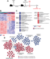
Transcriptomic analysis of CD8+ T cells from vaccinated and unvaccinated mice following LCMV Cl-13 challenge. (A) Schematic outlining the experimental setup. (B) Heat map at day 8 following challenge showing the log2 expression levels of each gene transformed to a Z-score, where the average expression level of each gene was subtracted and divided by its standard deviation across all samples, using a LIMMA F test (P < 0.05) accounting for all genes differentially expressed in at least one contrast. (C) Heat map representation of the GSEA-normalized enrichment scores of selected pathways with increased or decreased expression levels in vaccinated versus unvaccinated animals (nominal P value of <0.05 and FDR of <0.20). Pathways that show increased or decreased expression levels in vaccinated animals compared to unvaccinated animals following treatment with an isotype control antibody are indicated. IL-2, interleukin-2; TLR, Toll-like receptor; IFR7, interferon regulatory factor 7; TGF, transforming growth factor; TNF, tumor necrosis factor; IGF1, insulin-like growth factor 1; PLK1, polo-like kinase 1; EGR, early growth response. (D) Gene interaction networks showing the leading genes with increased or decreased expression levels in vaccinated versus unvaccinated isotype control-treated animals. Networks were generated by using the Cytoscape DyNet plug-in, and interactions were inferred from GeneMania (four mice per group).
FIG 6
FIG 6
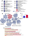
Transcriptomic analysis of CD8+ T cells from unvaccinated mice with or without NK cell depletion following LCMV Cl-13 challenge. (A) Heat map representation of the GSEA-normalized enrichment scores of selected pathways with increased or decreased expression levels in NK cell-depleted animals compared to isotype control-treated animals at day 8 postchallenge (four mice per group). PI3K, phosphatidylinositol 3-kinase; CTLA-4, cytotoxic-T lymphocyte-associated antigen 4. (B) Gene interaction networks showing the leading genes with increased and decreased expression levels in unvaccinated animals following NK cell depletion. Networks were generated by using the Cytoscape DyNet plug-in, and interactions were inferred from GeneMania. (C) Expression of PD-1 on GP33-specfic CD8+ T cells in unvaccinated animals (n = 8 per group). (D) Scaled representation showing the log2 fold changes in gene expression levels for modules associated with T cell and immune activation at day 8 post-LCMV challenge, comparing vaccinated and unvaccinated animals following NK cell depletion. All genes shown are significant, with a corrected P value cutoff of 0.05.

Similar articles

Cited by

References

    1. Jost S, Altfeld M. 2013. Control of human viral infections by natural killer cells. Annu Rev Immunol 31:163–194. doi: 10.1146/annurev-immunol-032712-100001. - DOI - PubMed
    1. Biron CA, Nguyen KB, Pien GC, Cousens LP, Salazar-Mather TP. 1999. Natural killer cells in antiviral defense: function and regulation by innate cytokines. Annu Rev Immunol 17:189–220. doi: 10.1146/annurev.immunol.17.1.189. - DOI - PubMed
    1. Waggoner SN, Cornberg M, Selin LK, Welsh RM. 2011. Natural killer cells act as rheostats modulating antiviral T cells. Nature 481:394–398. doi: 10.1038/nature10624. - DOI - PMC - PubMed
    1. Rydyznski C, Daniels KA, Karmele EP, Brooks TR, Mahl SE, Moran MT, Li C, Sutiwisesak R, Welsh RM, Waggoner SN. 2015. Generation of cellular immune memory and B-cell immunity is impaired by natural killer cells. Nat Commun 6:6375. doi: 10.1038/ncomms7375. - DOI - PMC - PubMed
    1. Cook KD, Whitmire JK. 2013. The depletion of NK cells prevents T cell exhaustion to efficiently control disseminating virus infection. J Immunol 190:641–649. doi: 10.4049/jimmunol.1202448. - DOI - PMC - PubMed

Publication types

MeSH terms