. 2018 Apr 2;215(4):1187-1203.
doi: 10.1084/jem.20172246. Epub 2018 Mar 7.

CDK2 inhibitors as candidate therapeutics for cisplatin- and noise-induced hearing loss

Affiliations

CDK2 inhibitors as candidate therapeutics for cisplatin- and noise-induced hearing loss

Tal Teitz et al. J Exp Med. .

Abstract

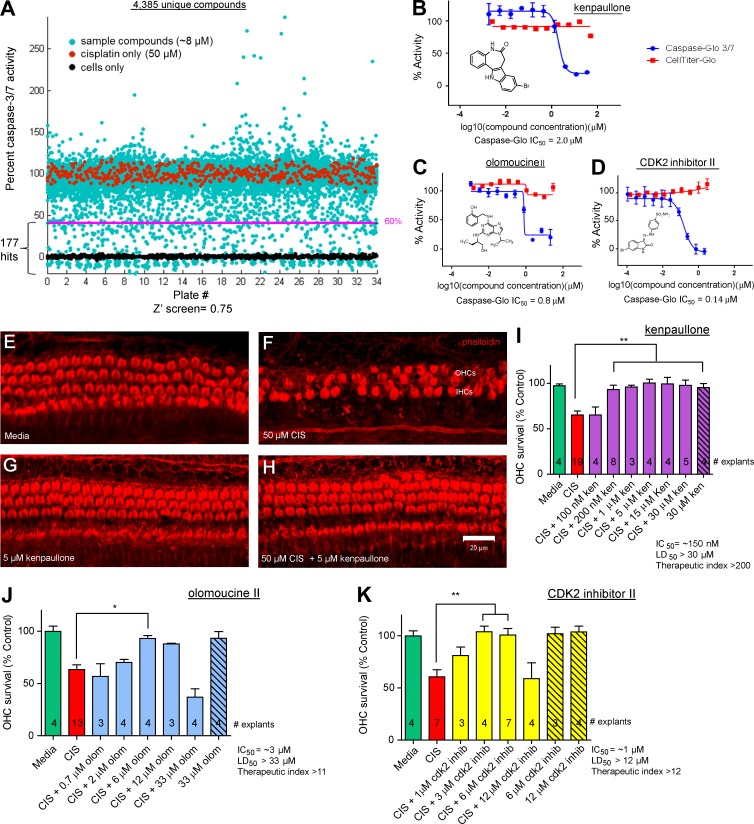
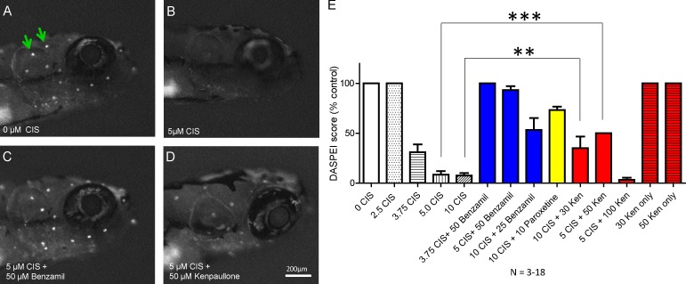
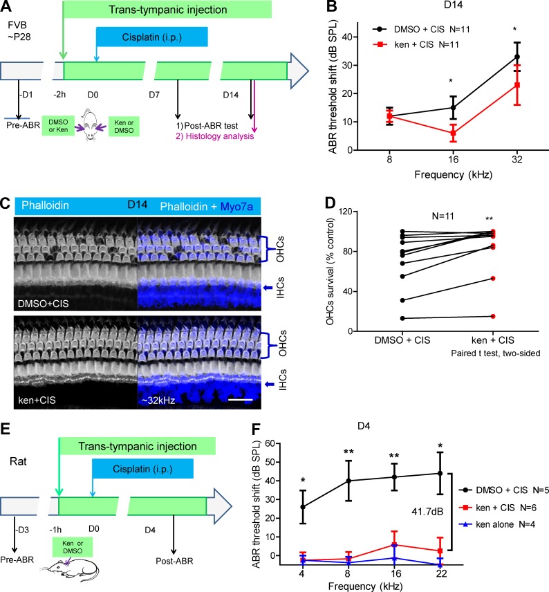
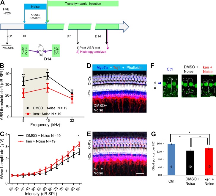
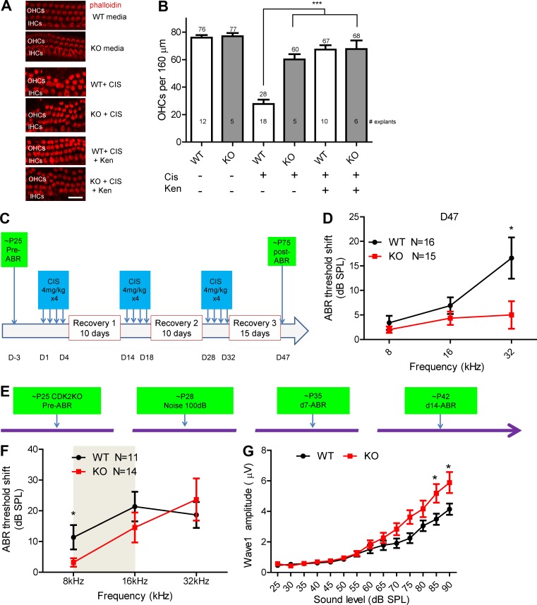
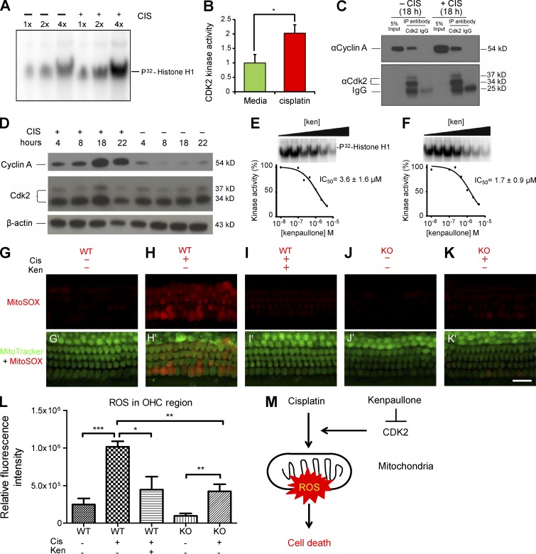
Hearing loss caused by aging, noise, cisplatin toxicity, or other insults affects 360 million people worldwide, but there are no Food and Drug Administration-approved drugs to prevent or treat it. We screened 4,385 small molecules in a cochlear cell line and identified 10 compounds that protected against cisplatin toxicity in mouse cochlear explants. Among them, kenpaullone, an inhibitor of multiple kinases, including cyclin-dependent kinase 2 (CDK2), protected zebrafish lateral-line neuromasts from cisplatin toxicity and, when delivered locally, protected adult mice and rats against cisplatin- and noise-induced hearing loss. CDK2-deficient mice displayed enhanced resistance to cisplatin toxicity in cochlear explants and to cisplatin- and noise-induced hearing loss in vivo. Mechanistically, we showed that kenpaullone directly inhibits CDK2 kinase activity and reduces cisplatin-induced mitochondrial production of reactive oxygen species, thereby enhancing cell survival. Our experiments have revealed the proapoptotic function of CDK2 in postmitotic cochlear cells and have identified promising therapeutics for preventing hearing loss.


Figures

Figure 1.
Figure 1.
Screening and identification of kenpaullone and CDK2 inhibitors that protect against cisplatin toxicity in HEI-OC1 cells. (A) Screening of a bioactive compound library of 4,385 unique compounds, including 845 FDA-approved drugs, in HEI-OC1 cells. Cells treated with 50 µM cisplatin (red dots) were assigned 100% caspase-3/7 activity. Cells not treated with cisplatin, i.e., those grown in medium only (black dots) were assigned 0% caspase-3/7 activity. Each compound was added to a final concentration of 8 µM in the presence of 50 µM cisplatin (cyan dots). The cell-based screen mean Z′ was 0.75, the signal window was 12, and the signal fold change was 4.9. In all, 177 compounds (those on the pink line or below it) were found to decrease cisplatin-induced caspase-3/7 activity by 60% or more and were further analyzed for dose response and toxicity. (B–D) Dose–response curves for kenpaullone (B), olomoucine II (C), and CDK2 inhibitor II (D), determined using the Caspase-Glo 3/7 assay (compound + 50 µM cisplatin, blue curves) and the CellTiter-Glo assay for cell viability (compound only, red curves) after incubation for 22 h in culture in HEI-OC1 cells. The caspase-3/7 IC50 activity and structure are indicated for each compound. Error bars: SEM. (E–K) Kenpaullone (ken; E–I), olomoucine II (olom; J), and CDK2 inhibitor II (K) protected against cisplatin-induced HC loss in mouse cochlear explants. Confocal images of whole-mount middle turn cochlear explants that were treated with medium alone (E), 50 µM cisplatin (F), 5 µM kenpaullone (G), or 50 µM cisplatin and 5 µM kenpaullone (H) for 24 h are shown. Phalloidin labels the HCs. (I–K) OHC survival (percentage; mean ± SEM) after treatment with various concentrations of compounds and/or cisplatin (CIS). The number of cochlear explants analyzed is indicated in each bar. *, P < 0.05 and **, P < 0.01 by one-way ANOVA followed by a Bonferroni comparison.
Figure 2.
Figure 2.
Kenpaullone protects against cisplatin-induced HC loss in zebrafish lateral lines in vivo. (A–D) Neuromasts (white dots; green arrows) in the zebrafish head were visualized under identical microscopic conditions by staining with 0.005% DASPEI vital dye after treatment for 20 h with medium alone (A), 5 µM cisplatin (B), 5 µM cisplatin and 50 µM benzamil (C), or 5 µM cisplatin and 50 µM kenpaullone. (E) The DASPEI intensity score (mean ± SEM) with various concentrations (micromolar) of kenpaullone (Ken), benzamil, paroxetine, and cisplatin (CIS). Benzamil and paroxetine protect against cisplatin-induced HC loss in zebrafish (Vlasits et al., 2012). The number of zebrafish tested for each condition ranged from three to 13. ** P, < 0.01 and *** P, < 0.001 by one-way ANOVA followed by a Bonferroni comparison.
Figure 3.
Figure 3.
Locally delivered kenpaullone (ken) protects against cisplatin (CIS)-induced HC loss and hearing loss in adult mice and rats in vivo. (A) Experimental design. One ear of each FVB WT mouse at P28 was trans-tympanically injected with compound (250 µM ken in 0.5% DMSO in a volume of 5 µl), and the other ear was injected with 0.5% DMSO only, in a random order. 2 h later, the mice were injected i.p. with CIS at 30 mg/kg body weight, which was expected to damage the OHCs in both ears equally. ABR thresholds were recorded for each ear before or at D7 or D14 after ken or DMSO treatment with CIS administration, and cochlear histology was examined at D14. All analysis was performed in a double-blinded manner. (B) ABR threshold shifts in ken-treated ears (red) versus DMSO-treated control ears (black) of 11 mice at D14. Results are presented as the mean ± SEM. P = 0.0221 at 16 kHz, and P = 0.0215 at 32 kHz. (C) Representative confocal images of cochleae at the 32-kHz region double stained with phalloidin with an antibody to the HC marker myosin 7a (Myo7a) at D14. Bar, 20 µM. (D) Comparison of the percentage of OHC survival in the 32-kHz region in ken- and DMSO-treated ears after CIS administration in all 11 mice in four cohorts. Each line links the two cochleae of one mouse. P = 0.0028 by the paired two-tailed Student’s t test. (E and F) The left ear of each Wistar rat (body weight 306–354 g) was trans-tympanically injected with compound (310 µM ken in 0.5% DMSO; red curve) or carrier (0.5% DMSO; black curve) in a volume of 30 µl. 1 h later, the rat was injected i.p. with CIS at 13 mg/kg body weight. ABR thresholds were recorded 1 d before and 4 d after CIS administration. Error bars: SEM. N, number of rats tested in each group (two cohorts). As controls, four rats were injected trans-tympanically with 310 µM ken only, without CIS treatment (blue curve). All analysis was performed in a double-blinded manner. Note that significant differences in protection (28.5, 41.7, 36.2, and 41.5 dB at 4, 8, 16, and 22 kHz, respectively) were detected between ken/CIS and DMSO/CIS rats, whereas no significant differences were detected between ken/CIS and ken alone rats at these four frequencies. *, P < 0.05 and **, P < 0.01 by the unpaired two-tailed Student’s t test.
Figure 4.
Figure 4.
Kenpaullone (ken) protects against noise-induced hearing loss in adult mice when delivered locally in vivo. (A) Experimental design. Adult FVB mice at P28 were exposed to noise (8–16 kHz at 100 dB for 2 h). Immediately afterward, 5 µl ken (250 µM ken in 0.5% DMSO) was delivered to one ear of each mouse via trans-tympanic injection, and DMSO (0.5% in PBS) was delivered to the other ear, in a random order. ABR thresholds were recorded for each ear before and at D7 and D14 after noise exposure. Cochlear histology was examined at 14 d after exposure. All analysis was double blinded. (B) ABR threshold shifts of 19 mice at D14 in DMSO-treated (black) and ken-treated (red) ears. The gray area indicates the noise octave bandwidth between 8 and 16 kHz. (C) Amplitudes of ABR wave 1 at 16 kHz were significantly higher in ken-injected ears than in DMSO-injected ears. *, P < 0.05 at the 90-dB stimulus level by paired two-tailed Student’s t test. (D and E) Confocal images of similar cochlear regions (approximately the 16-kHz regions) of ken- and DMSO-treated ears triple stained with phalloidin, Tuj1, and myosin 7a (Myo7a). HCs and spiral ganglion neuron fiber innervation were intact in the 16-kHz region. Bar, 20 µM. (F and G) Comparison of the number of Ctbp2 puncta per IHC around the 16-kHz region in ken (ken+noise)- and DMSO (DMSO+noise)-treated ears with 100-dB noise damage and in untreated control ears (Ctrl). Cochleae analyzed were from 10 mice randomly chosen from the 19 mice analyzed in B and C; in each cochlea, 12–18 IHCs from 16-kHz regions were analyzed. Representative confocal images are shown in F, and puncta quantification of IHCs is demonstrated in G. IHCs are outlined with dashed lines in F. Bar, 10 µm. * P, < 0.05 and **, P < 0.01 by paired two-tailed Student’s t test. Results are presented as the mean ± SEM.
Figure 5.
Figure 5.
Germline CDK2 KO cochlear explants exhibit resistance to cisplatin (CIS) treatment, and kenpaullone (ken) administration phenocopies CDK2 KO exhibit resistance to CIS ototoxicity. All analysis here was double blinded. (A) Middle turn organs of Corti showing actin staining (with phalloidin) in CDK2 WT and KO cochleae with no treatment, with 50 µM CIS treatment, and with 50 µM CIS and 5 µM ken treatment. Bar, 20 µm. (B) The numbers of actin-labeled OHCs per 160 µm of the middle turn cochleae were counted. The numbers of explants tested in each condition are indicated in the bars. ***, P < 0.001 by one-way ANOVA with Bonferroni’s multiple comparison test. (C) Experimental design: CDK2 KO and WT littermate mice were treated for 47 d with three rounds of treatment, each consisting of daily i.p. injections of 4 mg/kg cisplatin for 4 d followed by recovery for 10–15 d. (D) ABR threshold shifts at D47. *, P < 0.05 by unpaired two-tailed Student’s t test. Note that 11.6 dB was detected between WT and CDK2 KO mice at 32 kHz. (E) Experimental design: CDK2 KO mice at P28 were exposed to noise (8–16 kHz at 100 dB for 2 h), and ABR thresholds were recorded before and at D7 and D14 after noise exposure. Cochlear histology was examined at D14. (F) Comparison of ABR threshold shifts at D14 in control mice (black) and CDK2 KO mice. The gray area indicates the noise octave bandwidth between 8 and 16 kHz. (G) Comparison of ABR wave 1 amplitudes at 8 kHz at D14 in WT and CDK2 KO mice. *, P < 0.05 by unpaired two-tailed Student’s t test. Error bars represent SEM.
Figure 6.
Figure 6.
CDK2 kinase activity is up-regulated after cisplatin (CIS) treatment; kenpaullone (ken) inhibits CDK2 kinase activity in HEI-OC1 cells and reduces CIS-induced, mitochondrial ROS production in cochlear explants. (A) CDK2 kinase activity is up-regulated after CIS treatment in HEI-OC1 cells. Protein CDK2 was immunoprecipitated from HEI-OC1 cells treated with 50 µM CIS for 18 h with a CDK2-specific antibody (M2) and bound to Protein A agarose beads. The protein solution eluted from the beads was used in a kinase assay using [32P]histone H1 as the substrate, and different volumes (labeled 1×, 2×, and 4×) of the kinase reactions were loaded on an SDS-PAGE gel. (B) The CDK2 kinase activity reactions were performed in triplicate, and the intensities of the [32P]histone H1 bands for CIS-treated cells and untreated cells (green bar) were compared. (C) Cyclin A protein coimmunoprecipitated with CDK2 protein in HEI-OC1 cells treated and untreated with CIS. CDK2 was immunoprecipitated with a specific antibody (M2) from HEI-OC1 lysates treated or untreated with CIS for 18 h. Rabbit IgG was used as negative control. Equal amounts of immunoprecipitates and 5% of input were run on an SDS-PAGE gel; cyclin A and CDK2 were identified by Western blot analysis with specific antibodies. (D) Cyclin A is up-regulated in HEI-OC1 cells after CIS treatment. Cells were treated with 50 µM CIS or untreated and harvested at different time points. Cyclin A and CDK2 protein levels were compared by Western blot analysis with specific antibodies. β-Actin served as the loading control. (E and F) Ken inhibits CDK2–cyclin A kinase activity in HEI-OC1 cells untreated (E) and treated (F) with CIS. The dose response for ken inhibition of CDK2–cyclin A kinase was measured in the HEI-OC1 cells after 18 h in culture in medium as described in A, using [32P]histone H1 as the substrate. The IC50 was determined by curve fitting in triplicate experiments. The IC50 values are presented as the mean ± SD. (G–K′) Confocal images of the middle turns of organ of Corti explants labeled with MitoSOX red (G–K) or MitoTracker green and MitoSOX red (G'–K') under various conditions. CIS concentration was 150 µM. Bar, 20 µm. (L) Quantification of relative fluorescence intensities of MitoSOX ROS for each condition. CIS concentration was 150 µM. Data are presented as means ± SEM; *, P < 0.05; **, P < 0.01; and ***, P < 0.001. At least three different samples were analyzed for each condition. (M) Diagram of the mechanism by which ken and CDK2 KO protect against CIS-induced cell loss. CIS treatment induces mitochondrial ROS formation and thereby cell death. CDK2 KO or ken treatment decreases the ROS level induced by CIS treatment, further reducing cell loss.

References

    1. Altun Z.S., Güneş D., Aktaş S., Erbayraktar Z., and Olgun N.. 2010. Protective effects of acetyl-l-carnitine on cisplatin cytotoxicity and oxidative stress in neuroblastoma. Neurochem. Res. 35:437–443. 10.1007/s11064-009-0076-8 - DOI - PubMed
    1. Barbieri E., Mehta P., Chen Z., Zhang L., Slack A., Berg S., and Shohet J.M.. 2006. MDM2 inhibition sensitizes neuroblastoma to chemotherapy-induced apoptotic cell death. Mol. Cancer Ther. 5:2358–2365. 10.1158/1535-7163.MCT-06-0305 - DOI - PubMed
    1. Breglio A.M., Rusheen A.E., Shide E.D., Fernandez K.A., Spielbauer K.K., McLachlin K.M., Hall M.D., Amable L., and Cunningham L.L.. 2017. Cisplatin is retained in the cochlea indefinitely following chemotherapy. Nat. Commun. 8:1654 10.1038/s41467-017-01837-1 - DOI - PMC - PubMed
    1. Buck L.M., Winter M.J., Redfern W.S., and Whitfield T.T.. 2012. Ototoxin-induced cellular damage in neuromasts disrupts lateral line function in larval zebrafish. Hear. Res. 284:67–81. 10.1016/j.heares.2011.12.001 - DOI - PubMed
    1. Burgess A., Vigneron S., Brioudes E., Labbé J.C., Lorca T., and Castro A.. 2010. Loss of human Greatwall results in G2 arrest and multiple mitotic defects due to deregulation of the cyclin B-Cdc2/PP2A balance. Proc. Natl. Acad. Sci. USA. 107:12564–12569. 10.1073/pnas.0914191107 - DOI - PMC - PubMed

Publication types

MeSH terms