. 2018 May 1;29(9):1089-1099.
doi: 10.1091/mbc.E17-08-0516. Epub 2018 Mar 22.

A newly characterized vacuolar serine carboxypeptidase, Atg42/Ybr139w, is required for normal vacuole function and the terminal steps of autophagy in the yeast Saccharomyces cerevisiae

Affiliations

A newly characterized vacuolar serine carboxypeptidase, Atg42/Ybr139w, is required for normal vacuole function and the terminal steps of autophagy in the yeast Saccharomyces cerevisiae

Katherine R Parzych et al. Mol Biol Cell. .

Abstract

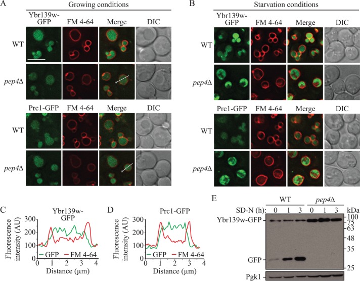
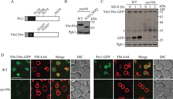
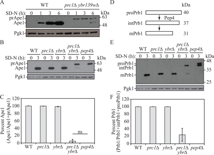
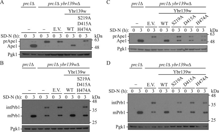
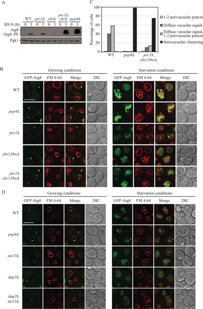
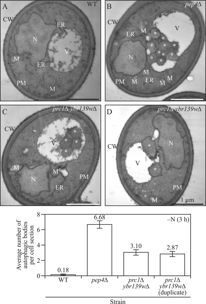
Macroautophagy (hereafter autophagy) is a cellular recycling pathway essential for cell survival during nutrient deprivation that culminates in the degradation of cargo within the vacuole in yeast and the lysosome in mammals, followed by efflux of the resultant macromolecules back into the cytosol. The yeast vacuole is home to many different hydrolytic proteins and while few have established roles in autophagy, the involvement of others remains unclear. The vacuolar serine carboxypeptidase Y (Prc1) has not been previously shown to have a role in vacuolar zymogen activation and has not been directly implicated in the terminal degradation steps of autophagy. Through a combination of molecular genetic, cell biological, and biochemical approaches, we have shown that Prc1 has a functional homologue, Ybr139w, and that cells deficient in both Prc1 and Ybr139w have defects in autophagy-dependent protein synthesis, vacuolar zymogen activation, and autophagic body breakdown. Thus, we have demonstrated that Ybr139w and Prc1 have important roles in proteolytic processing in the vacuole and the terminal steps of autophagy.


Figures

FIGURE 1:
FIGURE 1:
Ybr139w is a soluble vacuolar protein. The localization of Ybr139w-GFP and Prc1-GFP was examined in wild-type (KPY382 and KPY384) and pep4∆ (KPY383 and KPY385) cells in (A) growing and (B) starvation conditions. FM 4-64 was used to label the vacuole limiting membrane. DIC, differential interference contrast. Scale bar: 5 µm. (C, D) Line profile plot of fluorescence intensity along the line in the Ybr139w-GFP pep4∆ and Prc1-GFP pep4∆ strains from the “merge” panels in A; the circle indicates the line profile starting point. (E) GFP is cleaved from Ybr139w-GFP in a PEP4-dependent manner. Wild-type (KPY382) and pep4∆ (KPY383) cells expressing chromosomally tagged Ybr139w-GFP were grown to mid–log phase in YPD and then shifted to starvation conditions for the indicated times. Protein extracts were analyzed by Western blot using antibodies to YFP. Pgk1 is used as a loading control.
FIGURE 2:
FIGURE 2:
Ybr139w is a glycoprotein dependent on Vps10 for vacuolar delivery. (A) Schematic representation of Prc1 and Ybr139w. Gray box, signal peptide; black box, propeptide; numbers, glycosylated residues; *, predicted. (B) pep4∆ (TVY1) cells expressing wild-type (WT; pKP105) Ybr139w-PA (Ybr-PA) or Ybr139wN163,242Q-PA (N163,242Q; pKP110) on plasmids were grown to mid–log phase in SMD-uracil (URA), cells were harvested, and protein extracts were analyzed by Western blot using antibodies to protein A. (C) GFP is cleaved from Ybr139w-GFP in a VPS10-dependent manner. Wild-type (KPY382) and vps10∆ (KPY424) cells expressing chromosomally tagged Ybr139w-GFP were grown to mid–log phase in YPD and then shifted to starvation conditions for the indicated times. Protein extracts were analyzed by Western blot using antibodies to YFP. (D) The localization of Ybr139w-GFP and Prc1-GFP was examined in wild-type (KPY382 and KPY384) and vps10∆ (KPY424 and KPY426) cells in growing conditions. FM 4-64 was used to label the vacuole limiting membrane. DIC, differential interference contrast. Scale bar: 5 µm.
FIGURE 3:
FIGURE 3:
Vacuolar function is impaired in cells lacking PRC1 and YBR139W. (A) Wild-type (SEY6210) and prc1∆ ybr139w∆ (KPY325) cells were grown to mid–log phase in YPD and then shifted to starvation conditions for the indicated times. Protein extracts were analyzed by Western blot using antiserum to Ape1. The positions of precursor (pr) and mature Ape1 are indicated. (B, E) Wild-type (SEY6210), prc1∆ (KPY301), ybr139w∆ (KPY323), prc1∆ ybr139w∆ (KPY325), and pep4∆ (TVY1) cells were grown to mid–log phase in YPD and then shifted to starvation conditions for 3 h. Cells were harvested and protein extracts were analyzed by Western blot using antiserum to Ape1 (B) or Prb1 (E). The positions of the precursor (pro), intermediate (int), and mature forms of Prb1 are indicated. (C) Quantification of results in B. Percentage of Ape1 was calculated as amount of Ape1/total Ape1 (Ape1 + prApe1). Average of three experiments. Error bars, SD; ns, not significant. (D) Schematic representation of Prb1 processing in the vacuole. See text for details. (F) Quantification of results in E. Average of three experiments. Percentage of Prb1 was calculated as amount of Prb1/total Prb1 (Prb1 + intPrb1 + proPrb1). Average of three experiments. Error bars, SD.
FIGURE 4:
FIGURE 4:
Ybr139w is a serine carboxypeptidase. (A, B) prc1∆ (KPY301), prc1∆ ybr139w∆ (KPY325), and prc1∆ ybr139w∆ cells with integrated empty vector (KPY332), Ybr139w (KPY336), or Ybr139wS219,D415,H474A (KPY418) were grown to mid–log phase in YPD and then shifted to starvation conditions for 3 h. Cells were harvested and protein extracts were analyzed by Western blot using antiserum to Ape1 (A) or Prb1 (B). (C, D) prc1∆ (KPY301) and prc1∆ ybr139w∆ cells with integrated empty vector (KPY332), or expressing Ybr139w (KPY336), Ybr139wS219A (KPY404), Ybr139wD415A (KPY416), or Ybr139wH474A (KPY406) were grown to mid–log phase in YPD and then shifted to starvation conditions for 3 h. Cells were harvested and protein extracts were analyzed by Western blot using antiserum to Ape1 (C) or Prb1 (D).
FIGURE 5:
FIGURE 5:
Cells lacking PRC1 and YBR139W are defective in the terminal steps of autophagy. (A) Wild-type (SEY6210), prc1∆ (KPY301), ybr139w∆ (KPY323), prc1∆ ybr139w∆ (KPY325), and pep4∆ (TVY1) cells were grown to mid–log phase in YPD and then shifted to starvation conditions for 3 h. Cells were harvested and protein extracts were analyzed by Western blot using antiserum to Atg8. (B) Wild-type (SEY6210), pep4∆ (TVY1), prc1∆ (KPY301), ybr139w∆ (KPY323), and prc1∆ ybr139w∆ (KPY325) cells expressing GFP-Atg8 from a plasmid were grown in SMD-TRP to mid–log phase. Cells were stained with FM 4-64 for 30 min to label the vacuole and chased in either SMD-TRP for 1 h (growing) or SD-N for 2 h (starvation) before imaging. DIC, differential interference contrast. Scale bar: 5 µm. (C) Quantification of results in B. Cells with GFP-Atg8-positive vacuoles were divided into four categories based on the appearance of the GFP signal as indicated. Wild-type, n = 311 cells; pep4∆, n = 481 cells; prc1∆ ybr139w∆, n = 391 cells. (D) Wild-type (SEY6210), pep4∆ (TVY1), ste13∆ (KPY428), dap2∆ (KPY442), and dap2∆ ste13∆ (KPY443) cells expressing GFP-Atg8 from a plasmid were grown in SMD-TRP to mid–log phase. Cells were stained with FM 4-64 for 30 min to label the vacuole and chased in either SMD-TRP for 1 h (growing) or SD-N for 2 h (starvation) before imaging. DIC, differential interference contrast. Scale bar: 5 µm.
FIGURE 6:
FIGURE 6:
The prc1∆ ybr139w∆ double-knockout strain accumulated autophagic bodies. (A) Wild-type (SEY6210), (B) pep4∆ (FRY143), and (C, D) prc1∆ ybr139w∆ (KPY440 and KPY441 [an independent isolate]) cells were grown in YPD and shifted to SD-N for 3 h. Cells were then prepared for TEM analysis as described under Materials and Methods. Counting was done on three independent grids per condition and 100 cells per condition. Scale bars: 1 µm. *, autophagic body; CW, cell wall; ER, endoplasmic reticulum; M, mitochondria; N, nucleus; PM, plasma membrane; V, vacuole.

References

    1. Abeliovich H, Zhang C, Dunn WA, Shokat KM, Klionsky DJ. (2003). Chemical genetic analysis of Apg1 reveals a non-kinase role in the induction of autophagy. Mol Biol Cell , 477–490. - PMC - PubMed
    1. Backues SK, Chen D, Ruan J, Xie Z, Klionsky DJ. (2014). Estimating the size and number of autophagic bodies by electron microscopy. Autophagy , 155–164. - PMC - PubMed
    1. Baxter SM, Rosenblum JS, Knutson S, Nelson MR, Montimurro JS, Di Gennaro JA, Speir JA, Burbaum JJ, Fetrow JS. (2004). Synergistic computational and experimental proteomics approaches for more accurate detection of active serine hydrolases in yeast. Mol Cell Proteomics , 209–225. - PubMed
    1. Bech LM, Breddam K. (1989). Inactivation of carboxypeptidase Y by mutational removal of the putative essential histidyl residue. Carlsberg Res Commun , 165–171. - PubMed
    1. Caesar R, Blomberg A. (2004). The stress-induced Tfs1p requires NatB-mediated acetylation to inhibit carboxypeptidase Y and to regulate the protein kinase A pathway. J Biol Chem , 38532–38543. - PubMed

Publication types

MeSH terms

Substances