. 2018 Mar;30(3):717-736.
doi: 10.1105/tpc.17.00632. Epub 2018 Mar 7.

RecQ Helicases Function in Development, DNA Repair, and Gene Targeting in Physcomitrella patens

Affiliations

RecQ Helicases Function in Development, DNA Repair, and Gene Targeting in Physcomitrella patens

Gertrud Wiedemann et al. Plant Cell. 2018 Mar.

Abstract

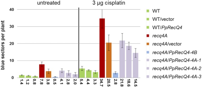
RecQ DNA helicases are genome surveillance proteins found in all kingdoms of life. They are characterized best in humans, as mutations in RecQ genes lead to developmental abnormalities and diseases. To better understand RecQ functions in plants we concentrated on Arabidopsis thaliana and Physcomitrella patens, the model species predominantly used for studies on DNA repair and gene targeting. Phylogenetic analysis of the six P. patens RecQ genes revealed their orthologs in humans and plants. Because Arabidopsis and P. patens differ in their RecQ4 and RecQ6 genes, reporter and deletion moss mutants were generated and gene functions studied in reciprocal cross-species and cross-kingdom approaches. Both proteins can be found in meristematic moss tissues, although at low levels and with distinct expression patterns. PpRecQ4 is involved in embryogenesis and in subsequent development as demonstrated by sterility of ΔPpRecQ4 mutants and by morphological aberrations. Additionally, ΔPpRecQ4 displays an increased sensitivity to DNA damages and an increased rate of gene targeting. Therefore, we conclude that PpRecQ4 acts as a repressor of recombination. In contrast, PpRecQ6 is not obviously important for moss development or DNA repair but does function as a potent enhancer of gene targeting.


Figures

Figure 1.
Figure 1.
Cladogram of the RecQ Family. Midpoint-rooted maximum-likelihood tree based on a multiple sequence alignment of RecQ CDS sequences identified in the Viridiplantae, H. sapiens, S. cerevisiae, and S. pombe. The cladogram was calculated with RAxML using the GTRGAMMA nucleotide substitution model and 1000 rapid bootstrap samples. Tip labels show the corresponding species abbreviation for each sequence. The six colored clades highlight the RecQ subfamilies.
Figure 2.
Figure 2.
Tissue-Specific Localization of PpRecQ4 and PpRecQ6 Visualized by GUS Staining of Reporter Lines. The GUS signal is detected in specific tissues throughout the life cycle by expression of the GUS-tagged endogenous PpRecQ4 ([A] to [I]) or PpRecQ6 ([J] to [R]) by fusion of the gene with the GUS coding sequence by insertion directly in front of the native stop codon. (A) and (J) Young gametophores: arrows point to the GUS signal at the apex. Bars = 50 µm. (B) and (K) Single gametophore prepared from a colony with GUS signal at the apex in gametangia highlighted by the box in (B) and arrow in (K). Bars = 1 mm. (C) and (L) Close-up (C) of apex highlighted by box in (B) (bar = 200 µm) and close-up (L) of the basal region of PpRecQ6:GUS with stained initials for side branches highlighted with the box in (K) (bar = 100 µm). (D) and (M) The developing antheridia ([D], left; [M], top) are completely stained, while in mature antheridia before release of sperm cells with the tip cell still intact ([D], right, [M], bottom), no GUS signal was detected. Bars = 25 µm. (E) and (N) Young archegonia with closed neck were completely stained. Bars = 25 µm. (F) and (O) Archegonia ready for fertilization with an opened neck showed accumulation of the GUS signal in the lower part; arrows point to the egg cell. Bars = 25 µm. (G) No GUS signal was detected in a developing PpRecQ4:GUS embryo and the surrounding tissue of the archegonium. Bar = 100 µm. (H) and (P) Gametophores carrying spore capsules. While the spore capsules of PpRecQ6:GUS did not show a signal, the foot of the sporophyte (highlighted by box) and the rhizoids showed GUS staining (box in [P]). Bars = 1 mm. (Q) Close-up of the PpRecQ6:GUS sporophyte foot. Bar = 50 µm. (I) and (R) Germinating spores after 5 d did not show any GUS staining. Bars = 50 µm.
Figure 3.
Figure 3.
Plant Growth Assay Using Different Concentrations of Cisplatin and MMS with Complementation Lines Containing PpRecQ4 Controlled by the 4A or 4B Promoter of Arabidopsis RecQ4 in recq4A Mutant Background. Fresh weight of 10 plantlets per line at each cisplatin or MMS concentration was measured and put into relation to the untreated plants of the same line. Each assay was performed at least five times, and the mean values including sd are shown. The color code of the lines is as follows: Three wild-type lines containing either no new construct, the empty vector, or the PpRecQ4 construct under the control of the AtRecQ4B promoter are in green, two fully complementing lines containing PpRecQ4 under control of the AtRecQ4B promoter are depicted in blue, and three only partially complementing lines containing PpRecQ4 under control of the AtRecQ4A promoter are shown in lilac. The original Atrecq4A mutant of Arabidopsis and the same mutant containing the empty vector are shown in red and orange, respectively.
Figure 4.
Figure 4.
Homologous Recombination Assay without or with Cisplatin Treatment in Complementation Lines Containing PpRecQ4 Controlled by the AtRecQ4A or AtRecQ4B Promoter in the recq4A Mutant Background. The capability of HR in Arabidopsis lines carrying different complementation constructs is shown. On the y axis the number of blue sectors is shown as a scale, and on the x axis the number of blue sectors is shown as average resulting from several experiments. The color code of the lines is as follows: Three wild-type lines containing either no new construct, the empty vector, or the PpRecQ4 construct under the control of the AtRecQ4B promoter are in green, two fully complementing lines containing PpRecQ4 under control of the AtRecQ4B promoter are depicted in blue, and three only partially complementing lines containing PpRecQ4 under control of the AtRecQ4A promoter are shown in lilac. The original Atrecq4A mutant of Arabidopsis and the same mutant containing the empty vector are shown in red and orange, respectively.
Figure 5.
Figure 5.
Morphology of Colonies, Gametophores, and Leaves Altered in ΔPpRecQ4 Compared with Wild-Type and AtRecQ4A_ΔPpRecQ4 Plants. (A) Colonies of wild type, ΔPpRecQ4-1RecQ4), ΔPpRecQ6-1RecQ6), and AtRecQ4A_ΔPpRecQ4-1#2 (AtRecQ4A_ΔRecQ4) were grown from protonema spot inocula on solid medium for 26 d. Bars = 2 mm. (B) Single gametophores of wild type, ΔPpRecQ4-1RecQ4), ΔPpRecQ6-1RecQ6), and AtRecQ4A_ΔPpRecQ4-1#1 (AtRecQ4A_ΔRecQ4) have been prepared from colonies grown from protonema spot inocula on solid medium for two months. Bars = 2 mm. (C) Single leaves prepared from gametophores grown on solid medium for 2 months of wild type (wild type), ΔPpRecQ6-1RecQ6), ΔPpRecQ4-1RecQ4), and AtRecQ4APpRecQ4-1#1 (AtRecQ4A_ΔRecQ4). Bars = 0.5 mm.
Figure 6.
Figure 6.
Bleomycin and Cisplatin Treatments Result in Dose-Dependent Growth Impairments. ΔPpRecQ4 plants are affected most severely while the expression of AtRecQ4A in ΔPpRecQ4 background leads to an intermediate phenotype compared with wild-type and ΔPpRecQ6 plants. Protonema of the wild type, ΔPpRecQ4-1RecQ4), ΔPpRecQ6-1RecQ6), and AtRecQ4APpRecQ4-1#2 (AtRecQ4ARecQ4) was subjected to treatment with 50 µg/mL (middle) or 200 µg/mL (bottom) bleomycin (1.5–2.0 units/mg) or incubated in Knop medium as a control (top) for 24 h (A) or 0.33 mM (middle), 0.66 mM (bottom) cisplatin, or Knop medium with 0.66% DMSO (corresponding amount of DMSO to the treatment with 0.66 mM cisplatin; top) for 48 h (B). After washing with minimal medium, spot inocula were transferred to solid medium to regenerate for 4 weeks. Bars = 1 mm.
Figure 7.
Figure 7.
Citrine-Based Reporter Construct to Assay Gene Targeting Rates. (A) Schematic representation of the genomic locus of carbonic anhydrase 2 (Pp1s43_118V6.1/ Pp3c1_19190C1.2) (top), the targeting construct (middle), and the genomic region upon successful integration of the construct via homologous integration into the moss genome (bottom). Dark gray boxes and lines represent exons and introns, respectively, for the gene model Pp3c1_19190C1.2, while light-gray boxes represent 5′- and 3′-UTRs, light-gray bars adjacent to intergenic genomic regions. The targeting construct consists of the citrine marker (yellow) including a nos terminator (orange), followed by a selection marker (nptII, hpt, BSD; light green) under the control of the 35S promoter and terminator (dark green) flanked at both sides by ∼1000-bp sequence homologous to the genomic sequence (black) for insertion via HR. The insertion of the transgene via HR takes place directly in front of the stop codon without deletion of any endogenous sequence information. If the construct is inserted via gene targeting into the genomic locus as depicted, the promoterless citrine is expressed under the control of the native promoter of the carbonic anhydrase 2. (B) Citrine fluorescence can be observed upon gene targeting of this construct in regenerating protoplasts 7 d after the transformation process (left panel, bar = 50 µm) as well as in protonema (middle; bar = 500 µm) and gametophores (right; bar = 500 µm) of plants surviving selection.
Figure 8.
Figure 8.
Rate of GT and Proportion of GT versus Untargeted Transgene Integration (IR) Is Higher in ΔPpRecQ4 Compared with the Wild Type but Reduced in ΔPpRecQ6 and Lines Expressing AtRecQ4A in ΔPpRecQ4 Background. Gene targeting in the different transgenic lines is assayed as percentage of colonies with citrine fluorescence from all regenerated colonies after selection using the test construct allowing selection on hygromycin (HR-hpt) for the wild type, ΔPpRecQ4RecQ4: ΔPpRecQ4-1 and ΔPpRecQ4-2, data pooled) and ΔPpRecQ6-2RecQ6) with three independent rounds of transformation for each line (A) or the version of the test construct allowing selection on BSDS (HR-BSD) for the wild type, ΔPpRecQ4-1RecQ4), and AtRecQ4APpRecQ4 (AtRecQ4ARecQ4: AtRecQ4A_ΔPpRecQ4-1#1 and #2, data pooled) with at least four independent rounds of transformations (C). The total number of surviving colonies with citrine fluorescence (light gray) in which the test construct was integrated via GT into the moss genome compared with colonies without fluorescence indicating IR (dark gray) for HR-hpt transformations into the wild type, ΔPpRecQ4RecQ4: ΔPpRecQ4-1 and ΔPpRecQ4-2, data pooled) and ΔPpRecQ6-2RecQ6) (B) or HR-BSD into the wild type, ΔPpRecQ4-1RecQ4), and AtRecQ4APpRecQ4 (AtRecQ4ARecQ4: AtRecQ4A_ΔPpRecQ4-1#1 and #2) (D).
None

References

    1. Altschul S.F., Gish W., Miller W., Myers E.W., Lipman D.J. (1990). Basic local alignment search tool. J. Mol. Biol. 215: 403–410. - PubMed
    1. Bagherieh-Najjar M.B., de Vries O.M.H., Kroon J.T.M., Wright E.L., Elborough K.M., Hille J., Dijkwel P.P. (2003). Arabidopsis RecQsim, a plant-specific member of the RecQ helicase family, can suppress the MMS hypersensitivity of the yeast sgs1 mutant. Plant Mol. Biol. 52: 273–284. - PubMed
    1. Bagherieh-Najjar M.B., de Vries O.M.H., Hille J., Dijkwel P.P. (2005). Arabidopsis RecQI4A suppresses homologous recombination and modulates DNA damage responses. Plant J. 43: 789–798. - PubMed
    1. Baumann P., West S.C. (1998). Role of the human RAD51 protein in homologous recombination and double-stranded-break repair. Trends Biochem. Sci. 23: 247–251. - PubMed
    1. Beike A.K., von Stackelberg M., Schallenberg-Rüdinger M., Hanke S.T., Follo M., Quandt D., McDaniel S.F., Reski R., Tan B.C., Rensing S.A. (2014). Molecular evidence for convergent evolution and allopolyploid speciation within the Physcomitrium-Physcomitrella species complex. BMC Evol. Biol. 14: 158. - PMC - PubMed

Publication types

LinkOut - more resources