

Progressive neurodegeneration following spinal cord injury: Implications for clinical trials
- PMID: 29514946
- PMCID: PMC5890610
- DOI: 10.1212/WNL.0000000000005258
Progressive neurodegeneration following spinal cord injury: Implications for clinical trials
Abstract
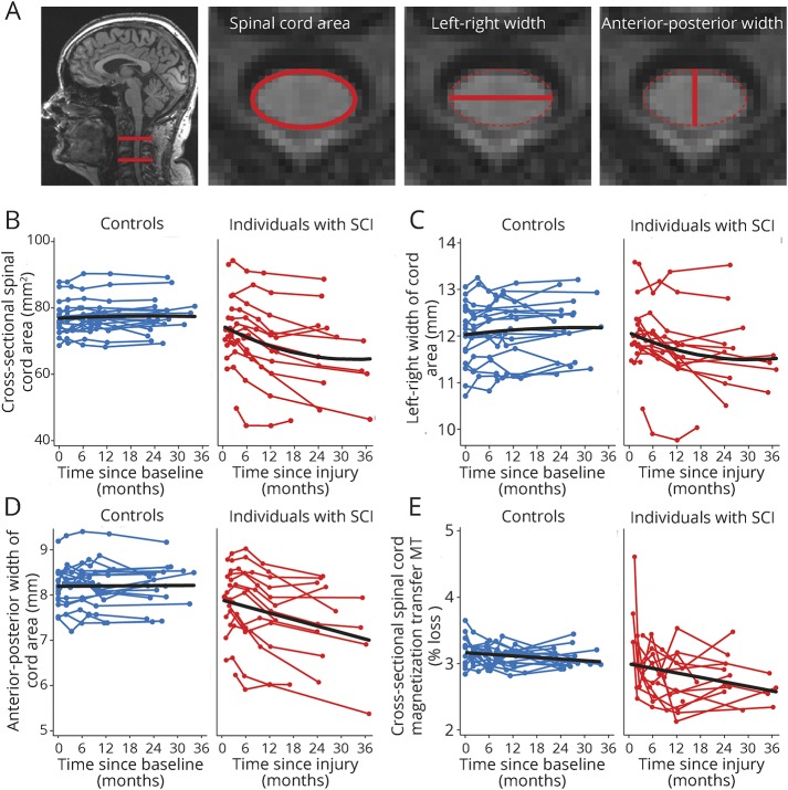
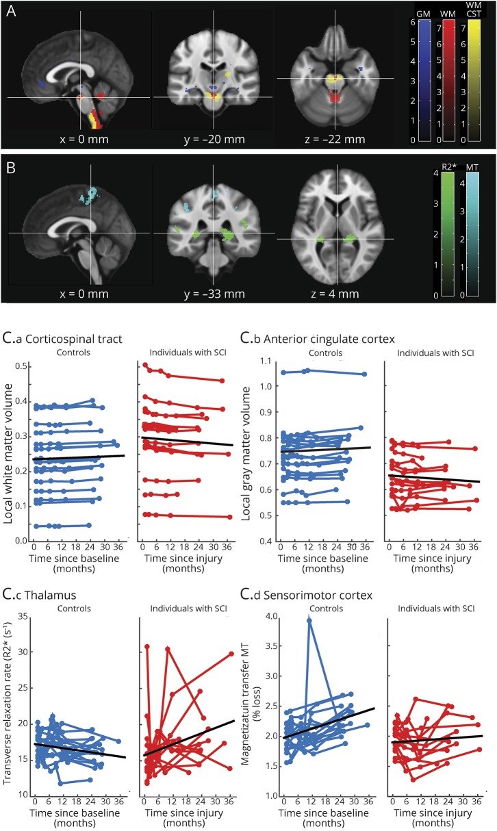
Objective: To quantify atrophy, demyelination, and iron accumulation over 2 years following acute spinal cord injury and to identify MRI predictors of clinical outcomes and determine their suitability as surrogate markers of therapeutic intervention.
Methods: We assessed 156 quantitative MRI datasets from 15 patients with spinal cord injury and 18 controls at baseline and 2, 6, 12, and 24 months after injury. Clinical recovery (including neuropathic pain) was assessed at each time point. Between-group differences in linear and nonlinear trajectories of volume, myelin, and iron change were estimated. Structural changes by 6 months were used to predict clinical outcomes at 2 years.
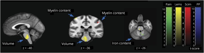
Results: The majority of patients showed clinical improvement with recovery stabilizing at 2 years. Cord atrophy decelerated, while cortical white and gray matter atrophy progressed over 2 years. Myelin content in the spinal cord and cortex decreased progressively over time, while cerebellar loss decreases decelerated. As atrophy progressed in the thalamus, sustained iron accumulation was evident. Smaller cord and cranial corticospinal tract atrophy, and myelin changes within the sensorimotor cortices, by 6 months predicted recovery in lower extremity motor score at 2 years. Whereas greater cord atrophy and microstructural changes in the cerebellum, anterior cingulate cortex, and secondary sensory cortex by 6 months predicted worse sensory impairment and greater neuropathic pain intensity at 2 years.
Conclusion: These results draw attention to trauma-induced neuroplastic processes and highlight the intimate relationships among neurodegenerative processes in the cord and brain. These measurable changes are sufficiently large, systematic, and predictive to render them viable outcome measures for clinical trials.
Copyright © 2018 The Author(s). Published by Wolters Kluwer Health, Inc. on behalf of the American Academy of Neurology.
Figures
Comment in
-
Reader response: Progressive neurodegeneration following spinal cord injury: Implications for clinical trials.Neurology. 2018 Nov 20;91(21):984-985. doi: 10.1212/WNL.0000000000006541. Neurology. 2018. PMID: 30455259 No abstract available.
-
Author response: Progressive neurodegeneration following spinal cord injury: Implications for clinical trials.Neurology. 2018 Nov 20;91(21):985. doi: 10.1212/WNL.0000000000006540. Neurology. 2018. PMID: 30455260 No abstract available.
References
-
- Fawcett JW, Curt A, Steeves JD, et al. . Guidelines for the conduct of clinical trials for spinal cord injury as developed by the ICCP panel: spontaneous recovery after spinal cord injury and statistical power needed for therapeutic clinical trials. Spinal Cord 2007;45:190–205. - PubMed
-
- Cadotte DW, Fehlings MG. Will imaging biomarkers transform spinal cord injury trials? Lancet Neurol 2013;12:843–844. - PubMed
Publication types
MeSH terms
Grants and funding
LinkOut - more resources
Full Text Sources
Other Literature Sources
Medical
Research Materials